

Subsets of CD1c+ DCs: Dendritic Cell Versus Monocyte Lineage
- PMID: 33101275
- PMCID: PMC7554627
- DOI: 10.3389/fimmu.2020.559166
Subsets of CD1c+ DCs: Dendritic Cell Versus Monocyte Lineage
Abstract
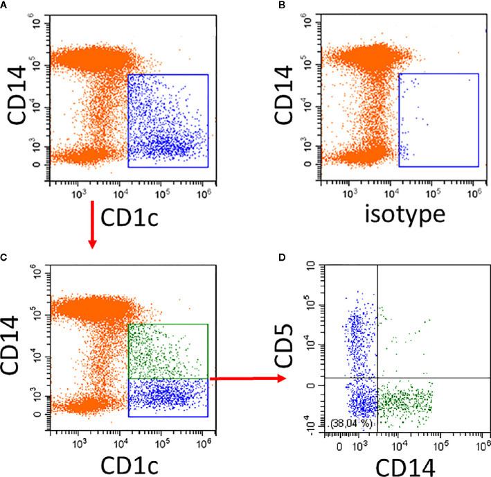
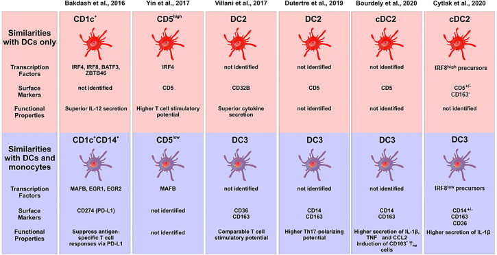
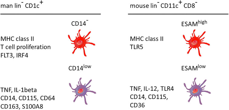
Currently three bona fide dendritic cell (DC) types are distinguished in human blood. Herein we focus on type 2 DCs (DC2s) and compare the three defining markers CD1c, CD172, and CD301. When using CD1c to define DC2s, a CD14+ and a CD14- subset can be detected. The CD14+ subset shares features with monocytes, and this includes substantially higher expression levels for CD64, CD115, CD163, and S100A8/9. We review the current knowledge of these CD1c+CD14+ cells as compared to the CD1c+CD14- cells with respect to phenotype, function, transcriptomics, and ontogeny. Here, we discuss informative mutations, which suggest that two populations have different developmental requirements. In addition, we cover subsets of CD11c+CD8- DC2s in the mouse, where CLEC12A+ESAMlow cells, as compared to the CLEC12A-ESAMhigh subset, also express higher levels of monocyte-associated markers CD14, CD3, and CD115. Finally, we summarize, for both man and mouse, the data on lower antigen presentation and higher cytokine production in the monocyte-marker expressing DC2 subset, which demonstrate that the DC2 subsets are also functionally distinct.
Keywords: CD14; CD172; CD1c; CD301; DC subsets; DC2; dendritic cells.
Copyright © 2020 Heger, Hofer, Bigley, de Vries, Dalod, Dudziak and Ziegler-Heitbrock.
Figures
Similar articles
-
Development of an Inflammatory CD14+ Dendritic Cell Subset in Humanized Mice.Front Immunol. 2021 Mar 15;12:643040. doi: 10.3389/fimmu.2021.643040. eCollection 2021. Front Immunol. 2021. PMID: 33790912 Free PMC article.
-
Transcriptional and Functional Analysis of CD1c+ Human Dendritic Cells Identifies a CD163+ Subset Priming CD8+CD103+ T Cells.Immunity. 2020 Aug 18;53(2):335-352.e8. doi: 10.1016/j.immuni.2020.06.002. Epub 2020 Jun 30. Immunity. 2020. PMID: 32610077 Free PMC article.
-
Non-small Cell Lung Cancer Cells Modulate the Development of Human CD1c+ Conventional Dendritic Cell Subsets Mediated by CD103 and CD205.Front Immunol. 2019 Dec 10;10:2829. doi: 10.3389/fimmu.2019.02829. eCollection 2019. Front Immunol. 2019. PMID: 31921114 Free PMC article.
-
Human dendritic cell subsets and function in health and disease.Cell Mol Life Sci. 2015 Nov;72(22):4309-25. doi: 10.1007/s00018-015-2005-0. Epub 2015 Aug 5. Cell Mol Life Sci. 2015. PMID: 26243730 Free PMC article. Review.
-
Human dendritic cell subsets.Immunology. 2013 Sep;140(1):22-30. doi: 10.1111/imm.12117. Immunology. 2013. PMID: 23621371 Free PMC article. Review.
Cited by
-
Unbiased high-dimensional flow cytometry identified NK and DC immune cell signature in Luminal A-type and triple negative breast cancer.Oncoimmunology. 2023 Dec 22;13(1):2296713. doi: 10.1080/2162402X.2023.2296713. eCollection 2024. Oncoimmunology. 2023. PMID: 38170155 Free PMC article.
-
Secretome profiling reveals acute changes in oxidative stress, brain homeostasis, and coagulation following short-duration spaceflight.Nat Commun. 2024 Jun 11;15(1):4862. doi: 10.1038/s41467-024-48841-w. Nat Commun. 2024. PMID: 38862464 Free PMC article.
-
A new era in melanoma immunotherapy: focus on DCs metabolic reprogramming.Cancer Cell Int. 2025 Apr 15;25(1):149. doi: 10.1186/s12935-025-03781-3. Cancer Cell Int. 2025. PMID: 40234886 Free PMC article. Review.
-
Maturation of Monocyte-Derived DCs Leads to Increased Cellular Stiffness, Higher Membrane Fluidity, and Changed Lipid Composition.Front Immunol. 2020 Nov 27;11:590121. doi: 10.3389/fimmu.2020.590121. eCollection 2020. Front Immunol. 2020. PMID: 33329576 Free PMC article.
-
Prostate cancer cancer-associated fibroblasts with stable markers post-androgen deprivation therapy associated with tumor progression and castration resistant prostate cancer.Cancer Sci. 2024 Sep;115(9):2893-2907. doi: 10.1111/cas.16267. Epub 2024 Jul 5. Cancer Sci. 2024. PMID: 38970292 Free PMC article.
References
-
- Heidkamp GF, Lehmann CHK, Heger L, Baranska A, Hoffmann A, Lühr J, et al. Functional Specialization of Dendritic Cell Subsets. Encyclopedia Cell Biol (2016) 3:588–604. 10.1016/B978-0-12-394447-4.30076-1 - DOI
Publication types
MeSH terms
Substances
Grants and funding
LinkOut - more resources
Full Text Sources
Other Literature Sources
Research Materials