. 2020 Sep 25:11:573040.
doi: 10.3389/fimmu.2020.573040. eCollection 2020.

4C3 Human Monoclonal Antibody: A Proof of Concept for Non-pathogenic Proteinase 3 Anti-neutrophil Cytoplasmic Antibodies in Granulomatosis With Polyangiitis

Affiliations

4C3 Human Monoclonal Antibody: A Proof of Concept for Non-pathogenic Proteinase 3 Anti-neutrophil Cytoplasmic Antibodies in Granulomatosis With Polyangiitis

Jérôme Granel et al. Front Immunol. .

Abstract

Granulomatosis with polyangiitis (GPA) is a severe autoimmune vasculitis associated with the presence of anti-neutrophil cytoplasmic antibodies (ANCA) mainly targeting proteinase 3 (PR3), a neutrophilic serine proteinase. PR3-ANCA binding to membrane-bound PR3 on neutrophils induce their auto-immune activation responsible for vascular lesions. However, the correlation between PR3-ANCA level and disease activity remains inconsistent, suggesting the existence of non-pathogenic PR3-ANCA. In order to prove their existence, we immortalized B lymphocytes from blood samples of GPA patients in remission having persistent PR3-ANCA to isolate non-activating PR3-ANCA. We obtained for the first time a non-activating human IgG1κ anti-PR3 monoclonal antibody (mAb) named 4C3. This new mAb binds soluble PR3 with a high affinity and membrane-bound PR3 on an epitope close to the PR3 hydrophobic patch and in the vicinity of the active site. 4C3 is able to bind FcγRIIA and FcγRIIIB and has a G2F glycosylation profile on asparagine 297. 4C3 did not induce activation of neutrophils and could inhibit human polyclonal PR3-ANCA-induced activation suggesting that 4C3 is non-pathogenic. This characteristic relies on the recognized epitope on PR3 rather than to the Fc portion properties. The existence of non-pathogenic PR3-ANCA, which do not activate neutrophils, could explain the persistence of high PR3-ANCA levels in some GPA patients in remission and why PR3-ANCA would not predict relapse. Finally, these results offer promising perspectives particularly regarding the understanding of PR3-ANCA pathogenicity and the development of new diagnostic and therapeutic strategies in GPA.

Keywords: anti-neutrophil cytoplasmic antibodies; epitope; granulomatosis with polyangiitis; human neutrophils; proteinase 3.


Figures

FIGURE 1
FIGURE 1
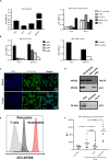
Identification and characterization of an anti-proteinase 3 (PR3) monoclonal human antibody 4C3. (A) Identification of a monoclonal human antibody specific to PR3. Binding of native PR3 by ELISA (left panel) in the presence of three antibodies (4C3, 4C5, and 5D11) obtained by immortalization of P2 B cells or serum from P2 containing PR3-ANCA used as a positive control (IgG P2). Similar results were obtained in four independent experiments. Specificity of 4C3 and 5D11 was checked by ELISA (right panel) in the presence of different antigens (PR3, OVA, Peanut, Alpha Gal). n = 5. (B) 4C3 is an IgG1κ. Subclass (left panel) and nature of light chain (right panel) of 4C3 were determined by ELISA compared to IgG P2. Similar results were obtained in three independent experiments. (C) 4C3 has a PR3-ANCA immunofluorescence pattern. Human neutrophils from healthy donors were purified and then fixed with ethanol (upper line) or formol (lower line) before staining with DAPI and 4C3-AF488 (1/100e). Objective x60. Superposition of fluorescence with ImageJ (Merged). Results from one of three experiments are presented. (D) 4C3 specifically recognizes intracellular PR3. Protein lysates (10 μg) from human neutrophils and HeLa cells were loaded before incubation with anti-Hsc70 or 4C3 antibodies for western blotting analysis. PR3 was used as a positive control, elastase and CatG (5 μg) as negative controls. Similar results were obtained in three independent experiments. (E) 4C3 has a membrane staining compatible with mbPR3 staining. Blood from a healthy donor was stained with a mixture containing CD45-APC H7/CD3-BV786/CD14-VioBlue/CD15-PE/4C3-AF488. Analysis of 4C3 staining at the surface of T cells (gated on CD3 positive cells, gray histogram), of monocytes (gated on CD14 positive cells, black histogram), and of neutrophils (gated on CD15 positive cells, red histogram). Similar results were obtained in five independent experiments. (F) 4C3 binds mbPR3 on quiescent and activated neutrophils. Human purified neutrophils from nine independent healthy donors were primed (TNFα) or not (w/o TNFα) with TNFα at 2 ng/ml for 15 min before staining with 4C3-AF488 (20 μg/ml). Alpha 1 anti-trypsin (α1AT) was used to solubilize mbPR3 by modifying its hydrophobic patch which is involved in its interaction with the membrane. PMA-ICa was used as a positive control of neutrophil activation. Each symbol represents one healthy donor. n = 9. NS: Non-significant; *p < 0.05 and **p < 0.005.
FIGURE 2
FIGURE 2
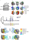
4C3 binds PR3 on a new epitope near the hydrophobic patch and close to the active site. (A) 4C3 preferentially recognizes a conformational epitope. Recognition of native (upper gel) or reduced PR3 (lower gel) used at different quantities in western blots after incubation of the membrane with 4C3. Ovalbumin was used as a negative control of binding. (B) Prediction of the epitope recognized by 4C3 on PR3. View of the top 30 ranked predicted conformations for the complex between 4C3 and PR3. The target is shown as a gray surface and the antibody in color (first line). The residues that probably belong to the epitope are shown on the structure of PR3 in four categories from purple for the highest probability to light blue for the lowest but still significant probability (second line). Four groups of validation peptides were designed and for each group, the targeted region of 15 residues is presented (third line). (C) 4C3 preferentially binds peptides 3.2 and 4.1 of PR3. Analysis by HTRF of the binding of 4C3 with PR3 predicted peptides. Results are expressed as means ± SEM of three independent experiments and each condition was tested in triplicates. Statistical differences are indicated on the graph (*p < 0.05; ***p < 0.0005). (D) 4C3 recognizes an epitope near the hydrophobic patch and close to the active site of PR3. The amino acids corresponding to the positive peptides (3.2 in orange and 4.1 in yellow) are indicated on the surface of PR3 (in gray). The catalytic triad (active site) of PR3 is shown in purple while the hydrophobic patch is represented in green. Two possible predicted conformations for the complex 4C3 (in blue) and PR3 (in gray) among the top 30 conformations are presented.
FIGURE 3
FIGURE 3
4C3 does not allow activation of human neutrophils and inhibits PR3-ANCA-induced activation. Purified neutrophils from eight independent healthy donors were primed with TNFα (2 ng/ml) for 15 min at 37°C (white columns) before incubation for 45 min with 4C3 (gray columns), separate (non-pooled) IgG preparations from two healthy donors (hatched columns) or from four active GPA patients at diagnosis (checkered columns) or IgG from patient P2 (vertical lines). (A) 4C3 does not induce production of reactive oxygen species (ROS) by neutrophils. ROS production was assessed by measuring the fluorescence (MFI) of DHR 123 by flow cytometry. (B) 4C3 does not induce degranulation. The degranulation of neutrophils was assessed by CD63 expression represented in percentage of positive cells. (C, D) 4C3 does not increase the adhesion phenotype of neutrophils. The adhesion criteria of neutrophils was assessed by measuring CD11b (C) and CD18 (D) surface expressions by flow cytometry. Results are expressed as mean ± SEM obtained in eight independent experiments with a circle representing one experiment. NS: Non-significant; *p < 0.05; **p < 0.005; and ***p < 0.0005. (E) 4C3 is able to inhibit ROS production induced by polyclonal PR3-ANCA. After priming, neutrophils from five independent healthy donors were first incubated for 45 min with 4C3 (gray column) or 6H4 (non-relevant mAb obtained under the same conditions as 4C3; black column) before addition of separate IgG preparations purified from five independent active GPA patients (IgG GPA at 200 μg/ml). Each symbols represents one healthy donor. ROS production was assessed by measuring the fluorescence (MFI) of DHR 123 by flow cytometry. The percentage of inhibition induced by 4C3 in ROS production is indicated on the graph. NS: Non-significant; *p < 0.05. n = 5.
FIGURE 4
FIGURE 4
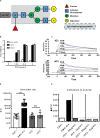
Non-activation of neutrophils by 4C3 is independent of a defect in the Fc portion. (A) 4C3 has a G2F profile of glycosylation with an increase in galactose residues. Glycosylation of 4C3 in asparagine 297, obtained by mass spectrometry, is presented compared to classical glycosylation of IgG (dotted line). (B) The recombinant form of 4C3 (r4C3) has the same ability to bind PR3 as native 4C3. Binding of 4C3 (black columns) and recombinant 4C3 (r4C3; gray columns) to PR3 by ELISA. BSA (0 μg of PR3) was used as a negative control of binding. Results are expressed in Optical Density. The results of one experiment out of three are presented. (C) r4C3 has an affinity for PR3 comparable to that of 4C3. Increasing concentrations of PR3 (1.25 to 20 nM) were injected on 4C3 and r4C3 coated flow cells for 500 min. Binding was monitored as an increase in SPR signal expressed in resonance units (RU). The results of one experiment out of three are presented. (D) r4C3 does not induce ROS production by neutrophils. Purified neutrophils from nine independent healthy donors were primed with TNFα (2 ng/ml) for 15 min at 37°C (white histogram) before incubation for 45 min with PMA-ICa (black column), 4C3 (dark gray column) or r4C3 (light gray column). ROS production was assessed by measuring the fluorescence (MFI) of DHR 123 by flow cytometry. n = 9. NS: Non-significant. ***p < 0.0005. (E) r4C3 does not increase cathepsin G activity. Purified neutrophils from three independent healthy donors were primed with TNFα (2 ng/ml; white histogram) for 15 min at 37°C before incubation for 45 min with 4C3 (dark gray columns) or r4C3 (light gray columns) at 2 and 20 μg/ml or PMA-ICa (black column). Cathepsin G activity was assessed after adding the substrate to the supernatants of neutrophils and reading the fluorescence (ΔRFU) by spectrofluorimetry. The results of one experiment out of three are presented.

References

    1. Jennette JC, Falk RJ, Bacon PA, Basu N, Cid MC, Ferrario F, et al. 2012 revised International Chapel Hill consensus conference nomenclature of vasculitides. Arthritis Rheum. (2013) 65:1–11. 10.1002/art.37715 - DOI - PubMed
    1. Lamprecht P, Kerstein A, Klapa S, Schinke S, Karsten CM, Yu X, et al. Pathogenetic and clinical aspects of anti-neutrophil cytoplasmic autoantibody-associated vasculitides. Front Immunol. (2018) 9:680. 10.3389/fimmu.2018.00680 - DOI - PMC - PubMed
    1. Flossmann O, Berden A, de Groot K, Hagen C, Harper L, Heijl C, et al. Long-term patient survival in ANCA-associated vasculitis. Ann Rheum Dis. (2011) 70:488–94. 10.1136/ard.2010.137778 - DOI - PubMed
    1. Korkmaz B, Lesner A, Letast S, Mahdi YK, Jourdan M-L, Dallet-Choisy S, et al. Neutrophil proteinase 3 and dipeptidyl peptidase I (cathepsin C) as pharmacological targets in granulomatosis with polyangiitis (Wegener granulomatosis). Semin Immunopathol. (2013) 35:411–21. 10.1007/s00281-013-0362-z - DOI - PubMed
    1. Damoiseaux J, Csernok E, Rasmussen N, Moosig F, van Paassen P, Baslund B, et al. Detection of antineutrophil cytoplasmic antibodies (ANCAs): a multicentre European Vasculitis Study Group (EUVAS) evaluation of the value of indirect immunofluorescence (IIF) versus antigen-specific immunoassays. Ann Rheum Dis. (2017) 76:647–53. 10.1136/annrheumdis-2016-209507 - DOI - PubMed

Publication types

MeSH terms