. 2020 Sep 25:11:585530.
doi: 10.3389/fimmu.2020.585530. eCollection 2020.

Maresin-1 and Resolvin E1 Promote Regenerative Properties of Periodontal Ligament Stem Cells Under Inflammatory Conditions

Affiliations

Maresin-1 and Resolvin E1 Promote Regenerative Properties of Periodontal Ligament Stem Cells Under Inflammatory Conditions

Emmanuel Albuquerque-Souza et al. Front Immunol. .

Abstract

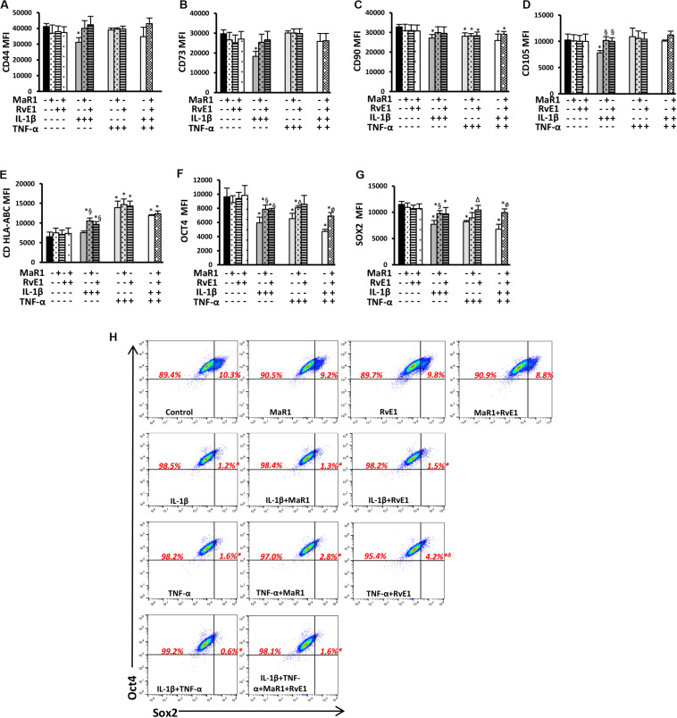
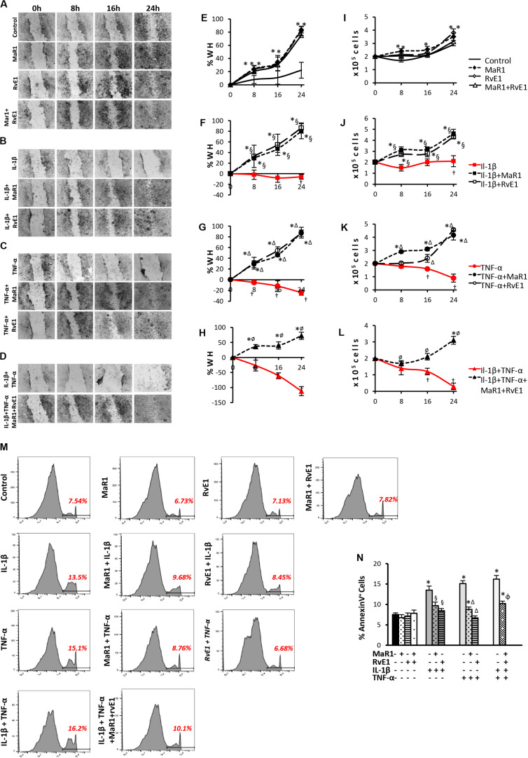
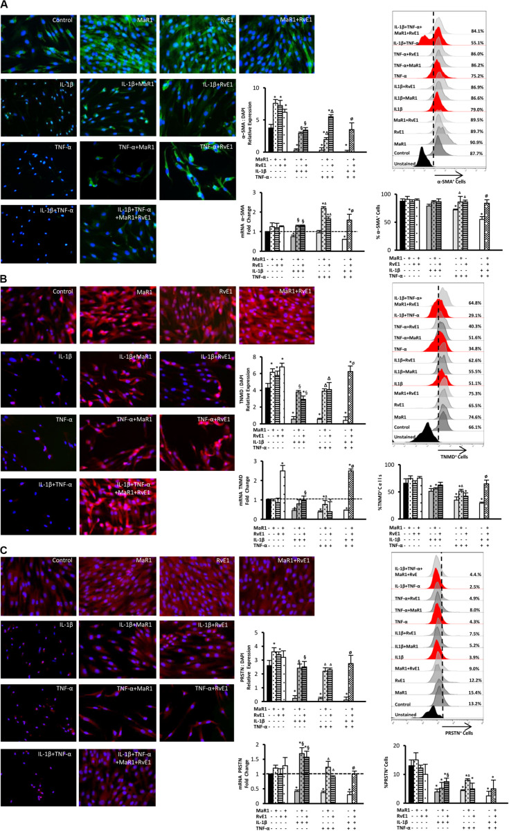
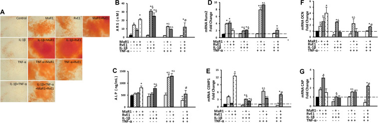
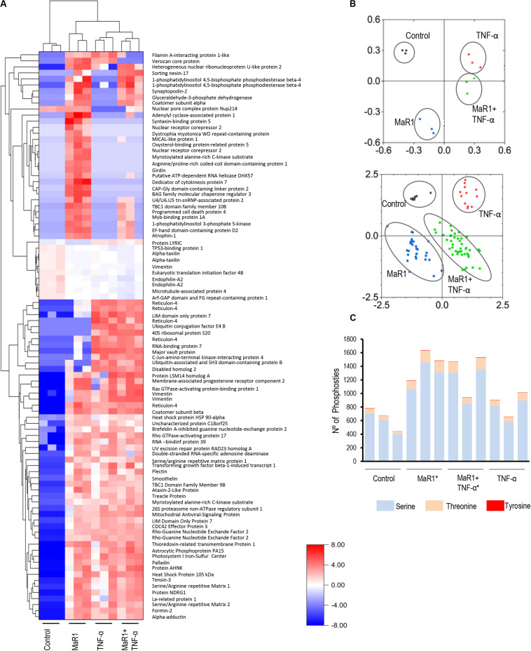
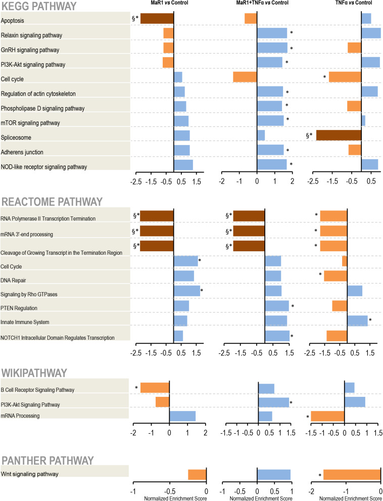
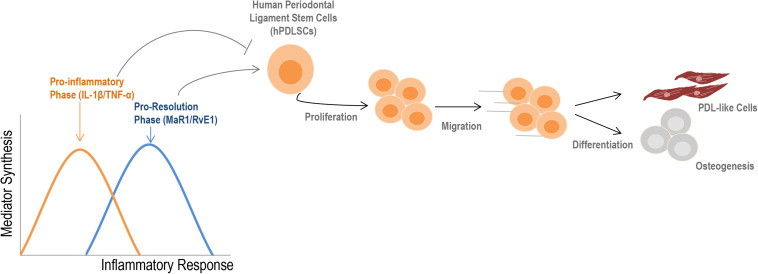
Maresin-1 (MaR1) and Resolvin E1 (RvE1) are specialized pro-resolving lipid mediators (SPMs) that regulate inflammatory processes. We have previously demonstrated the hard and soft tissue regenerative capacity of RvE1 in an in vivo model of the periodontal disease characterized by inflammatory tissue destruction. Regeneration of periodontal tissues requires a well-orchestrated process mediated by periodontal ligament stem cells. However, limited data are available on how SPMs can regulate the regenerative properties of human periodontal ligament stem cells (hPDLSCs) under inflammatory conditions. Thus, we measured the impact of MaR1 and RvE1 in an in vitro model of hPDLSC under stimulation with IL-1β and TNF-α by evaluating pluripotency, migration, viability/cell death, periodontal ligament markers (α-smooth muscle actin, tenomodulin, and periostin), cementogenic-osteogenic differentiation, and phosphoproteomic perturbations. The data showed that the pro-inflammatory milieu suppresses pluripotency, viability, and migration of hPDLSCs; MaR1 and RvE1 both restored regenerative capacity by increasing hPDLSC viability, accelerating wound healing/migration, and up-regulating periodontal ligament markers and cementogenic-osteogenic differentiation. Protein phosphorylation perturbations were associated with the SPM-induced regenerative capacity of hPDLSCs. Together, these results demonstrate that MaR1 and RvE1 restore or improve the regenerative properties of highly specialized stem cells when inflammation is present and offer opportunities for direct pharmacologic treatment of lost tissue integrity.

Keywords: Maresin 1; Omega 3 (n–3) polyunsaturated fatty acids; Resolvin E1; inflammation; stem cells.


Figures

FIGURE 1
FIGURE 1
(A–G) Mean fluorescence intensity (MFI) analysis of positive surface and pluripotency markers (A–G). (H) Representative dot plots displaying percentages of Sox2+ and Oct4+ hPDLSCs. (*) p < 0.05 vs control (non-stimulated); (§) p < 0.05 vs IL-1β treated group; (Δ) p < 0.05 vs TNF-α treated group; (ø) p < 0.05 vs IL-1β +TNF-α treated group. ANOVA with post-hoc test Tukey was used to determine statistical differences between experimental groups at the 0.05 level. Results are given as mean ± SEM of three independent experiments.
FIGURE 2
FIGURE 2
Analysis of hPDLSCs migration, viability, and apoptosis in wound closure assay using scratches in monolayers with ∼2 × 10⁵ cells unstimulated (control) or stimulated under different experimental conditions. (A–D) representative images in 10X magnification at each time-point (Baseline/0, 8, 16, and 24 h). (E–H) percentage of wound healing at each time-point. Positive values correspond to the reduction of the wound area, and negative values correspond to an increase in this area. (I–L) values relative to the total number of viable cells using MTT assay at each time-point. (M) representative histograms of flow cytometry analysis using AnnexinV set in unstained cells to analyze positive populations. (N) Percentage of AnnexinV+ hPDLSCs (early apoptotic cells) after 24 h treatment under different conditions. (*) p < 0.05 vs control (non-stimulated); (§) p < 0.05 vs IL-1β treated group; (Δ) p < 0.05 vs TNF-α treated group; and (ø) p < 0.05 vs IL-1β +TNF-α treated group. ANOVA with post-hoc test Tukey was used to determine statistical differences between experimental groups at the 0.05 level. Results are given as mean ± SEM of three independent experiments.
FIGURE 3
FIGURE 3
Analysis of the periodontal ligament-like cell markers: α-SMA (A), tenomodulin (TNMD), (B), and periostin (PRSTN; C) after 24 h of stimuli with mediators. Immunofluorescence representative images in 40X magnification. Protein expression was analyzed by immunofluorescence intensity relative expression in relation to DAPI. Relative gene expression (mRNA) was evaluated by qPCR using GAPDH as endogenous control and the ΔΔCT method. Flow cytometry analysis was used to measure the percentage of positive hPDLSCs for each marker, and unstained cells were used to set positive cell populations. (*) p < 0.05 vs control (unstimulated); (§) p < 0.05 vs IL-1β treated group; (Δ) p < 0.05 vs TNF-α treated group; and (ø) p < 0.05 vs IL-1β +TNF-α treated group. ANOVA with post-hoc test Tukey was used to determine statistical differences between experimental groups at the 0.05 level. Results are presented as mean ± SEM of three independent experiments.
FIGURE 4
FIGURE 4
Analysis of hPDLSCs cementogenic-osteogenic differentiation after 7-day treatment in the osteogenic medium under different experimental conditions. (A) Representative figures for each condition showing ARS calcified deposits. (B) quantification (in nM) of extracted ARS calcified deposits. (C) the concentration of alkaline phosphatase (ALP; in ng/mL) measured at supernatants. (D–G) relative gene expression. (mRNA) of cementogenic-osteogenic markers (Runx2, Osteocalcin/OCN, CEMP1, and CAP) evaluated by qPCR using GAPDH as endogenous control and the ΔΔCT method. (*) p < 0.05 vs control (non-stimulated); (§) p < 0.05 vs IL-1β treated group; (Δ) p < 0.05 vs TNF-α treated group; and (ø) p < 0.05 vs IL-1β +TNF-α treated group.
FIGURE 5
FIGURE 5
Identification and quantification of protein phosphorylation in control and the presence of MaR1, TNF-α, and MaR1/TNF-α. For each of the four experimental conditions, the phosphoproteome data from three independent biological replicates were collected. All phosphopeptides with FDR-adjusted p-values of ≤ 0.05 (ANOVA) passed the threshold criteria for statistical significance. (A) Hierarchical cluster analysis of the differentially expressed phosphoproteins and log2-fold changes between treatments and control. (B) Principal component analysis (PCA) showing a loading plot that visualizes the clustering of all biological replicates from the respective experimental groups (top) and a score plot that illustrates the grouping of statistically significant phosphopeptides (bottom). (C) Illustration of the total number of serine, threonine, and tyrosine phosphosites that were detectable in each experimental condition in three independent biological replicates. (*) p < 0.05 vs control (non-stimulated).
FIGURE 6
FIGURE 6
Pathway analysis. After identification, the protein accession list was scored and ranked by adjusted p-values to be analyzed using the WEB-based GEne SeT AnaLysis Toolkit and the Gene Set Enrichment Analysis (GSEA) method. The pathways identification was performed using different functional databases: KEGG, Reactome, PANTHER, and Wikipathway. (*) Significant difference using t-test; (§) Significant difference after adjustment by Bonferroni post-hoc test.
FIGURE 7
FIGURE 7
Summary of the findings. During an inflammatory response, two distinct phases can be observed according to the type of mediators that are synthesized. Initially, an increase in the release of cytokines such as IL-1β and TNF-α into the extracellular milieu typically describe the pro-inflammatory phase. Subsequently, a metabolic shift initiates the pro-resolution phase, which provides the synthesis of specialized pro-resolving lipid mediators (e.g., RvE1, MAR1) to restore the homeostatic state (2, 3). The initial pro-inflammatory response reduces regenerative-related activities of hPDLSCs; SPMs counter these effects by increasing viability, migration, and the cementogenic-osteogenic potential of cells, besides ensuring a PDL-like phenotype. Altogether, these findings indicate that an imbalance of mediators during the inflammatory response can profoundly alter the properties of stem cells, such as hPDLSCs.

References

    1. Hajishengallis G, Darveau RP, Curtis MA. The keystone-pathogen hypothesis. Nat Rev Microbiol. (2012) 10:717–25. 10.1038/nrmicro2873 - DOI - PMC - PubMed
    1. Van Dyke TE. Proresolving lipid mediators: potential for prevention and treatment of periodontitis. J Clin Periodontol. (2011) 38(Suppl 11):119–25. 10.1111/j.1600-051X.2010.01662.x - DOI - PubMed
    1. Serhan CN. Pro-resolving lipid mediators are leads for resolution physiology. Nature. (2014) 510:92–101. 10.1038/nature13479 - DOI - PMC - PubMed
    1. Herrera BS, Hasturk H, Kantarci A, Freire MO, Nguyen O, Kansal S, et al. Impact of resolvin E1 on murine neutrophil phagocytosis in type 2 diabetes. Infect Immun. (2015) 83:792–801. 10.1128/IAI.02444-14 - DOI - PMC - PubMed
    1. Lee CT, Teles R, Kantarci A, Chen T, McCafferty J, Starr JR, et al. Resolvin E1 reverses experimental periodontitis and dysbiosis. J Immunol. (2016) 197:2796–806. 10.4049/jimmunol.1600859 - DOI - PMC - PubMed

Publication types

MeSH terms

Substances