

Plasmatic Membrane Expression of Adhesion Molecules in Human Cardiac Progenitor/Stem Cells Might Explain Their Superior Cell Engraftment after Cell Transplantation
- PMID: 33101423
- PMCID: PMC7569451
- DOI: 10.1155/2020/8872009
Plasmatic Membrane Expression of Adhesion Molecules in Human Cardiac Progenitor/Stem Cells Might Explain Their Superior Cell Engraftment after Cell Transplantation
Erratum in
-
Corrigendum to "Plasmatic Membrane Expression of Adhesion Molecules in Human Cardiac Progenitor/Stem Cells Might Explain Their Superior Cell Engraftment after Cell Transplantation".Stem Cells Int. 2022 Mar 4;2022:9786160. doi: 10.1155/2022/9786160. eCollection 2022. Stem Cells Int. 2022. PMID: 35283998 Free PMC article.
Abstract
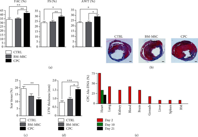
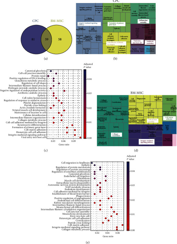
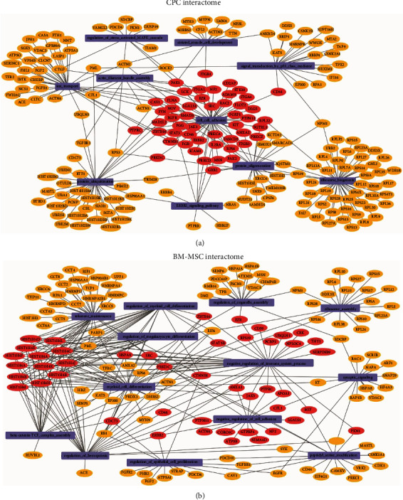
Human bone marrow mesenchymal stem cells (BM-MSCs) and cardiac progenitor/stem cells (CPCs) have been extensively studied as a potential therapeutic treatment for myocardial infarction (MI). Previous reports suggest that lower doses of CPCs are needed to improve cardiac function relative to their bone marrow counterparts. Here, we confirmed this observations and investigated the surface protein expression profile that might explain this effect. Myocardial infarction was performed in nude rats by permanent ligation of the left coronary artery. Cardiac function and infarct size before and after cell transplantation were evaluated by echocardiography and morphometry, respectively. The CPC and BM-MSC receptome were analyzed by proteomic analysis of biotin-labeled surface proteins. Rats transplanted with CPCs showed a greater improvement in cardiac function after MI than those transplanted with BM-MSCs, and this was associated with a smaller infarct size. Analysis of the receptome of CPCs and BM-MSCs showed that gene ontology biological processes and KEGG pathways associated with adhesion mechanisms were upregulated in CPCs compared with BM-MSCs. Moreover, the membrane protein interactome in CPCs showed a strong relationship with biological processes related to cell adhesion whereas the BM-MSCs interactome was more related to immune regulation processes. We conclude that the stronger capacity of CPCs over BM-MSCs to engraft in the infarcted area is likely linked to a more pronounced cell adhesion expression program.
Copyright © 2020 Imelda Ontoria-Oviedo et al.
Conflict of interest statement
The authors declare no competing conflicts of interest.
Figures
Similar articles
-
C-Kit Positive Cardiac Stem Cells and Bone Marrow-Derived Mesenchymal Stem Cells Synergistically Enhance Angiogenesis and Improve Cardiac Function After Myocardial Infarction in a Paracrine Manner.J Card Fail. 2017 May;23(5):403-415. doi: 10.1016/j.cardfail.2017.03.002. Epub 2017 Mar 8. J Card Fail. 2017. PMID: 28284757
-
Experimental study of bone marrow-derived mesenchymal stem cells combined with hepatocyte growth factor transplantation via noninfarct-relative artery in acute myocardial infarction.Gene Ther. 2006 Nov;13(22):1564-8. doi: 10.1038/sj.gt.3302820. Epub 2006 Jun 29. Gene Ther. 2006. PMID: 16810195
-
Mechanisms of improvement of left ventricle remodeling by trans-planting two kinds of autologous bone marrow stem cells in pigs.Chin Med J (Engl). 2008 Dec 5;121(23):2403-9. Chin Med J (Engl). 2008. PMID: 19102957
-
Pericardial Grafting of Cardiac Progenitor Cells in Self-Assembling Peptide Scaffold Improves Cardiac Function After Myocardial Infarction.Cell Transplant. 2023 Jan-Dec;32:9636897231174078. doi: 10.1177/09636897231174078. Cell Transplant. 2023. PMID: 37191272 Free PMC article.
-
Allogeneic transplantation of fetal membrane-derived mesenchymal stem cell sheets increases neovascularization and improves cardiac function after myocardial infarction in rats.Transplantation. 2013 Oct 27;96(8):697-706. doi: 10.1097/TP.0b013e31829f753d. Transplantation. 2013. PMID: 23912174
Cited by
-
Corrigendum to "Plasmatic Membrane Expression of Adhesion Molecules in Human Cardiac Progenitor/Stem Cells Might Explain Their Superior Cell Engraftment after Cell Transplantation".Stem Cells Int. 2022 Mar 4;2022:9786160. doi: 10.1155/2022/9786160. eCollection 2022. Stem Cells Int. 2022. PMID: 35283998 Free PMC article.
-
Extracellular vesicles from dental pulp mesenchymal stem cells modulate macrophage phenotype during acute and chronic cardiac inflammation in athymic nude rats with myocardial infarction.Inflamm Regen. 2024 May 28;44(1):25. doi: 10.1186/s41232-024-00340-7. Inflamm Regen. 2024. PMID: 38807194 Free PMC article.
-
Modeling Transposition of the Great Arteries with Patient-Specific Induced Pluripotent Stem Cells.Int J Mol Sci. 2021 Dec 9;22(24):13270. doi: 10.3390/ijms222413270. Int J Mol Sci. 2021. PMID: 34948064 Free PMC article.
References
-
- Quevedo H. C., Hatzistergos K. E., Oskouei B. N., et al. Allogeneic mesenchymal stem cells restore cardiac function in chronic ischemic cardiomyopathy via trilineage differentiating capacity. Proceedings of the National Academy of Sciences of the United States of America. 2009;106(33):14022–14027. doi: 10.1073/pnas.0903201106. - DOI - PMC - PubMed
-
- Collantes M., Pelacho B., García-Velloso M. J., et al. Non-invasive in vivo imaging of cardiac stem/progenitor cell biodistribution and retention after intracoronary and intramyocardial delivery in a swine model of chronic ischemia reperfusion injury. Journal of Translational Medicine. 2017;15(1) doi: 10.1186/s12967-017-1157-0. - DOI - PMC - PubMed
-
- Crisostomo V., Baez C., Abad J. L., et al. Dose-dependent improvement of cardiac function in a swine model of acute myocardial infarction after intracoronary administration of allogeneic heart-derived cells. Stem Cell Research & Therapy. 2019;10(1):p. 152. doi: 10.1186/s13287-019-1237-6. - DOI - PMC - PubMed
-
- Crisostomo V., Baez-Diaz C., Maestre J., et al. Delayed administration of allogeneic cardiac stem cell therapy for acute myocardial infarction could ameliorate adverse remodeling: experimental study in swine. Journal of Translational Medicine. 2015;13(1) doi: 10.1186/s12967-015-0512-2. - DOI - PMC - PubMed
