Review
. 2020 Sep 11:85:e509-e531.
doi: 10.5114/pjr.2020.99415. eCollection 2020.

Magnetic resonance imaging of the knee

Affiliations
Review

Magnetic resonance imaging of the knee

Alexander Chien et al. Pol J Radiol. .

Abstract

Knee pain is frequently seen in patients of all ages, with a wide range of possible aetiologies. Magnetic resonance imaging (MRI) of the knee is a common diagnostic examination performed for detecting and characterising acute and chronic internal derangement injuries of the knee and helps guide patient management. This article reviews the current clinical practice of MRI evaluation and interpretation of meniscal, ligamentous, cartilaginous, and synovial disorders within the knee that are commonly encountered.

Keywords: cartilage; knee; ligament; magnetic resonance imaging; meniscus; patellar instability.


Conflict of interest statement

The authors report no conflict of interest.

Figures

Figure 1
Figure 1
Meniscal anatomy. The medial meniscus is larger in the AP dimen- sion than the lateral meniscus. The posterior horn of the medial meniscus is also broader than the anterior horn of the medial meniscus. Relative inser- tion sites of the meniscal root attachments are shown
Figure 2
Figure 2
Normal medial and lateral menisci. A) Sagittal proton density- weighted (PDW) magnetic resonance (MR) image in a 41-year-old male with normal anterior and posterior horns of the medial meniscus. B) Sagit- tal PDW fat-saturated MR image in a 38-year-old female with normal anterior and posterior horns of the lateral meniscus, also demonstrating in- tact anteroinferior and posterosuperior popliteomeniscal fascicles (arrows)
Figure 3
Figure 3
Discoid meniscus. Sagittal proton density-weighted magnetic resonance image in an 11-year-old female with a discoid lateral meniscus with extensive intrameniscal signal (arrow)
Figure 4
Figure 4
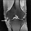
Meniscal tear, para-meniscal cyst. Coronal proton density-weighted fat-saturated magnetic resonance image in a 55-year-old female with small parameniscal cyst (black arrow) adjacent to the body of the lateral meniscus with an associated meniscal tear (white arrow)
Figure 5
Figure 5
Meniscal ossicle. Sagittal proton density-weighted magnetic res- onance image in a 40-year-old male showing a meniscal ossicle (arrow) in the region of the posterior horn medial meniscal root
Figure 6
Figure 6
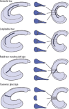
Horizontal, longitudinal, radial, and transverse tears. Horizontal, longitudinal, radial, and transverse tears depicted in three-dimensional and cross-sectional drawings
Figure 7
Figure 7
Horizontal meniscal tear. Coronal proton density-weighted fat- saturated magnetic resonance image in a 63-year-old female with linear intrameniscal signal (closed arrow) extending to the undersurface of the body of the lateral meniscus, consistent with tear. High signal (open arrow) in the contralateral, medial meniscus does not extend to the meniscal arti- cular surface and is consistent with mucoid degeneration
Figure 8
Figure 8
Horizontal meniscal tear with displaced fragment. Coronal proton densi- ty-weighted fat-saturated magnetic resonance image in a 53-year-old male with tear of the medial meniscus and displaced meniscal fragment/flap (arrow) locat- ed between the medial collateral ligament and the medial tibial plateau. There is subchondral bone marrow oedema-like signal in the adjacent tibia
Figure 9
Figure 9
Meniscal tears with displaced fragments. Parrot beak, bucket han- dle, and flap tears in three-dimensional drawings
Figure 10
Figure 10
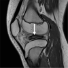
Longitudinal vertical tear. Sagittal proton density-weighted magnetic resonance image in a 27-year-old male with longitudinal vertical tear (arrow) in the posterior horn of the medial meniscus
Figure 11
Figure 11
Displaced bucket handle tears. A) Sagittal and B) coronal proton density-weighted fat-saturated magnetic resonance images in a 34-year-old male with a bucket handle tear show a displaced meniscal fragment adjacent the posterior cruciate ligament (PCL) (white arrow), known as the “double PCL” sign, as well as a displaced meniscal fragment in the intercondylar notch (black arrow). C) Sagittal proton density-weighted fat-saturated magnetic resonance image in a 66-year-old male with a bucket handle tear shows the fragment displaced anteriorly (white arrow), adjacent the anterior horn, known as the “double anterior horn” sign
Figure 12
Figure 12
Radial tear of meniscal root. A) Coronal and B) sagittal proton density-weighted fat-saturated magnetic resonance images in a 65-year- old female with a radially oriented tear of the root of the medial meniscus, with an absent posterior meniscal root attachment (white arrow in A), cre- ating the “ghost meniscus” sign (open arrow in B)
Figure 13
Figure 13
Parrot beak tear. A) Coronal and B) axial proton density-weight- ed fat-saturated magnetic resonance images show a vertical tear that progresses from a radially oriented tear (open arrow in A) at the anterior horn of the medial meniscus. This tear has a curved appearance similar to a parrot’s beak on axial images (white arrow in B)
Figure 14
Figure 14
Magnetic resonance arthrography. A) Coronal T1 fat-saturat- ed magnetic resonance image post intra-articular contrast injection in an 18-year-old female with lateral meniscus linear signal that is intermedi- ate in signal intensity (white arrow), compared to bright contrast injected into joint space (open arrow), probably representing granulation tissue in a previously surgically repaired lateral meniscus. B) Coronal T1 fat-satu- rated magnetic resonance image post intra-articular contrast injection in a 19-year-old male demonstrating a contrast-bright intensity defect (black arrow) in the medial meniscus, representing a radial tear
Figure 15
Figure 15
Meniscal transplant. Coronal proton density-weighted non-fat- saturated contrast enhanced magnetic resonance (MR) images in a 39-year-old female following medial meniscal transplant. A) Postoperative MR image taken 3 months after meniscal transplant shows the meniscal transplant (arrow) and the posterior tibial tunnel (arrowhead). B) MR image obtained two years later, after the patient started experiencing worsening medial knee pain, shows new tearing of the posterior root ligament (arrow) and heterogeneous undersurface signal along the periphery of the body/posterior horn junction (arrowhead), which was found to represent a developing peripheral meniscal tear
Figure 16
Figure 16
Normal anterior cruciate ligament (ACL). A) Coronal and B) sagittal magnetic resonance (MR) images in a skeletally immature patient, and C) axial proton density-weighted fat-saturated MR image in a 35-year-old male demonstrate the normal appearance of a normal ACL (arrow in C), including the anteromedial bundle (thin arrow in A and B) and posterolateral bundle (open arrow in A and B)
Figure 17
Figure 17
Anterior cruciate ligament (ACL) tears: single bundle; partial thickness. A) Coronal and B) sagittal proton density-weighted (PDW) fat-satu- rated (FS) magnetic resonance (MR) images in a 62-year-old male with an anteromedial bundle ACL tear (open arrows). C) Sagittal PDW FS MR image in a 28-year-old male with a partial thickness tear (arrow) shows attenuated and wavy fibres of the ACL with partial discontinuity at its proximal attachment
Figure 18
Figure 18
Full thickness anterior cruciate ligament (ACL) tear and recon- struction. A) Sagittal proton density-weighted fat-saturated magnetic resonance image in a 23-year-old female demonstrates complete discon- tinuity of the proximal to mid ACL (arrow). B) Arthroscopic image confirms complete ACL tear, with ACL stumps visible. C) Subsequent arthroscopic image following ligament graft reconstruction shows an intact hamstring autograft reconstruction
Figure 19
Figure 19
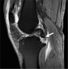
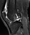
Mucoid degeneration. Sagittal proton density-weighted fat-satu- rated magnetic resonance image in a 43-year-old male with increased signal within the anterior cruciate ligament (open arrow) with intact fibres repre- sents mucoid degeneration. A cruciate ligament ganglion is incidentally noted posteriorly at the posterior cruciate ligament insertion (closed arrow)
Figure 20
Figure 20
Secondary signs of anterior cruciate ligament (ACL) tear. A) Sagittal proton density-weighted (PDW) fat-saturated (FS) magnetic resonance (MR) image in a 25-year-old male shows the characteristic osseous contusion pat- tern in ACL injuries from a pivot-shift mechanism, with contusion/impaction along the terminal sulcus of the lateral femoral condyle (open arrow) and the posterior tibial plateau (closed arrow). B) Sagittal PDW FS MR image from a different patient with an ACL injury shows a Segond fracture (black arrow) and injury to the underlying anterolateral ligament (thin, white arrow)
Figure 21
Figure 21
Single bundle anterior cruciate ligament (ACL) reconstruction. A) Coronal and B) sagittal proton density-weighted fat-saturated magnetic resonance images in a 41-year-old female post ACL reconstruction, with intact graft. Note the tibial interference screw (closed arrow) and the fem- oral Endobutton susceptibility artifact (open arrow) related to graft fixation
Figure 22
Figure 22
Anterior cruciate ligament (ACL) reconstruction complications. A) Sagittal proton density-weighted (PDW) fat-saturated (FS) magnetic resonance (MR) image in a 51-year-old male with completely absent ACL graft reconstruction representing tear of ACL reconstruction. There is an abnormally vertically oriented tibial tunnel interference screw present (closed arrow). Note the buckled appearance of the posterior cruciate ligament (open arrow). B) Sagittal PDW FS MR image in an 18-year-old female who is 3 years status post ACL reconstruction, with arthrofibrosis at the anterior aspect of the intercondylar notch, called a “cyclops lesion” (black arrow). A femoral interference screw is partially visible, and there is a thin halo of intermediate signal surrounding it (small arrow), which may represent granulation reaction that can form adjacent to intraosseous device constructed from bioabsorbable materials
Figure 23
Figure 23
Anterior cruciate ligament (ACL) reconstruction complications. A) Sagittal proton density-weighted (PDW) fat-saturated (FS) magnetic resonance (MR) image following ACL reconstruction, with an anteriorly positioned tibial tunnel (black arrow), associated with impingement of the ACL graft along the intercondylar roof (open arrow). B) Sagittal PDW FS MR image in a different patient following ACL reconstruction shows cystic de- generative changes along the tibial tunnel (white arrow)
Figure 24
Figure 24
Normal posterior cruciate ligament. Sagittal proton density- weighted fat-saturated image in a 15-year-old female with normal posterior cruciate ligament (arrow). The ligament is relatively uniform in thickness and dark in signal
Figure 25
Figure 25
Posterior cruciate ligament (PCL): partial tear and mucoid degeneration. A) Sagittal proton density-weighted (PDW) fat-saturated (FS) magnetic resonance (MR) image in an 84-year-old male with increased signal in midportion of PCL representing prior partial PCL tear (arrow). B) Sagittal PDW FS magnetic resonance image in a different patient shows “tram track” appearance (arrow) of PCL consistent with mucoid degeneration
Figure 26
Figure 26
Posterior cruciate ligament (PCL): osseous avulsion. Sagittal proton density-weighted fat-saturated magnetic resonance image in a 54-year-old male shows a complete avulsion of the PCL from its tibial insertion (arrow)
Figure 27
Figure 27
Posterior cruciate ligament (PCL): complete tear and secondary signs of injury. Sagittal proton density-weighted fat-saturated magnetic resonance image in a 44-year-old male with characteristic osseous contusions in the an- terior proximal tibia (black arrow) and distal femur (open arrow), which occur with PCL injuries. The PCL is thickened, with increased signal and indistinct fibres, and a complete proximal tear from its femoral origin (thin, white arrows)
Figure 28
Figure 28
Medial collateral ligament (MCL) injuries. Coronal proton density-weighted fat-saturated magnetic resonance images in a: A) 20-year-old female with oedema surrounding the MCL without disruption of the ligament fibres (arrow), consistent with a low-grade injury/grade I MCL sprain, B) 21-year-old female with oedema and thickening of the MCL fibres (arrow), consistent with partial-thickness tear/grade II MCL sprain, C) 45-year-old male with oedema and discontinuity of the proximal MCL fibres (arrow), consistent with a full-thickness tear/grade III MCL sprain
Figure 29
Figure 29
Iliotibial band syndrome. Coronal proton density-weighted fat-saturated magnetic resonance image of a 34-year-old male with ilio- tibial band friction syndrome, with fluid and oedema adjacent to the lateral femoral condyle and surrounding the iliotibial band (arrows)
Figure 30
Figure 30
Posterolateral corner (PLC) injury. A) Sagittal T1W non-fat-satu- rated and (B) coronal proton density-weighted fat-saturated magnetic res- onance images in a 24-year-old male with a PLC injury. An “arcuate” fracture is present with osseous avulsion of the fibular head styloid process (white arrow), and there is a complete avulsion of the lateral collateral ligament from its fibular attachment with proximal retraction close to the joint line (black arrow)
Figure 31
Figure 31
Quadriceps tendon tear. Sagittal proton density-weighted fat- saturated magnetic resonance image in a 59-year-old male with a quadri- ceps tendon tear, with complete discontinuity and retraction of quadriceps tendon stump (arrow), and associated patella baja
Figure 32
Figure 32
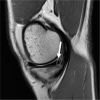
Patellar tendinosis. Sagittal proton density-weighted fat-satu- rated magnetic resonance image in a 49-year-old male with severe patellar tendinosis with interstitial tearing (arrow)
Figure 33
Figure 33
Lateral patellar dislocation. Axial proton density-weighted fat-saturated magnetic resonance image in a 13-year-old female with prior lateral patellar dislocation, currently with lateral subluxation. Note the dysplastic trochlear groove (thin, white arrows) and bone contusion in the lateral femoral condyle (thicker, white arrow). Cartilage injuries (open arrows) are present along the patella. The medial patellofemoral ligament is avulsed from its patellar attachment (black arrow)
Figure 34
Figure 34
Cartilage defect. Coronal proton density-weighted fat-saturat- ed magnetic resonance image in a 36-year-old male with a full thickness (grade 4) lateral femoral condyle cartilaginous defect (arrow) and adjacent subchondral bone marrow oedema-like signal
Figure 35
Figure 35
Osteochondritis dissecans. Sagittal proton density-weighted fat-saturated magnetic resonance image in a 23-year-old female with a large osteochondral defect of the lateral femoral condyle (closed arrow) with adjacent marrow oedema-like signal abnormality. The osteochondral fragment is displaced into the suprapatellar recess (open arrow)
Figure 36
Figure 36
Mosaicplasty. A) Sagittal proton density-weighted (PDW) non- fat-saturated and (B) axial PDW fat-saturated images in a 41-year-old male with cylindrically shaped osteochondral grafts/bone plugs (closed arrows in A and B). Also note the intra-articular bodies present (open arrows in A and B)
Figure 37
Figure 37
Subchondral insufficiency fracture. Coronal proton density- weight ed fat-saturated magnetic resonance image in a 68-year-old female with linear low signal intensity (arrow) beneath the weight-bearing articular surface of the medial femoral condyle, consistent with fracture with adjacent subchondral bone marrow oedema-like signal. The overlying cartilage is intact with the exception of moderate grade pre-existing chondral degeneration
Figure 38
Figure 38
Plica. Axial proton density-weighted fat-saturated magnetic resonance image in a 30-year-old male with linear low-signal intensity non-thickened tissue connected to the synovial lining and surrounded by joint fluid, compatible with a medial patellar plica (arrow)
Figure 39
Figure 39
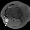
Pes anserine bursa and popliteal cyst. Axial proton density-weight- ed fat-saturated magnetic resonance image in a 62-year-old female with fluid in both the pes anserine bursa (closed arrow), located between the distal tendons of the sartorius, gracilis, and semitendinosus muscles, as well as within a popliteal (Baker’s) cyst, with fluid between the medial head of the gastrocnemius muscle and the semimembranosus tendon (open arrow)
Figure 40
Figure 40
Intraneural ganglion cyst. Axial proton density-weighted fat-sat- urated magnetic resonance image in a 46-year-old female with an intran- eural ganglion cyst (open arrow) along the course of the peroneal nerve (closed arrow), just caudal to the proximal tibiofibular joint

References

    1. Qi ZH, Li CF, Li ZF, et al. . Preliminary study of 3T 1H MR spectroscopy in bone and soft tissue tumors. Chin Med J 2009; 122: 39-43. - PubMed
    1. Chhabra A, Lee PP, Bizzell C, et al. . 3 Tesla MR neurography–technique, interpretation, and pitfalls. Skeletal Radiol 2011; 40: 1249-1260. - PubMed
    1. Koff MF, Burge AJ, Koch KM, et al. . Imaging near orthopedic hardware. J Magn Reson Imaging 2017; 46: 24-39. - PMC - PubMed
    1. Miller JD, Nazarian S, Halperin HR. Implantable Electronic Cardiac Devices and Compatibility With Magnetic Resonance Imaging. J Am Coll Cardiol 2016; 68: 1590-1598. - PubMed
    1. American College of Radiology. ACR–SPR–SSR practice parameter for the performance and interpretation of magnetic resonance imaging (MRI) of the knee. Available from: https://www.acr.org/-/media/ACR/Files/Practice-Parameters/MR-Knee.pdf. - PubMed