. 2020 Oct 17;9(10):e1194.
doi: 10.1002/cti2.1194. eCollection 2020.

Integrative immunogenomic analysis of gastric cancer dictates novel immunological classification and the functional status of tumor-infiltrating cells

Affiliations

Integrative immunogenomic analysis of gastric cancer dictates novel immunological classification and the functional status of tumor-infiltrating cells

Yasuyoshi Sato et al. Clin Transl Immunology. .

Abstract

Objectives: A better understanding of antitumor immunity will help predict the prognosis of gastric cancer patients and tailor the appropriate therapies in each patient. Therefore, we propose a novel immunological classification of gastric cancer.

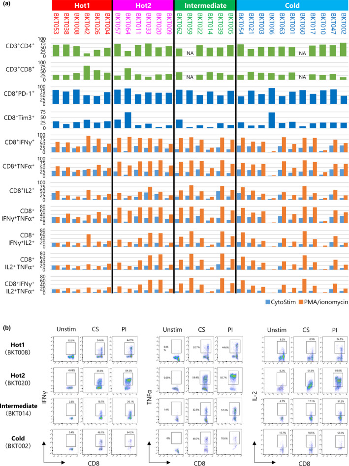
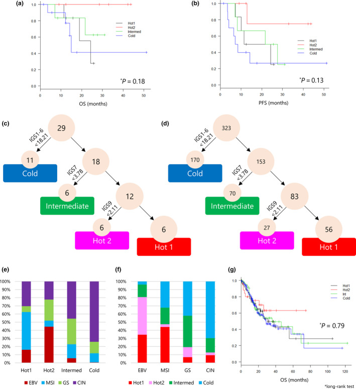
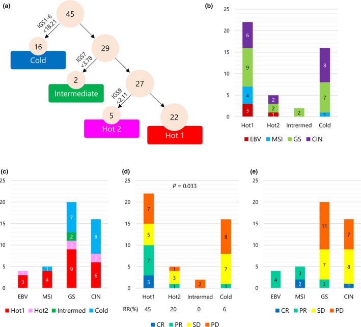
Methods: We performed whole-exome sequencing (WES), RNA-Seq and flow cytometry in 29 gastric cancer patients who received surgery. The TCGA data set of 323 gastric cancer patients and RNA-Seq data of 45 patients who received pembrolizumab (Kim et al. Nat Med 2018; 24: 1449-1458) were also analysed.

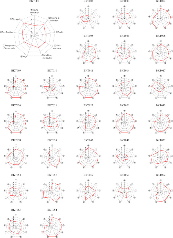
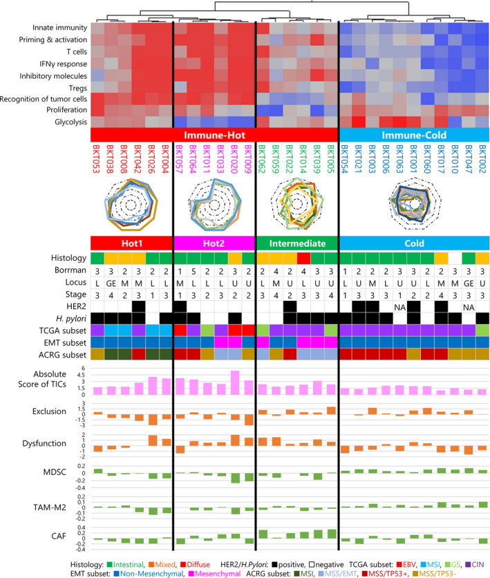
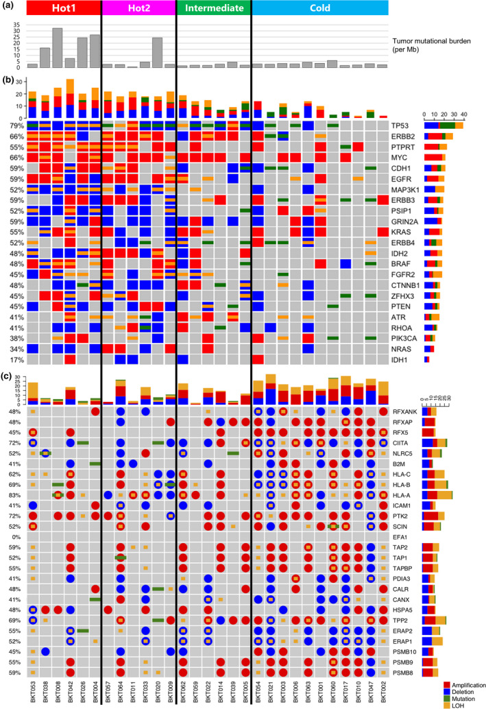
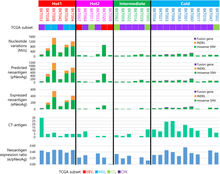
Results: Immunogram analysis of cancer-immunity interaction of gastric cancer revealed immune signatures of four main types, designated Hot1, Hot2, Intermediate and Cold. Immunologically hot tumors displayed a dysfunctional T-cell signature, while cold tumors had an exclusion signature. Ex vivo tumor-infiltrating lymphocyte analysis documented T-cell dysfunction with the expression of checkpoint molecules and impaired cytokine production. The T-cell function was more profoundly damaged in Hot1 than Hot2 tumors. Patients in Hot2 subtypes had better survival in our cohort and TCGA cohort. Although these immunological subtypes overlapped to some degree with the molecular subtypes in the TCGA, intratumoral immune responses cannot be predicted solely based on histological or molecular subtyping of gastric cancer. Molecular and immunological classifications complement each other to predict the responses to anti-PD-1 therapy and have the potential to be a biomarker for the treatment of gastric cancer.

Conclusion: The immunological classification of gastric cancer resulted in four subtypes. Hot tumors were further divided into two subtypes, between which the functional status of T cells was different.

Keywords: RNA‐Seq; T‐cell function; gastric cancer; immunogram; tumor immunity.


Conflict of interest statement

Dr Kakimi reports grants from TAKARA BIO Inc. and grants from MSD, outside the submitted work. In addition, Dr Kakimi has a patent Immunogram pending and the Department of Immunotherapeutics, The University of Tokyo Hospital, is an endowed department by TAKARA BIO Inc. Dr Sato reports personal fees from ONO Pharmaceutical Co., Ltd, Bristol‐Myers Squibb Company, MSD KK and TAIHO Pharmaceutical Co., Ltd, outside the submitted work. Dr Takahashi reports grants and personal fees from Bristol‐Myers Squibb KK, grants and personal fees from ONO Pharmaceutical Co., Ltd, grants and personal fees from MSD, grants and personal fees from AstraZeneca, grants and personal fees from Chugai, and grants and personal fees from BAYER, outside the submitted work. Drs Kakimi and Mineno have a patent Immunogram pending. The other authors have no competing interests to disclose.

Figures

Figure 1
Figure 1
Immunograms for cancer–immunity interactions in 29 patients with gastric cancer. Immunograms were generated using RNA‐Seq data. We selected nine gene sets, including innate immunity (for immunogram score 1, IGS1), priming and activation (IGS2), T cells (IGS3), IFN‐γ response (IGS4), inhibitory molecules (IGS5), Tregs (IGS6), recognition of tumor cells (IGS7) proliferation (IGS8) and glycolysis (IGS9). We performed a single‐sample gene set enrichment analysis (ssGSEA). The ssGSEA scores for each IGS were assessed, normalised and scored onto these axes of the immunogram, which was generated for each patient by integration onto a radar chart.
Figure 2
Figure 2
Immunological subtypes of gastric cancer. Immunogram analysis of 29 gastric cancer patients was performed. By hierarchical cluster analysis of the immunogram scores, gastric cancer cases were first divided into Immune‐Hot and Immune‐Cold subtypes. Then, Immune‐Hot subtype was further divided into Hot1, Hot2 and Intermediate subtypes. Clinical profiles with histology by the Lauren classification, macroscopic classification by the Borrmann classification, locus of the primary site, TNM clinical staging, overexpression of human epidermal growth factor receptor 2 (HER2) protein and the presence or absence of Helicobacter pylori infection are depicted. EBV, MSI, GS and CIN subtypes of molecular classification by TCGA 5 and MSI, MSS/EMT, MSS/TP53+ and MSS/TP53 subtypes of ACRG classification 6 are indicated by colour. Absolute scores of tumor‐infiltrating cells (TICs) were estimated by CIBERSORTx. 20 Scores for exclusion and dysfunction and signatures for MDSCs, TAM‐M2 and CAF were evaluated by TIDE. 21 The EMT subset was determined by a Mesenchymal or Non‐Mesenchymal signature. 19
Figure 3
Figure 3
Mutational analysis. (a) Tumor mutational burden (TMB) was calculated as the total number of nonsynonymous mutations divided by the actual number of bases analysed (per Mb). (b) Heatmap representation of the distribution of gene alterations in known driver genes. (c) Nucleotide and copy‐number variants found in the antigen presentation pathway. Stacked bar plot summarising the total numbers of amplification, deletion, mutation and loss of heterozygosity (LOH) per patient (longitudinal) or per gene (horizontal). Different colours represent different types of nucleotide variants, red for amplification, blue for deletion, green for mutation and yellow for LOH.
Figure 4
Figure 4
Tumor antigens. The number of single nucleotide variants (SNVs; green), indels (orange) and fusion genes (purple) is depicted in a stacked bar plot. The numbers of predicted neoantigens (pNeoAg), expressed neoantigens (eNeoAg), CT antigens (yellow) and neoantigen expression ratio (eNeoAg/pNeoAg) in each patient are depicted as bar graphs.
Figure 5
Figure 5
Summary of T‐cell phenotypes and functions. (a) Tumor‐infiltrating cells (TICs) were analysed by flow cytometry. Bar graphs show the percentage of CD3+CD4+ T cells, CD3+CD8+ T cells, CD8+PD‐1+ T cells, CD8+Tim‐3+ T cells, CD8+IFN‐γ+ T cells, CD8+IL‐2+ T cells, CD8+TNF‐α+ T cells, CD8+IFN‐γ+ T cells, CD8+IFN‐γ+IL‐2+ T cells, CD8+IFN‐γ+TNF‐α+ T cells, CD8+IL‐2+ TNF‐α+ T cells and CD8+ IFN‐γ+IL‐2+ TNF‐α+ T cells. TICs were stimulated with CytoStim (Blue) or PMA/ionomycin (orange); the percentages of cytokine‐producing cells were measured by the intracellular cytokine staining. (b) TICs were unstimulated (Unstim) or incubated with CytoStim (CS) or PMA/ionomycin (PI) for 4 h. Examples of staining patterns are shown. CD45+CD3+CD8+ T cells were gated. The percentage of IFN‐γ‐, TNF‐α‐ and IL‐2‐producing cells are shown.
Figure 6
Figure 6
Immunological subtypes of gastric cancer and associations with survival. (a, b) The association between immunological subtypes and overall survival (OS) and progression‐free survival (PFS) was analysed by the Kaplan–Meier method, and the log‐rank test was used to determine the statistical significance of the differences. (c) A decision tree for the immunological subtypes of gastric cancer was applied to 29 gastric cancer patients. The sum of IGS1 to IGS6 of < 18.21 was taken as the cut‐off value to identify the Cold subtype. Similarly, IGS7 < 3.78 was determined as the cut‐off value for Intermediate and IGS9 < 2.11 was used to discriminate Hot1 from Hot2 tumors. (d) A decision tree for the immunological subtypes of gastric cancer was applied to 323 gastric cancer patients in the TCGA cohort. (e) The frequency of molecular subtypes of TCGA in each immunological subtype. (f) The frequency of immunological subtypes in each molecular subtype of TCGA. (g) Kaplan–Meier analysis and log‐rank test for OS of 311 gastric cancer patients for the four immunological subtypes.
Figure 7
Figure 7
Immunological subtypes of gastric cancer and responses to anti‐PD‐1 therapy. (a) A decision tree for the immunological subtypes of gastric cancer was applied to 45 gastric cancer patients who received anti‐PD‐1 therapy from Kim et al. 9 (b) The number of patients by molecular subtypes of TCGA in each immunological subtype. (c) The number of patients by immunological subtypes in each molecular subtype of TCGA. (d) The number of patients by responses to anti‐PD‐1 therapy in each immunological subtype. (e) The number of patients by responses to anti‐PD‐1 therapy in each molecular subtype of TCGA.

References

    1. Bray F, Ferlay J, Soerjomataram I, Siegel RL, Torre LA, Jemal A. Global cancer statistics 2018: GLOBOCAN estimates of incidence and mortality worldwide for 36 cancers in 185 countries. CA Cancer J Clin 2018; 68: 394–424. - PubMed
    1. Muro K, Chung HC, Shankaran V et al Pembrolizumab for patients with PD‐L1‐positive advanced gastric cancer (KEYNOTE‐012): a multicentre, open‐label, phase 1b trial. Lancet Oncol 2016; 17: 717–726. - PubMed
    1. Kang YK, Boku N, Satoh T et al Nivolumab in patients with advanced gastric or gastro‐oesophageal junction cancer refractory to, or intolerant of, at least two previous chemotherapy regimens (ONO‐4538‐12, ATTRACTION‐2): a randomised, double‐blind, placebo‐controlled, phase 3 trial. Lancet 2017; 390: 2461–2471. - PubMed
    1. Wong SS, Kim KM, Ting JC et al Genomic landscape and genetic heterogeneity in gastric adenocarcinoma revealed by whole‐genome sequencing. Nat Commun 2014; 5: 5477. - PubMed
    1. Cancer Genome Atlas Research Network . Comprehensive molecular characterization of gastric adenocarcinoma. Nature 2014; 513: 202–209. - PMC - PubMed