. 2020 Oct 22:6:108.
doi: 10.1038/s41420-020-00336-5. eCollection 2020.

Cancer-testis antigens, semenogelins 1 and 2, exhibit different anti-proliferative effects on human lung adenocarcinoma cells

Affiliations

Cancer-testis antigens, semenogelins 1 and 2, exhibit different anti-proliferative effects on human lung adenocarcinoma cells

Oleg Shuvalov et al. Cell Death Discov. .

Abstract

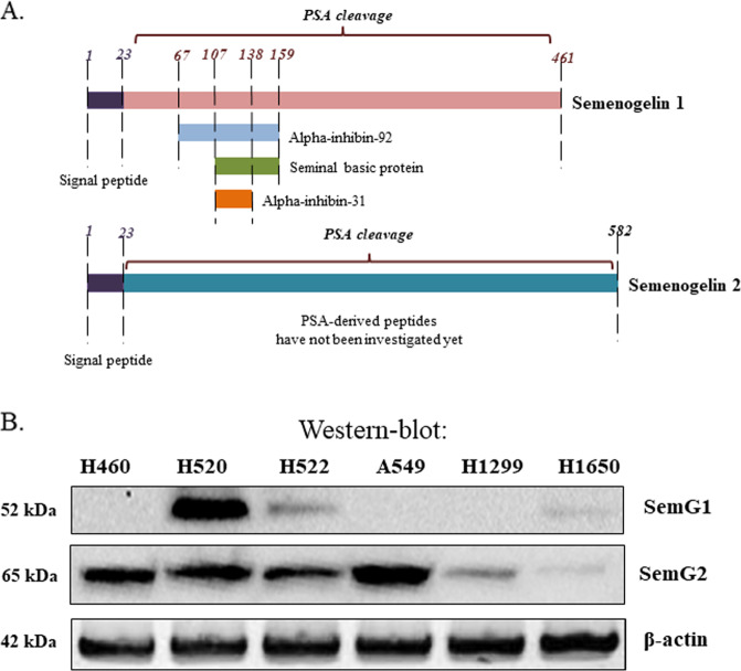
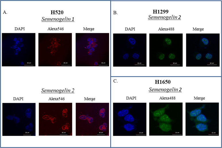
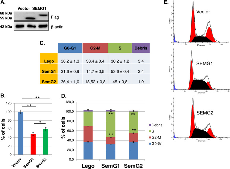
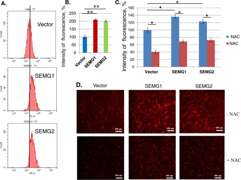
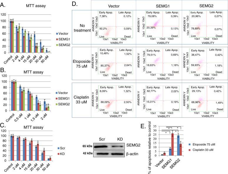
Сancer-testis antigens (CTAs) comprise proteins which are aberrantly expressed in various malignancies, yet under normal situation are restricted to only germ cells. Semenogelins 1 and 2 (SEMG1 and 2, respectively) belong to the family of non-X-linked (autosomal) cancer-testis antigens. They are the major protein ingredients of human semen and share 78% of similarity between them on the gene level. SEMG1/2 gene products regulate the motility and fertility of sperm, as well as provide sperm the antibacterial defense. Besides, SEMG1 and SEMG2 were detected in various malignancies including small cell lung cancer (SCLC). However, the biological role of both SEMG1 and 2 proteins in tumorigenesis has not been fully understood. We demonstrate here that SEMG1 and SEMG2 (SEMGs) exhibit different patterns of expression and sub-cellular localization in non-small cell lung cancer (NSCLC) cell lines. To elucidate the biological properties of SEMGs in NSCLC, we established H1299 cell lines that were stably transduced with either SEMGs-overexpressing or knockdown vectors, respectively. Using fluorescence-based dihydroethidium (DHE) assay we showed that both SEMGs augmented the production of reactive oxygen species (ROS) up to 2 times. Moreover, SEMGs (especially SEMG1) strongly increased the number of Annexin V-positive apoptotic cells manifesting an increased sensitivity to genotoxic drugs including doxorubicin, etoposide, and cisplatin. Taken our results together, SEMGs may arguably play a positive role in tumorigenesis by sensitizing NSCLCs to genotoxic therapy.

Keywords: Mechanisms of disease; Oncogenes.


Conflict of interest statement

Conflict of interestThe authors declare that they have no conflict of interest.

Figures

Fig. 1
Fig. 1. NSCLC cell lines express SEMG1 and SEMG2.
a Comparison of SEMG1 and SEMG2. PSA – prostate specific antigen, cleaves SEMG1 and SEMG2 to small peptides indicated. b Distribution of SEMG1 and SEMG2 in different NSCLC cell lines. Western-blot analysis.
Fig. 2
Fig. 2. Immunofluorescence of SEMGs.
a H520, b H1299 and c H1650 cells. Secondary antibodies conjugated with Alexa546 and Alexa 488 were used to visualize primary anti-SEMG1 and anti-SEMG2 antibodies.
Fig. 3
Fig. 3. Overexpression of SEMG1 and SEMG2 inhibits proliferation and cell cycle progression of H1299 cells.
a H1299 cells stably overexpressed SEMG1, SEMG2 or vehicle (western-blot). b Proliferation assay based on cell counting. Results are represented as mean ± SEM of five experiments. *P < 0.05; **P < 0.01; ce Cell cycle analysis of H1299 cells overexpressing SEMG1, SEMG2 or vehicle. Results are represented as mean ± SEM of three experiments. *P < 0.05; **P < 0.01.
Fig. 4
Fig. 4. Overexpression of SEMG1 and SEMG2 up-regulates production of ROS.
Cells were incubated with DHE and then analyzed by flow cytometry (a, b). Cells were pre-incubated or not with NAC, than were stained with DHE and analyzed by fluorescent microscopy (c, d) Results are represented as mean ± SEM of three experiments. **P < 0.01.
Fig. 5
Fig. 5. SEMGs induce apoptosis and sensitize cells to genotoxic drugs.
MTT assay of H1299 cells overexpressing vector, SEMG1, SEMG2 and incubated with different concentrations of a doxorubicin or b cisplatin. c MTT assay of H1299 cells with scramble or SEMG2 knockdown (KD) treated with cisplatin. Knockdown of SEMG2 is visualized by western-blotting. d Apoptosis profile of non-treated or treated with 30 μM of cisplatin or 75 μM of etoposide H1299 cells overexpressing vector, SEMG1 or SEMG2. e Percentage of apoptosis increase in H1299 cells with vector, SEMG1 or SEMG2 overexpressing after treatment with etoposide or cisplatin. Results are represented as mean ± SEM of three experiments. **P < 0.01.

Similar articles

Cited by

References

    1. Scanlan MJ, Simpson A, Old LJ. The cancer/testis genes: review, standardization, and commentary. Cancer Immun. 2004;4:1. - PubMed
    1. Prasad ML, et al. Expression and significance of cancer testis antigens in primary mucosal melanoma of the head and neck. Head Neck. 2004;26:1053–1057. doi: 10.1002/hed.20112. - DOI - PubMed
    1. Velazquez EF, et al. Expression of the cancer/testis antigen NY-ESO-1 in primary and metastatic malignant melanoma (MM)-correlation with prognostic factors. Cancer Immun. Arch. 2007;7:11. - PMC - PubMed
    1. Figueiredo DL, et al. Expression of cancer testis antigens in head and neck squamous cell carcinomas. Head Neck J. Sci. Specialties Head Neck. 2006;28:614–619. doi: 10.1002/hed.20380. - DOI - PubMed
    1. Fratta E, et al. The biology of cancer testis antigens: putative function, regulation and therapeutic potential. Mol. Oncol. 2011;5:164–182. doi: 10.1016/j.molonc.2011.02.001. - DOI - PMC - PubMed