. 2020 Sep 6;7(20):2001853.
doi: 10.1002/advs.202001853. eCollection 2020 Oct.

Polymeric Nanoparticles with ROS-Responsive Prodrug and Platinum Nanozyme for Enhanced Chemophotodynamic Therapy of Colon Cancer

Affiliations

Polymeric Nanoparticles with ROS-Responsive Prodrug and Platinum Nanozyme for Enhanced Chemophotodynamic Therapy of Colon Cancer

Ying Hao et al. Adv Sci (Weinh). .

Abstract

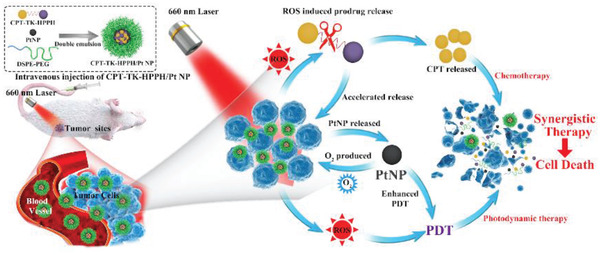
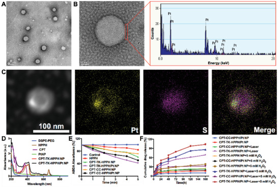
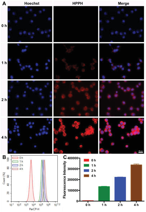
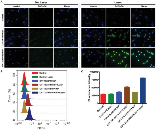
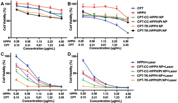
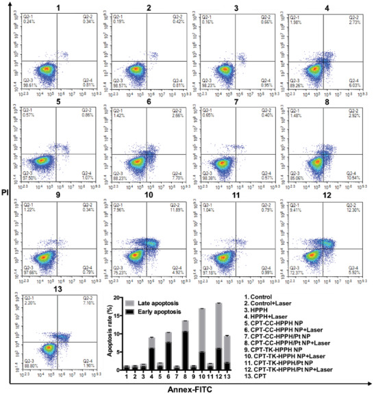
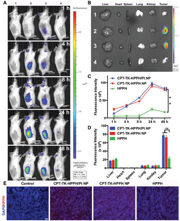
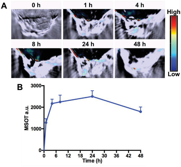
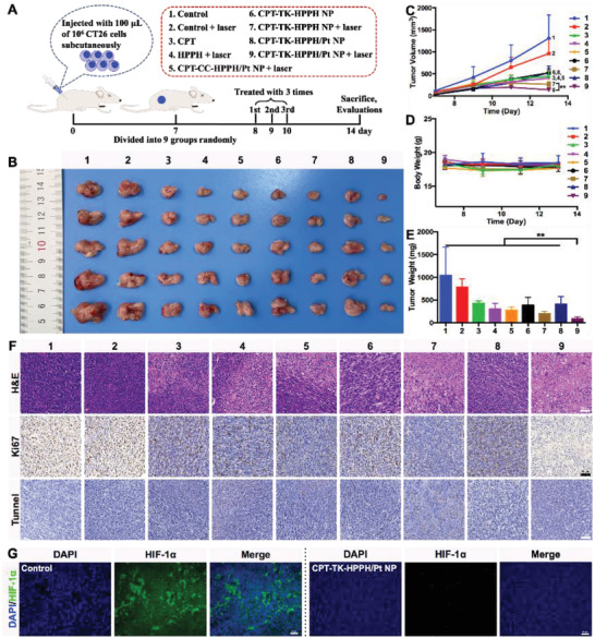
The combination of chemotherapy and photodynamic therapy (PDT) has promising potential in the synergistic treatment of cancer. However, chemotherapy and photodynamic synergistic therapy are impeded by uncontrolled chemotherapeutics release behavior, targeting deficiencies, and hypoxia-associated poor PDT efficacy in solid tumors. Here, a platinum nanozyme (PtNP) loaded reactive oxygen species (ROS)-responsive prodrug nanoparticle (CPT-TK-HPPH/Pt NP) is created to overcome these limitations. The ROS-responsive prodrug consists of a thioketal bond linked with camptothecin (CPT) and photosensitizer-2-(1-hexyloxyethyl)-2-devinyl pyropheophorbide-a (HPPH). The PtNP in CPT-TK-HPPH/Pt NP can efficiently catalyze the decomposition of hydrogen peroxide (H2O2) into oxygen to relieve hypoxia. The production of oxygen can satisfy the consumption of HPPH under 660 nm laser irradiation to attain the on-demand release of CPT and ensure enhanced photodynamic therapy. As a tumor diagnosis agent, the results of photoacoustic imaging and fluorescence imaging for CPT-TK-HPPH/Pt NP exhibit desirable long circulation and enhanced in vivo targeting. CPT-TK-HPPH/Pt NPs effectively inhibit tumor proliferation and growth in vitro and in vivo. CPT-TK-HPPH/Pt NP, with its excellent ROS-responsive drug release behavior and enhanced PDT efficiency can serve as a new cancer theranostic agent, and will further promote the research of chemophotodynamic synergistic cancer therapy.

Keywords: ROS‐responsive prodrugs; chemophotodynamic therapy; colon cancer; platinum nanozymes; polymeric nanoparticles.


Conflict of interest statement

The authors declare no conflict of interest.

Figures

Figure 1
Figure 1
Schematic illustration of colon cancer treatment using CPT‐TK‐HPPH/Pt NP under 660 nm laser irradiation for 5 min.
Figure 2
Figure 2
Characterization of CPT‐TK‐HPPH/Pt NP. A) TEM image (scale bar: 100 nm), B) magnified TEM image (scale bar: 20 nm) and energy dispersive X‐ray spectroscopy of the local area of a CPT‐TK‐HPPH/Pt NP, C) dark‐field TEM image and corresponding TEM elemental mappings of the Pt and S signals of CPT‐TK‐HPPH/Pt NP (scale bar: 100 nm). D) UV–vis absorbance spectra of DSPE‐PEG, HPPH, CPT, PtNP, CPT‐TK‐HPPH NP, and CPT‐TK‐HPPH/Pt NP, E) ABDA absorbance of control, HPPH, CPT‐TK‐HPPH NP, CPT‐TK‐HPPH/Pt NP, CPT‐CC‐HPPH NP, and CPT‐CC‐HPPH/Pt NP groups, F) release behavior of CPT‐TK‐HPPH NP, CPT‐TK‐HPPH/Pt NP, and CPT‐CC‐HPPH/Pt NP under different conditions. Data in (D) and (E) are presented as mean ± SD (n = 3).
Figure 3
Figure 3
Cellular uptake ability of CT26 cells. A) Fluorescence images incubated with CPT‐TK‐HPPH/Pt NP from 0 to 4 h (blue channel: Hoechst, red channel: HPPH, scale bar: 20 µm). B) Flow cytometry analysis of cellular uptake ability. C) Quantitative analysis of fluorescence intensity. All quantitative data are presented as mean ± SD (n = 3).
Figure 4
Figure 4
A) The fluorescence images of intracellular ROS generation using DCFH‐DA as the sensor (Blue channel: Hoechst, Green channel: DCFH‐DA, scale bar: 20 µm). B) The flow cytometry analysis and C) the quantitative analysis of intracellular ROS generation. All quantitative data are presented as mean ± SD (n = 3).
Figure 5
Figure 5
Cell viability of CT26 cells after treatment with CPT, HPPH, CPT‐CC‐HPPH NP, CPT‐CC‐HPPH/Pt NP, CPT‐TK‐HPPH NP, and CPT‐TK‐HPPH/Pt NP for A) 24 and B) 48 h. Cell viability of HPPH, CPT‐CC‐HPPH NP, CPT‐CC‐HPPH/Pt NP, CPT‐TK‐HPPH NP, and CPT‐TK‐HPPH/Pt NP with 660 nm laser irradiation for incubation for C) 24 and D) 48 h. All quantitative data are presented as mean ± SD (n = 6).
Figure 6
Figure 6
Flow cytometric analysis of CT26 cell apoptosis induced by different treatments. All quantitative data are presented as mean ± SD (n = 3).
Figure 7
Figure 7
A) In vivo time‐dependent fluorescence image of tumors in CT26 tumor‐bearing mice. B) In vitro imaging of the livers, hearts, spleens, lungs, kidneys, and tumors excised from CT26 tumor‐bearing mice after 48 h (1. Control group, 2. CPT‐TK‐HPPH/Pt NP, 3. CPT‐TK‐HPPH NP, 4. HPPH). Quantitative fluorescence intensity of C) in vivo and ex vivo tumor tissues measured by RIO value. D) Fluorescence images of ex vivo tissues (blue channel: DAPI, red channel: HPPH, scale bar: 100 µm). All quantitative data are presented as mean ± SD (n = 3). “**” means the P < 0.01.
Figure 8
Figure 8
A) In vivo PA imaging of CT26 tumor‐bearing mice before and after injection with CPT‐TK‐HPPH/Pt NP. B) MSOT intensity variation of tumor tissues at 680 nm. All quantitative data are presented as mean ± SD (n = 3).
Figure 9
Figure 9
A) Schematic illustration of in vivo antitumor efficacy of CPT‐TK‐HPPH/Pt NP, B) photograph, C) growth curves, D) body weight, and E) tumor weight of CT26 tumor‐bearing mice in each group. F) Representative H&E stained images, Ki‐67 immune histochemical images, and tunnel histochemical images of CT26 tumors (scale bar: 50 µm). G) Representative immune‐fluorescence imaging of situation of hypoxia in tumors (scale bar: 50 µm). All quantitative data are presented as mean ± SD (n = 5). “**” means the P <0.01. (1. Control, 2. Control+Laser, 3. CPT, 4. HPPH+Laser, 5. CPT‐CC‐HPPH/Pt NP+Laser, 6. CPT‐TK‐HPPH NP, 7. CPT‐TK‐HPPH NP+Laser, 8. CPT‐TK‐HPPH/Pt NP, 9. CPT‐TK‐HPPH/Pt NP+Laser.).

References

    1. Miller K. D., Nogueira L., Mariotto A. B., Rowland J. H., Yabroff K. R., Alfano C. M., Jemal A., Kramer J. L., Siegel R. L., Ca‐Cancer J. Clin. 2019, 69, 363. - PubMed
    1. Liu R., Yu M., Yang X., Umeshappa C. S., Hu C., Yu W., Qin L., Huang Y., Gao H., Adv. Funct. Mater. 2019, 29, 1808462.
    1. a) Shan L., Fan W., Wang W., Tang W., Yang Z., Wang Z., Liu Y., Shen Z., Dai Y., Cheng S., Jacobson O., Zhai K., Hu J., Ma Y., Kiesewetter D. O., Gao G., Chen X., ACS Nano 2019, 13, 8903; - PubMed
    2. b) Zhang M. K., Li C. X., Wang S. B., Liu T., Song X. L., Yang X. Q., Feng J., Zhang X. Z., Small 2018, 14, 1803602;
    3. c) Hao Y., Dong M., Zhang T., Peng J., Jia Y., Cao Y., Qian Z., ACS Appl. Mater. Interfaces 2017, 9, 15317; - PubMed
    4. d) Zhang X., Li L., Liu Q., Wang Y., Yang J., Qiu T., Zhou G., J. Biomed. Nanotechnol. 2019, 15, 204. - PubMed
    1. Wang Z., Zhang F., Shao D., Chang Z., Wang L., Hu H., Zheng X., Li X., Chen F., Tu Z., Li M., Sun W., Chen L., Dong W. F., Adv. Sci. 2019, 6, 1901690. - PMC - PubMed
    1. a) Chen J., Fan T., Xie Z., Zeng Q., Xue P., Zheng T., Chen Y., Luo X., Zhang H., Biomaterials 2020, 237, 119827; - PubMed
    2. b) Hu D., Pan M., Yu Y., Sun A., Shi K., Qu Y., Qian Z., VIEW 2020, 1, e6.

LinkOut - more resources