. 2020 Oct 6:7:1356-1365.
doi: 10.1016/j.toxrep.2020.10.001. eCollection 2020.

Evaluation of acute and sub-acute toxicity of selected traditional antiurolithiatic medicinal plant extracts in Wistar albino rats

Affiliations

Evaluation of acute and sub-acute toxicity of selected traditional antiurolithiatic medicinal plant extracts in Wistar albino rats

Tilahun Alelign et al. Toxicol Rep. .

Abstract

Introduction: Achyranthes aspera, Chenopodium murale, Satureja punctata, Rumex abyssinicus and Aloe pulcherrima are traditionally used to treat urolithiasis in Ethiopia. However, there are limited reports on toxicity studies.

Objective: This study was intended to evaluate the acute and sub-acute toxicity effects of plants.

Materials and methods: The crude extracts of A. aspera and C. murale leaves, S. punctata aerial parts, R. abyssinicus rhizomes, and A. Pulcherrima gel were prepared using 70 % ethanol. In acute toxicity, 125, 500 and 2000 mg/kg were tested in a stepwise manner; whereas 2000 mg/kg administrated to female rats using gavage during sub-acute toxicity. On day 14 and 28, blood samples were collected from retro-orbital sinus; liver and kidneys of each animal were collected under anaesthesia. Data were analyzed using one-way ANOVA, Dunnett's comparison test of the Graph Pad Prism.

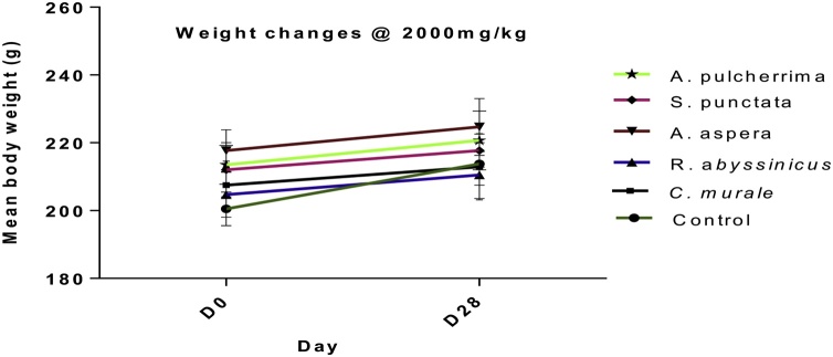
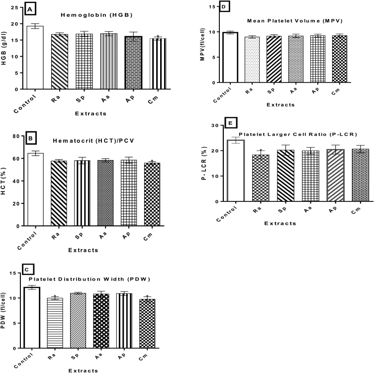
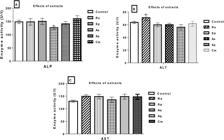
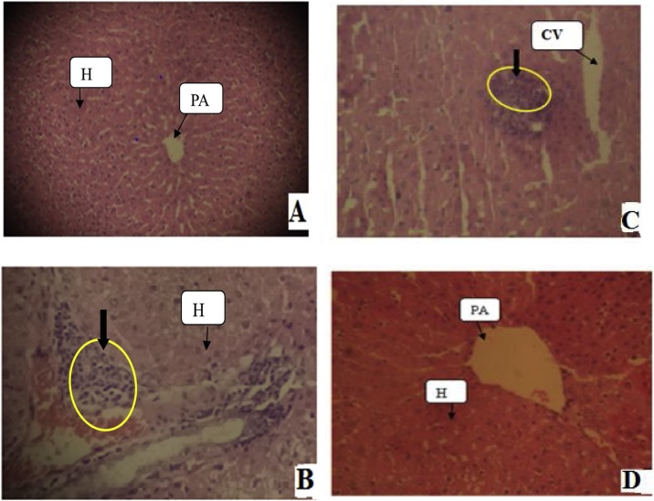
Results: No mortality and significant weight loss for all extracts in both toxicity tests. In acute toxicity, C. murale extract significantly reduced hemoglobin and platelets (P < 0.01) compared with the control. Likewise, S. punctata (P < 0.05) and R. abyssinicus (P < 0.01) extracts revealed significant reduction in platelet count. An exposure to C. murale and R. abyssinicus extracts reduced the concentrations of platelet distribution width and platelet larger cell ratio (p < 0.05) during sub-acute toxicity test. The level of creatinine reduced due to A. aspera extract administrations(P < 0.05). Liver histopathological examinations revealed focal periportal hepatitis following sub-acute toxicity test of C. murale. Histopathological studies of liver demonstrated that R. abyssinicus, A. aspera and S. punctata extracts showed mild acute liver injury. A. pulcherrima was not associated with any toxicity.

Conclusion: C. murale extract showed hematological, and histopathological toxicity profiles in rats. Furthermore, chronic toxicity studies of A. aspera, S. punctata and R. abyssinicus extracts would be beneficial to ensure safety.

Keywords: Acute toxicity; Albino wistar female rats; Antiurolithiatic plant extracts; Sub-acute toxicity.


Conflict of interest statement

The authors report no declarations of interest.

Figures

Fig. 1
Fig. 1
Acute toxicity effects of plant extracts on body weight changes in non-pregnant female Wistar albino rats at dose (A) 125 mg/kg, (B) 500 mg/kg, and (C) 2000 mg/kg b.w. Note: D0 = day zero, and D14 = day 14. Comparisons were made with a control group before and after the experiment. The data illustrated the mean + SD of 3 rats per treatment groups (n = 3).
Fig. 2
Fig. 2
Effects of plant extracts on platelet concentrations in Wistar female rats exposed for a dose 2000 mg/kg in acute toxicity study. Note: Aa=A. aspera, Sp= S. punctata, Ra=R. abyssinicus, Cm=C. murale and Ap=A. pulcherrima. *P < 0.05, **p < 0.01,***p < 0.001 indicate significant changes in comparison with the normal control.
Fig. 3
Fig. 3
Effects of plant extracts on body weight changes in non-pregnant female Wistar rats during sub-acute toxicity study. Note: D0= day zero, and D28 = day 28. Comparisons were made with a control group before and after the experiment. Data illustrated the mean + SD of 6 rats per treatment groups (n = 6).
Fig. 4
Fig. 4
Hematological markers of toxicity in non-pregnant female Wistar albino rats exposed for 28 days. Values are expressed as mean ± SD (n = 6). (A) HGB, (B) PCV, (C) PDW, (D) MPV, and (E) P-LCR. Note: Aa=A. aspera leaves, Sp=S. punctata aerial parts, Ra=R. abyssinicus rhizome, Cm=C. murale leaves, and Ap=A. pulcherrima gel. *P < 0.05 statistically significant compared to the control group.
Fig. 5
Fig. 5
Estimation of (A) ALP, (B) ALT, and (C) AST in serum indicating the effects of plant extracts on liver functional indices of non-pregnant female Wistar rats exposed for 28 days. Note: ALP = Alkaline phosphatase, ALT = alanine aminotransferase, AST = aspartate aminotransferase, Aa=A. aspera leaves, Sp=S. punctata aerial parts, Ra=R. abyssinicus rhizomes, Cm=Chenopodium murale leaves, and Ap=A. pulcherrima gel. The data illustrated the mean + SD of six rats per treatment groups (n = 6). *P < 0.05 statistically significant compared to the control group.
Fig. 6
Fig. 6
Effects of plant extracts on liver tissues of non-pregnant female rats exposed for 28 days. (A) Liver tissues from control rats had normal architecture with hepatocytes arranged around the central vein; (B) Liver showing periportal hepatitis (C. murale extract); (C)Liver tissue revealed local inflammatory infiltrations (R. abyssinicus, A. aspera and S. punctata extracts), and (D) A. pulcherrima extracts with normal view. Photomicrographs were at 100x magnification using light Microscope, and 5 μm thick paraffin sections, Hematoxylene and Eosin stain. Arrows indicate on tissues with yellow circle shows lobular hepatitis. Note: PA = Portal Area; H= Hepatocytes; and CV = Central Vein.
Fig. 7
Fig. 7
Sub-acute toxic effects of plant extracts on kidney tissues of on-pregnant female rats after exposure for 28 days. (A) Kidney tissues of control group showing normal morphology of the glomeruli and tubules; (B) Mild tubular injury with congestions (C. murale extract); (C) Mild tubular inflammations (R. abyssinicus, A. aspera and S. punctata extracts), (D) A. pulcherrima extract with normal view. Photomicrographs were at 100x magnification using a light microscope, and 5 μm thick paraffin sections, Hematoxylene and Eosin stain. Arrows with yellow circles indicate tissues affected as a result of herbal extracts. Note: G = Glomerulus, BS = Bowman’s Space, DT = Distal Convoluted Tube.

References

    1. Ankur C., Amarchand P., Aadarsh C., Deepa I., Pawar R.S., Patil U.K. Potential of medicinal plants in kidney, gall and urinary stones. Int. J. Drug Dev. Res. 2010;2(2):431–447.
    1. Diallo D., Paulsen B.S., Hvemm B. Production of traditional medicine: preparations accepted as medicines in Mali. In: Hostettmann K., Chinyanganya F., Maillard M., Wolfender J.L., editors. Chemistry, Biological and Pharmacological Properties of African Medicinal Plants: Proceedings of the First International IOCD-Symposium. UZ Publications; Victoria Falls, Zimbabwe: 1996. pp. 235–243. February 25-28 Harare.
    1. Endashaw B. Study on actual situation of medicinal plants in Ethiopia. Japan Assoc. Int.Coll. Agric. Fores. 2007 http://www.jaicaf.or.jp/publications/ethiopiaac.pdf
    1. Rates S.M. Plants as source of drugs. Toxicon. 2001;39(5):603–613. - PubMed
    1. Kale O.E., Awodele O., Akindele A.J. Subacute and subchronic oral toxicity assessments of Acridocarpus smeathmannii (DC.) Guill. & Perr. Root in Wistar rats. Toxicol. Rep. 2019;6:161–175. - PMC - PubMed

LinkOut - more resources