Review
. 2020 Sep 25:10:577559.
doi: 10.3389/fcimb.2020.577559. eCollection 2020.

Emerging Evasion Mechanisms of Macrophage Defenses by Pathogenic Bacteria

Affiliations
Review

Emerging Evasion Mechanisms of Macrophage Defenses by Pathogenic Bacteria

Clarisse Leseigneur et al. Front Cell Infect Microbiol. .

Abstract

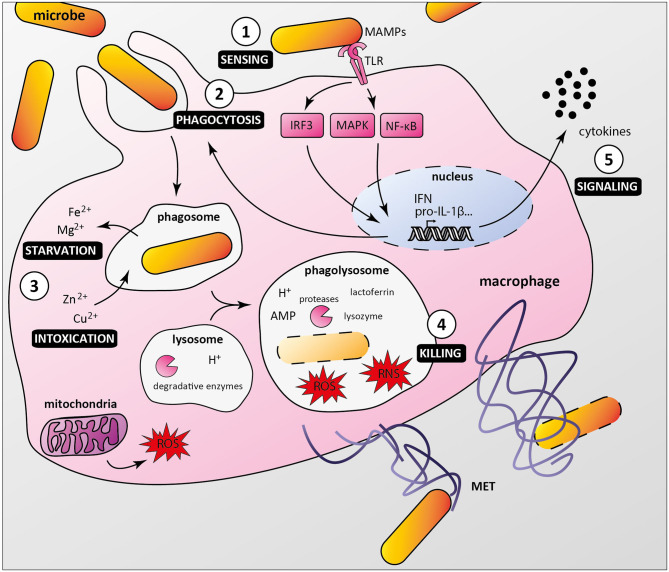
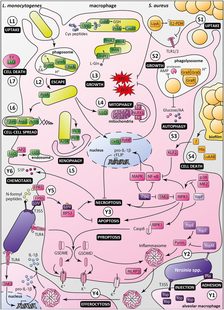
Macrophages participate to the first line of defense against infectious agents. Microbial pathogens evolved sophisticated mechanisms to escape macrophage killing. Here, we review recent discoveries and emerging concepts on bacterial molecular strategies to subvert macrophage immune responses. We focus on the expanding number of fascinating subversive tools developed by Listeria monocytogenes, Staphylococcus aureus, and pathogenic Yersinia spp., illustrating diversity and commonality in mechanisms used by microorganisms with different pathogenic lifestyles.

Keywords: immune escape; listeriosis; phagocyte; plague; staphylococcal infection; virulence; yersiniosis.


Figures

Figure 1
Figure 1
Macrophage anti-microbial mechanisms. (1) Bacteria are recognized by macrophage pattern recognition receptors (PRRs) such as Toll-like receptors (TLR), which bind conserved microbe-associated molecular patterns (MAMPs). (2) MAMP/PRR interaction triggers signaling cascades (e.g., IRF3, MAPKs, NF-κB) leading to macrophage responses, including formation of the phagocytic cup. (3) Internalized bacteria reside in phagosomes, from which nutrients and essential factors such as iron and magnesium are transported to the cytoplasm, restricting their supply to bacteria. Macrophages combine this starvation strategy with a poisoning mechanism involving phagosomal import of toxic amount of zinc and copper. (4) Phagosome maturation and fusion with lysosomes lead to acidification of the compartment lumen and activation of digestive enzymes such as proteases, which along with antimicrobial peptides (AMP), reactive oxygen and nitrogen species (ROS and RNS), lysozyme and lactoferrin contribute to bacterial killing. Macrophages can also undergo ETosis to release macrophage extracellular traps (MET) that immobilize and kill extracellular bacteria. (5) Additionally, infected macrophages secrete multiple cytokines to attract and activate other cells, which contribute to an effective immune response.
Figure 2
Figure 2
Macrophage evasion mechanisms by Listeria monocytogenes, Staphylococcus aureus and pathogenic Yersinia. Listeria monocytogenes: (L1) Upon uptake by the macrophage, Listeria monocytogenes is engulfed in a phagosome, in which ethanolamine uptake through EutH permease and activation of redox-responsive spxA1 and ohrA are required for its survival. (L2) In addition to secretion of LLO and phospholipases, processing of the PlpA lipoprotein is required for phagosomal escape. (L3) Once in the cytosol, bacterial growth and virulence are mediated by the master regulator PrfA, which is activated by CodY and glutathione (GSH). The Opp permease ensures importation of cysteine-containing oligopeptides to allow glutathione synthesis by GshF. Full expression of Listeria virulence genes requires appropriate amounts of L-glutamine imported by the high-affinity ABC transporter GlnPQ. PrfA triggers secretion of PrsA2 chaperone and HtrA chaperone/protease, whose functions are required for invasion and intracellular growth. (L4) Listeria induces mitophagy through the oligomerization of NLRX1 receptor by LLO, lowering ROS levels, and promoting bacterial survival. Listeria also controls ROS levels by secretion of the nucleomodulin OrfX, which interacts with the regulator RybP. (L5) ActA inhibits xenophagy along with PlcA and PlcB, which block LC3 lipidation. Listeria intracellular survival also depends on Ap2a2-mediated control of LLO cytotoxicity by restricting its cytosolic activity. (L6) Besides ActA, YjbH, and ArpJ are required for efficient bacterial spread from cell to cell. (L7) Listeria produces extracellular vesicles (EV) containing many virulence factors, including LLO, to promote macrophage death and control innate immunity response. Staphylococcus aureus: (S1) Staphylococcus aureus is phagocytosed by macrophages. The moonlighting metabolic protein pyruvate dehydrogenase, once lipoylated by LipA, suppresses macrophage activation by lipopeptides through binding to TLR1/2. LipA also decreases RONS production. (S2) Staphylococci reside and multiply in mature phagosomes, through sensing of acidification by GraXRS and activation of several genes allowing bacterial replication and resistance to antimicrobial peptides (AMP). (S3) S. aureus also modulates metabolic fluxes to induce a starvation-like state of macrophages, triggering autophagy. (S4) Secretion of alpha-toxin Hla and leukocidin AB (LukAB), besides having a direct cytotoxic effect, inhibits macrophage phagocytosis and promotes biofilm formation. Through a yet unknown intermediate, biofilm conditioned medium attenuates NF-κB activation by increasing KLF2 expression. Pathogenic Yersinia, i.e., Y. enterocolitica (light purple), Y. pseudotuberculosis (purple), and Y. pestis (dark purple): (Y1) In the lungs, Yersinia adheres to alveolar macrophages through Pla, which shows immunosuppressive properties. (Y2) Injection of Yops, virulence effectors, in macrophages through the T3SS allows manipulation of host cell pathways. In absence of YopM, YopE, and YopT activate the inflammasome by dephosphorylating pyrin. (Y3) Once translocated into the host cell, LcrV is glutathionylated, promoting binding to RPS3, suppressing apoptosis and increasing necroptosis. YopJ inhibition of TAK1 leads to activation of RIPK1 and induction of necroptosis or apoptosis of targeted macrophage. YopJ also inhibits MAPK and NF-κB pathways, inhibiting pro-IL-1β production and limiting pro-inflammatory response. YopP inhibits RIPK1 phosphorylation by p38MAPK/MK2, triggering macrophage apoptosis and activation of cell death effectors gasdermin D and E. (Y4) Macrophages intoxicated with low levels of YopJ can release IL-1β upon uptake of inflammasome from highly intoxicated dead cells, possibly by efferocytosis. (Y5) N-formylpeptides released by Y. pestis are recognized by host receptor FPR1, promoting immune cell chemotaxis toward bacteria. Adhesion of bacteria to macrophage is mediated by FPR1/LcrV interaction, which allows the assembly of type three secretion system. (Y6) Sphingosine-1-phosphate released from dead cells attracts new phagocytes, which in turn are targeted by Yersinia released from necroptotic cells, ultimately promoting infection.

References

    1. Ahmed J. K, Freitag N. E. (2016). Secretion chaperones PrsA2 and HtrA are required for Listeria monocytogenes replication following intracellular induction of virulence factor secretion. Infect. Immun. 84, 3034–3046. 10.1128/IAI.00312-16 - DOI - PMC - PubMed
    1. Alboslemy T., Yu B., Rogers T., Kima M. H. (2019). Staphylococcus aureus biofilm-conditioned medium impairs macrophage-mediated antibiofilm immune response by upregulating KLF2 expression. Infect. Immun. 87:e00643–e00618. 10.1128/IAI.00643-18 - DOI - PMC - PubMed
    1. Anderson C. J., Satkovich J., Köseoglu V. K., Agaisse H., Kendall M. M. (2018). The ethanolamine permease EutH promotes vacuole adaptation of Salmonella enterica and Listeria monocytogenes during macrophage infection. Infect. Immun. 86:e00172–e00118. 10.1128/IAI.00172-18 - DOI - PMC - PubMed
    1. Arifuzzaman M., Ang W. X. G., Choi H. W., Nilles M. L., St John A. L., Abraham S. N. (2018). Necroptosis of infiltrated macrophages drives Yersinia pestis dispersal within buboes. JCI Insight. 3:122188. 10.1172/jci.insight.122188 - DOI - PMC - PubMed
    1. Banerjee S. K., Huckuntod S. D., Mills S. D., Kurten R. C., Pechous R. D. (2019). Modeling pneumonic plague in human precision-cut lung slices highlights a role for the plasminogen activator protease in facilitating type 3 secretion. Infect. Immun. 87, e00175–e00119. 10.1128/IAI.00175-19 - DOI - PMC - PubMed

Publication types