. 2020 Sep 25:8:577557.
doi: 10.3389/fbioe.2020.577557. eCollection 2020.

Ceria Nanoparticles Decrease UVA-Induced Fibroblast Death Through Cell Redox Regulation Leading to Cell Survival, Migration and Proliferation

Affiliations

Ceria Nanoparticles Decrease UVA-Induced Fibroblast Death Through Cell Redox Regulation Leading to Cell Survival, Migration and Proliferation

Fabianne Martins Ribeiro et al. Front Bioeng Biotechnol. .

Abstract

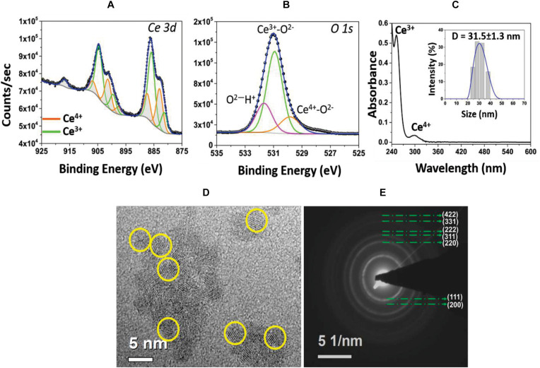
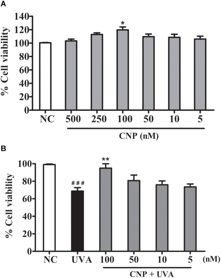
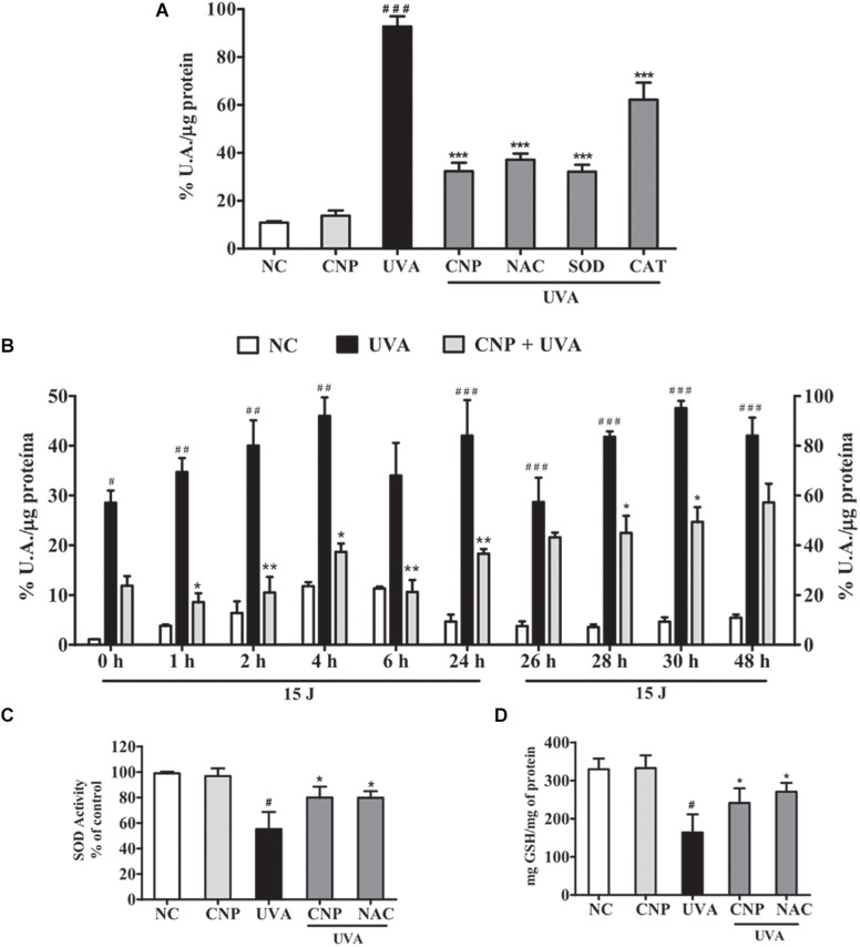
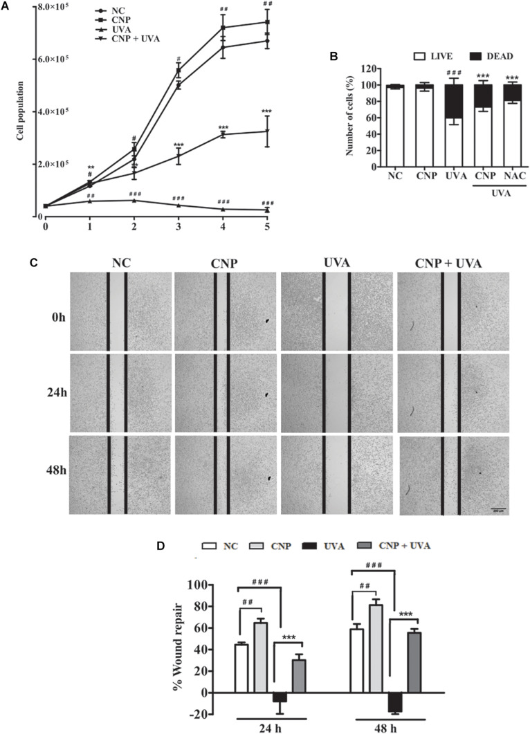
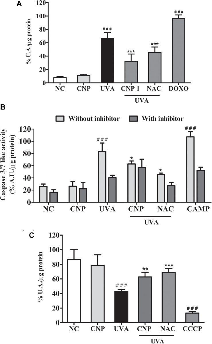
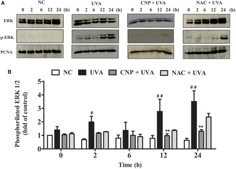
Exposure to ultraviolet radiation is a major contributor to premature skin aging and carcinogenesis, which is mainly driven by overproduction of reactive oxygen species (ROS). There is growing interest for research on new strategies that address photoaging prevention, such as the use of nanomaterials. Cerium oxide nanoparticles (nanoceria) show enzyme-like activity in scavenging ROS. Herein, our goal was to study whether under ultraviolet A rays (UVA)-induced oxidative redox imbalance, a low dose of nanoceria induces protective effects on cell survival, migration, and proliferation. Fibroblasts cells (L929) were pretreated with nanoceria (100 nM) and exposed to UVA radiation. Pretreatment of cells with nanoceria showed negligible cytotoxicity and protected cells from UVA-induced death. Nanoceria also inhibited ROS production immediately after irradiation and for up to 48 h and restored the superoxide dismutase (SOD) activity and GSH level. Additionally, the nanoceria pretreatment prevented apoptosis by decreasing Caspase 3/7 levels and the loss of mitochondrial membrane potential. Nanoceria significantly improved the cell survival migration and increased proliferation, over a 5 days period, as compared with UVA-irradiated cells, in wound healing assay. Furthermore, it was observed that nanoceria decreased cellular aging and ERK 1/2 phosphorylation. Our study suggests that nanoceria might be a potential ally to endogenous, antioxidant enzymes, and enhancing the redox potentials to fight against UVA-induced photodamage and consequently modulating the cells survival, migration, and proliferation.

Keywords: antioxidant; nanoceria; photoaging; ultraviolet radiation; wound healing.


Figures

FIGURE 1
FIGURE 1
(A) High resolution XPS spectrum of Ce 3d envelope. Deconvoluted and peak fitted spectrum shows variations in the multiplet components of Ce 3d5/2 and Ce 3d3/2 doublets. The characteristic peaks of Ce3+ (green lines) and Ce4+ (orange lines) oxidation states are identified. (B) Asymmetry in the O1s spectrum and deconvoluted peaks are attributed to ions associated with the Ce4+ and Ce3+ as well as surface hydroxyl species, OH/O2. (C) UV-Visible spectrum of CNP sample evidencing signature peaks at 253 nm, corresponding to Ce3+ population, and at 300 nm, for Ce4+ oxidation state. Inset: hydrodynamic size (∼31.5 nm diameter) of CNPs with narrow size distribution. (D) HRTEM image of CNP with the particle size of 3–5 nm and (E) SAED pattern illustrating the material’s nano-crystalline character.
FIGURE 2
FIGURE 2
Effect of CNP on viability of non-irradiated and irrradiated L929 cells. (A) Cells were treated with CNP (500, 250, 100, 50, 25, and 5 nM) for 24 h. (B) Cells were treated with CNP (100 nM), exposed to UVA radiation (30 J/cm2) and incubated for more 24 h. In both experiments, cell viability was assessed by MTT assay. NC (non–treated and non-irradiated cells), CNP (treated and non-irradiated cells), UVA (non-treated and irradiated cells) and CNP + UVA (treated and irradiated cells). *p < 0.05, significantly different from NC; **p < 0.01, significantly different from UVA; ###p < 0.001, significantly different from NC.
FIGURE 3
FIGURE 3
CNP antioxidant effect on UVA-irradiated L929 fibroblasts. (A,B) Detection of total ROS in L929 cells treated with CNP (100 nM) for 24 h and irradiated with UVA, using H2DCFDA. (A) Cells were exposed to UVA radiation (30 J/cm2) and the readings performed immediately after irradiation. (B) Cells were exposed to UVA radiation (15 J/cm2), and the readings performed in different times (0, 1, 2, 4, 6, and 24 h). After 24 h of incubation the cells were reirradiated with 15 J/cm2 and the readings performed in different times (26, 28, 30, and 48 h). The level of intracellular ROS is expressed as the percentage mean of DCF fluorescence intensity. (C) Detection of SOD activity and (D) GSH levels in L929 cells treated with CNP (100 nM) for 24 h and irradiated with UVA (30 J/cm2). The readings were performed after 1 h. SOD activity assessed by autoxidation of pyrogallol. GSH content was assayed by the o-phthalaldehyde method. NC (non-treated and non-irradiated cells), UVA (non-treated and irradiated cells), CNP + UVA (treated and irradiated cells), CNP (treated and non-irradiated cells), NAC + UVA (cells treated with NAC and irradiated). SOD + UVA (cells treated with superoxide dismutase and irradiated), CAT + UVA (cells treated with catalase and irradiated). *p < 0.05, significantly different from UVA; **p < 0.01, significantly different from UVA; ***p < 0.001, significantly different from UVA, #p < 0.05, significantly different from NC, ##p < 0.01, significantly different from NC; ###p < 0.001, significantly different from NC.
FIGURE 4
FIGURE 4
Evaluation of L929 cell proliferation and migration. (A,B) Effect of CNP on L929 cells proliferation. Cells were treated with CNP (100 nM) for 24 h followed by irradiation with UVA (30 J/cm2) and incubated for 1, 2, 3, 4 and 5 days. Every 24 h cells were counted using trypan blue dye exclusion method, for 5 days considering live cells. Dead and live cells were quantified after 24h incubation after irradiation (day 1) and counted by trypan blue dye exclusion method. (C,D) Wound healing assay. L929 cells were treated with 100 nM CNP for 24 h, irradiated with 30 J/cm2 UVA, and cells were scratched. Representative cell images from each group in the indicated time points after scratrch are shown. The area of the wound was measured at the 0, 24 and 48 h time points and compared in every group. Photomicrographs were taken at ×5 magnification in a light microscope. NC (non-treated and non-irradiated cells), UVA (non-treated and irradiated cells), CNP + UVA (treated and irradiated cells), CNP (treated and non-irradiated cells), NAC + UVA (cells treated with N-Acetylcysteine and irradiated). #p < 0.05: significantly different from NC; ##p < 0.01: significantly different from NC, ###p < 0.001: significantly different from NC, **p < 0.01: significantly different from UVA; ***p < 0.001: significantly different from UVA.
FIGURE 5
FIGURE 5
Assessment of senescence and cell death in L929 cells. (A) Senescence-associated β-galactosidase (SA-bG) activity measured based on fluorescein production. Cells were treated with 100 nM CNP and irradiated with to 10 J/cm2 for three consecutive days and incubated for 24 h. Fluorescence was measured after 24 h of incubation with FDG. (B) Caspases 3/7 like activity. Cells were treated with 100 nM CNP and irradiated with 30 mJ/cm2 UVA. Caspase-3/7 activity was measured using Z-DEVD-AMC substrate after 24 h of irradiation. (C) Measurement of mitochondrial dysfunction. Cells were treated with 100 nM CNP and irradiated with (30 J/cm2) After 2 h of incubation, cells were stained with the fluorescent probe (TMRE, 100 nM). NC (non-treated and non-irradiated cells), UVA (non-treated and irradiated cells), CNP + UVA (treated and irradiated cells), CNP (treated and non-irradiated cells), NAC + UVA (cells treated with N-acetylcysteine and irradiated), DOXO (cells treated with doxorubicin and non-irradiated), CAMP (cells treated with camptothecin and non-irradiated), CCCP (cells treated with carbonyl cyanide 3-chlorophenylhydrazone and non-irradiated. ###p < 0.001, significantly different from NC; ***p < 0.001, significantly different from UVA; **p < 0.01, significantly different from UVA; *p < 0.05, significantly different from UVA.
FIGURE 6
FIGURE 6
ERK phosphorylation and cell cycle analysis. (A,B) ERK 1/2 phosphorylation was performed by western blot. After 24 h of CNP (100 nM) treatment and UVA exposure (30 J/cm2) cell lysates were prepared for subsequent analysis by polyacrylamide gel electrophoresis followed by western blot analysis for ERK 1/2 (ERK total), phospho-ERK 1/2 (p-ERK 1/2) and PCNA. The density of each band was normalized with corresponding PCNA levels (bar graphs). NC (non-treated and non-irradiated cells), UVA (non-treated and irradiated cells), CNP + UVA (treated and irradiated cells), CNP (treated and non-irradiated cells), NAC + UVA (cells treated with N-acetylcysteine and irradiated). #p < 0.05, significantly different from NC; **p < 0.01, significantly different from UVA; ##p < 0.01, significantly different from NC.

Similar articles

Cited by

References

    1. Akhtar M. J., Ahamed M., Alhadlaq H. A., Khan M. A. M., Alrokayan S. A. (2015). Glutathione replenishing potential of CeO2 nanoparticles in human breast and fibrosarcoma cells. J. Colloid Interface Sci. 453 21–27. 10.1016/j.jcis.2015.04.049 - DOI - PubMed
    1. Asati A., Santra S., Kaittanis C., Perez J. M. (2010). Surface-charge-dependent cell localization and cytotoxicity of cerium oxide nanoparticles. ACS Nano 4 5321–5331. 10.1021/nn100816s - DOI - PMC - PubMed
    1. Bainbridge P., Healing W., Repair T. (2013). Wound healing and the role of fibroblasts. J. Wound Care 22 407–408. 10.12968/jowc.2013.22.8.407 - DOI - PubMed
    1. Bizanek M., Webster T. J., Roy A. K. (2018). Increased viability of fibroblasts when pretreated with ceria nanoparticles during serum deprivation. Int. J. Nanomed. 13 895–901. 10.2147/IJN.S148390 - DOI - PMC - PubMed
    1. Caputo F., De Nicola M., Sienkiewicz A., Giovanetti A., Bejarano I., Licoccia S., et al. (2015). Cerium oxide nanoparticles, combining antioxidant and UV shielding properties, prevent UV-induced cell damage and mutagenesis. Nanoscale 7 15643–15656. 10.1039/c5nr03767k - DOI - PubMed