

How Do Electric Fields Coordinate Neuronal Migration and Maturation in the Developing Cortex?
- PMID: 33102486
- PMCID: PMC7546860
- DOI: 10.3389/fcell.2020.580657
How Do Electric Fields Coordinate Neuronal Migration and Maturation in the Developing Cortex?
Abstract
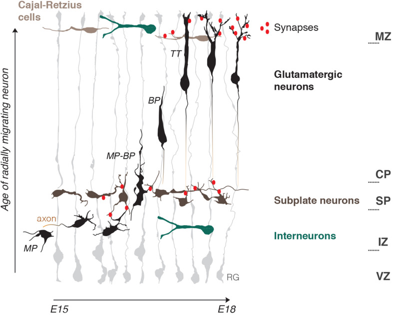
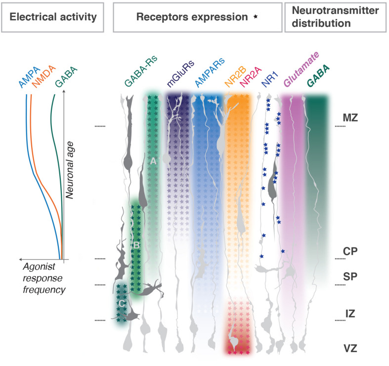
During development the vast majority of cells that will later compose the mature cerebral cortex undergo extensive migration to reach their final position. In addition to intrinsically distinct migratory behaviors, cells encounter and respond to vastly different microenvironments. These range from axonal tracts to cell-dense matrices, electrically active regions and extracellular matrix components, which may all change overtime. Furthermore, migrating neurons themselves not only adapt to their microenvironment but also modify the local niche through cell-cell contacts, secreted factors and ions. In the radial dimension, the developing cortex is roughly divided into dense progenitor and cortical plate territories, and a less crowded intermediate zone. The cortical plate is bordered by the subplate and the marginal zone, which are populated by neurons with high electrical activity and characterized by sophisticated neuritic ramifications. Neuronal migration is influenced by these boundaries resulting in dramatic changes in migratory behaviors as well as morphology and electrical activity. Modifications in the levels of any of these parameters can lead to alterations and even arrest of migration. Recent work indicates that morphology and electrical activity of migrating neuron are interconnected and the aim of this review is to explore the extent of this connection. We will discuss on one hand how the response of migrating neurons is altered upon modification of their intrinsic electrical properties and whether, on the other hand, the electrical properties of the cellular environment can modify the morphology and electrical activity of migrating cortical neurons.
Keywords: cerebral cortex; dendritogenesis; development; electric field; neuronal migration.
Copyright © 2020 Medvedeva and Pierani.
Figures
Similar articles
-
Changing patterns of synaptic input to subplate and cortical plate during development of visual cortex.J Neurophysiol. 1991 Dec;66(6):2059-71. doi: 10.1152/jn.1991.66.6.2059. J Neurophysiol. 1991. PMID: 1812236
-
The mode of migration of neurons to the hippocampus: a Golgi and electron microscopic analysis in foetal rhesus monkey.J Neurocytol. 1979 Dec;8(6):697-718. doi: 10.1007/BF01206671. J Neurocytol. 1979. PMID: 120417
-
Synaptophysin immunohistochemistry reveals inside-out pattern of early synaptogenesis in ferret cerebral cortex.J Comp Neurol. 1993 Apr 1;330(1):48-64. doi: 10.1002/cne.903300105. J Comp Neurol. 1993. PMID: 8468403
-
Patterns of neuronal migration in the embryonic cortex.Trends Neurosci. 2004 Jul;27(7):392-9. doi: 10.1016/j.tins.2004.05.001. Trends Neurosci. 2004. PMID: 15219738 Review.
-
Reelin Signaling Inactivates Cofilin to Stabilize the Cytoskeleton of Migrating Cortical Neurons.Front Cell Neurosci. 2017 May 23;11:148. doi: 10.3389/fncel.2017.00148. eCollection 2017. Front Cell Neurosci. 2017. PMID: 28588454 Free PMC article. Review.
Cited by
-
Calcium and activity-dependent signaling in the developing cerebral cortex.Development. 2022 Sep 1;149(17):dev198853. doi: 10.1242/dev.198853. Epub 2022 Sep 14. Development. 2022. PMID: 36102617 Free PMC article. Review.
-
Characterizing the Diversity of Layer 2/3 Human Neocortical Neurons in Pediatric Epilepsy.eNeuro. 2025 May 8;12(5):ENEURO.0247-24.2025. doi: 10.1523/ENEURO.0247-24.2025. Print 2025 May. eNeuro. 2025. PMID: 40246555 Free PMC article.
-
Orchestration of Ion Channels and Transporters in Neocortical Development and Neurological Disorders.Front Neurosci. 2022 Feb 14;16:827284. doi: 10.3389/fnins.2022.827284. eCollection 2022. Front Neurosci. 2022. PMID: 35237124 Free PMC article. Review.
-
Involvement of Calcium-Dependent Pathway and β Subunit-Interaction in Neuronal Migration and Callosal Projection Deficits Caused by the Cav1.2 I1166T Mutation in Developing Mouse Neocortex.Front Neurosci. 2021 Dec 8;15:747951. doi: 10.3389/fnins.2021.747951. eCollection 2021. Front Neurosci. 2021. PMID: 34955712 Free PMC article.
-
Biomolecular Basis of Cellular Consciousness via Subcellular Nanobrains.Int J Mol Sci. 2021 Mar 3;22(5):2545. doi: 10.3390/ijms22052545. Int J Mol Sci. 2021. PMID: 33802617 Free PMC article. Review.
References
Publication types
LinkOut - more resources
Full Text Sources