. 2020 Oct 9:2020:1605358.
doi: 10.1155/2020/1605358. eCollection 2020.

Characterization of the Oxidative Stress in Renal Ischemia/Reperfusion-Induced Cardiorenal Syndrome Type 3

Affiliations

Characterization of the Oxidative Stress in Renal Ischemia/Reperfusion-Induced Cardiorenal Syndrome Type 3

Wellington Caio-Silva et al. Biomed Res Int. .

Abstract

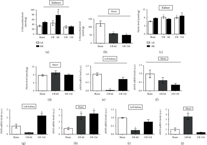
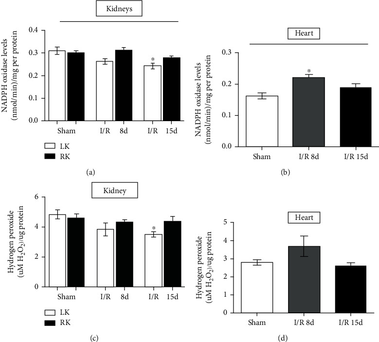
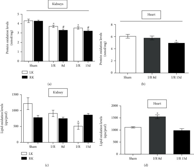
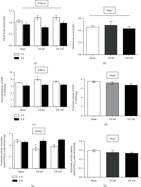
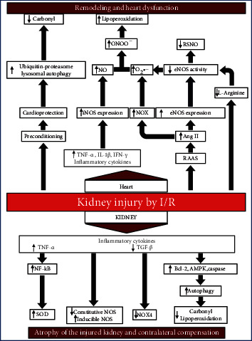
In kidney disease (KD), several factors released into the bloodstream can induce a series of changes in the heart, leading to a wide variety of clinical situations called cardiorenal syndrome (CRS). Reactive oxygen species (ROS) play an important role in the signaling and progression of systemic inflammatory conditions, as observed in KD. The aim of the present study was to characterize the redox balance in renal ischemia/reperfusion-induced cardiac remodeling. C57BL/6 male mice were subjected to occlusion of the left renal pedicle, unilateral, for 60 min, followed by reperfusion for 8 and 15 days, respectively. The following redox balance components were evaluated: catalase (CAT), superoxide dismutase (SOD), total antioxidant capacity (FRAP), NADPH oxidase (NOX), nitric oxide synthase (NOS), hydrogen peroxide (H2O2), and the tissue bioavailability of nitric oxide (NO) such as S-nitrosothiol (RSNO) and nitrite (NO2 -). The results indicated a process of renoprotection in both kidneys, indicated by the reduction of cellular damage and some oxidant agents. We also observed an increase in the activity of antioxidant enzymes, such as SOD, and an increase in NO bioavailability. In the heart, we noticed an increase in the activity of NOX and NOS, together with increased cell damage on day 8, followed by a reduction in protein damage on day 15. The present study concludes that the kidneys and heart undergo distinct processes of damage and repair at the analyzed times, since the heart is a secondary target of ischemic kidney injury. These results are important for a better understanding of the cellular mechanisms involved in CRS.


Conflict of interest statement

The authors declare that they have no conflict of interest.

Figures

Figure 1
Figure 1
S-Nitrosothiol, nitrite dosages, and mRNA levels in heart and kidney tissues. NO bioavailability in renal tissue. (a) Total tissue S-nitrosothiol concentration in the kidneys (n = 5). (b) Total tissue S-nitrosothiol concentration in the heart (n = 4). (c) Total tissue nitrite concentration in the kidneys (n = 5). (d) Total tissue nitrite concentration in the heart (n = 5). (e) Analysis of the gene expression of nNOS in the left kidney. (f) Analysis of the gene expression of nNOS in the heart. (g) Analysis of the gene expression of iNOS in the left kidney. (h) Analysis of the gene expression of iNOS in the heart. (i) Analysis of the gene expression of eNOS in the left kidney. (j) Analysis of the gene expression of eNOS in the heart. (sham, n = 5; I/R 8 d, n = 5; and I/R 15 d, n = 5). All samples were evaluated by real-time PCR. Data are expressed as the mean ± SEM. One-way ANOVA followed by the Bonferroni posttest for the selected pairs and number of animals within the bars. P < 0.05 with respect to the corresponding sham group.
Figure 2
Figure 2
Oxidant agents in heart and kidney tissues. (a) NADPH oxidase activity in the kidneys. (b) NADPH oxidase activity in the heart. (c) Hydrogen peroxide levels in the kidneys. (d) Hydrogen peroxide levels in the heart. Heart assays (sham, n = 10; I/R 8 d, n = 6; and I/R 15 d, n = 5) and kidney assays (sham, n = 6; I/R 8 d, n = 6; and I/R 15 d, n = 7). Data are expressed as the mean ± SEM. One-way ANOVA followed by the Bonferroni posttest for the selected pairs and number of animals within the bars. P < 0.05 with respect to the corresponding sham group.
Figure 3
Figure 3
Oxidative stress by protein and lipid damage in the heart and kidney tissues. (a) Protein oxidation by carbonyl groups in the kidneys. (b) Protein oxidation by carbonyl groups in the heart. (c) Lipid peroxidation by chemiluminescence in the kidneys. (d) Lipid peroxidation by chemiluminescence in the heart. Heart assays (sham, n = 8; I/R 8 d, n = 6; and I/R 15 d, n = 7) and kidney assays (sham, n = 7; I/R 8 d, n = 6; and I/R 15 d, n = 6). Data are expressed as the mean ± SEM. One-way ANOVA followed by the Bonferroni posttest for the selected pairs and number of animals within the bars. P < 0.05 with respect to the corresponding left kidney of the sham group. #P < 0.05 with respect to the corresponding right kidney of the sham group.
Figure 4
Figure 4
Antioxidant agents in heart and kidney tissues. (a) Total antioxidant activity by FRAP in the kidney. (b) Total antioxidant activity by FRAP in the heart. (c) Superoxide dismutase in the kidneys. (d) Superoxide dismutase in the heart. (e) Catalase activity in the kidneys. (f) Catalase activity in the heart. N = 6. Data are expressed as the mean ± SEM. One-way ANOVA followed by the Bonferroni posttest for the selected pairs and number of animals within the bars. P < 0.05 with respect to the corresponding sham group.
Figure 5
Figure 5
Flowchart of possible modulations in the model according to the bibliographic survey and data obtained here.

Similar articles

Cited by

References

    1. Di Lullo L., Reeves P. B., Bellasi A., Ronco C. Cardiorenal syndrome in acute kidney injury. Seminars in Nephrology. 2019;39(1):31–40. doi: 10.1016/j.semnephrol.2018.10.003. - DOI - PubMed
    1. Thurman J. M. Triggers of inflammation after renal ischemia/reperfusion. Clinical Immunology. 2007;123(1):7–13. doi: 10.1016/j.clim.2006.09.008. - DOI - PMC - PubMed
    1. Murphy S. P., Kakkar R., McCarthy C. P., Januzzi J. L. Inflammation in heart failure: JACC state-of-the-art review. Journal of the American College of Cardiology. 2020;75(11):1324–1340. doi: 10.1016/j.jacc.2020.01.014. - DOI - PubMed
    1. Li Q., Wei G., Tao T. Leukocyte immunoglobulin-like receptor B4 (LILRB4) negatively mediates the pathological cardiac hypertrophy by suppressing fibrosis, inflammation and apoptosis via the activation of NF-κB signaling. Biochemical and Biophysical Research Communications. 2019;509(1):16–23. doi: 10.1016/j.bbrc.2018.11.137. - DOI - PubMed
    1. Trentin-Sonoda M., da Silva R. C., Kmit F. V., et al. Knockout of toll-like receptors 2 and 4 prevents renal ischemia-reperfusion-induced cardiac hypertrophy in mice. PLoS One. 2015;10(10, article e0139350) doi: 10.1371/journal.pone.0139350. - DOI - PMC - PubMed

LinkOut - more resources