. 2020 Oct 10:2020:7161027.
doi: 10.1155/2020/7161027. eCollection 2020.

Ephrin-A5 Is Involved in Retinal Neovascularization in a Mouse Model of Oxygen-Induced Retinopathy

Affiliations

Ephrin-A5 Is Involved in Retinal Neovascularization in a Mouse Model of Oxygen-Induced Retinopathy

Wei Du et al. Biomed Res Int. .

Abstract

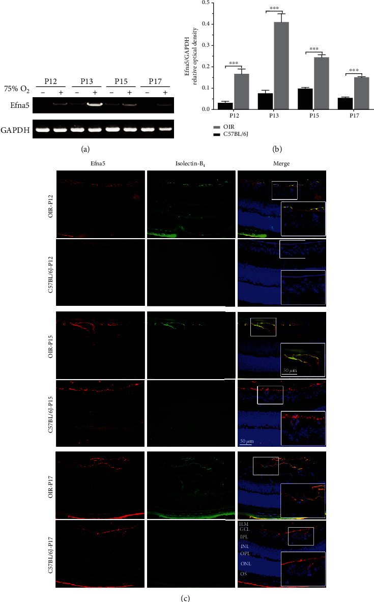
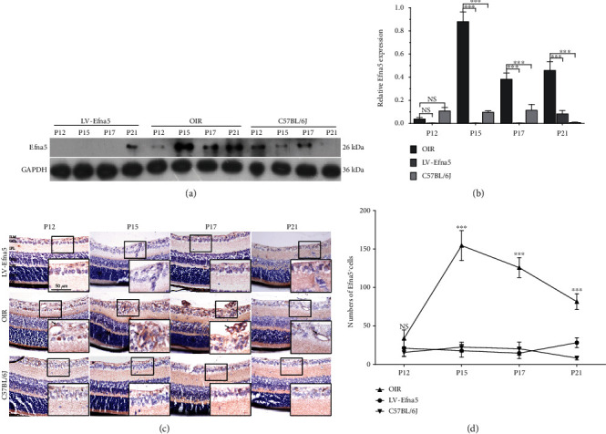
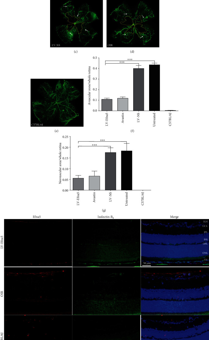
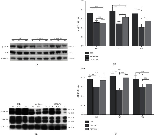
Retinal neovascularization (RNV) is an important pathological feature of vitreoretinopathy that can lead to severe vision loss. The purpose of this study was to identify the role of ephrin-A5 (Efna5) in RNV and to explore its mechanism. The expression pattern and biological significance of Efna5 were investigated in a mouse model of oxygen-induced retinopathy (OIR). The expression of Efna5 and downstream signaling pathway members was determined by RT-PCR, immunofluorescence, immunohistochemistry, and western blot analyses. shRNA was used to knockdown Efna5 in the retina of the OIR mouse model. Retinal flat mounts were performed to evaluate the impact of Efna5 silencing on the RNV process. We found that the Efna5 was greatly upregulated in the retina of OIR mice. Elevated Efna5 mainly colocalized with the retinal vessels and endothelial cells. We then showed that knockdown of Efna5 in OIR mouse retinas using lentivirus-mediated shRNA markedly decreased the expression of Efna5 and reduced the retinal neovascularization and avascular retina area. We further showed hypoxia stimulation dramatically increased both total and phosphorylation levels of ERK1/2 and the phosphorylation levels of Akt in OIR mice. More importantly, knockdown of Efna5 could inhibit the p-Akt and p-ERK signaling pathways. Our results suggested that Efna5 may regulate the RNV. This study suggests that Efna5 was significantly upregulated in the retina of OIR mice and closely involved in the pathological retinal angiogenesis.


Conflict of interest statement

The authors declare that there is no conflict of interest regarding the publication of this paper.

Figures

Figure 1
Figure 1
The expression patterns and distribution of Efna5 in OIR mice. (a) Retinas of OIR mice and C57BL/6J mice were harvested at P12, P13, P15, and P17, and the Efna5 mRNA level was measured with RT-PCR. The RT-PCR result was a representative of three independent experiments. (b) The relative expression level of Efna5 was calculated by normalizing the optical density of the Efna5 band to the GAPDH band. (c) Retinas of OIR mice and C57BL/6J mice were harvested at P12, P15, and P17. Immunofluorescence staining of Efna5 was performed with an anti-Efna5 antibody (red). Immunofluorescence staining of the endothelial cell was performed with Isolectin-B4 (green). Scale bars represent 50 μm; P: postnatal day; +: OIR mouse; -: C57BL/6J mouse; ILM: internal limiting membrane; GCL: ganglion cell layer; IPL: inner plexiform layer; INL: inner nuclear layer; OPL: outer plexiform layer; ONL: outer nuclear layer; OS: outer segment.
Figure 2
Figure 2
Intravitreal injection of LV-Efna5-shRNA effectively silenced Efna5 expression in OIR mouse retinas. (a) The protein level of Efna5 in the retina of OIR mice injected with LV-Efna5-shRNA (LV-Efna5), OIR mice, and C57BL/6J mice was determined by western blot analysis. (b) Relative quantification of protein bands from western blot film was performed (representative of three experiments). (c) Immunohistochemistry staining of Efna5 in the retina of OIR mice injected with LV-Efna5-shRNA (LV-Efna5), OIR mice, and C57BL/6J mice. (d) Counts of Efna5+ cells determined by IHC analysis (n = 4 animals per group). Scale bar represents 50 μm; P: postnatal day; ILM: internal limiting membrane; GCL: ganglion cell layer; IPL: inner plexiform layer; INL: inner nuclear layer; OPL: outer plexiform layer; ONL: outer nuclear layer; OS: outer segment; black arrow: Efna5+ cells; ∗∗∗P < 0.001; NS: not significant.
Figure 3
Figure 3
Intravitreal injection of LV-Efna5-shRNA markedly reduced the avascular area and neovascularization in the retinas. (a–e) After receiving intravitreal injections, mice from each group were perfused with fluorescein isothiocyanate- (FITC-) dextran (green) at P17; retinal flat mounts were prepared for each sample and analyzed by fluorescence microscopy. (f) Quantification of avascular areas in the retinas of OIR mice. Avascular areas were featured by a lack of fluorescence. (g) Quantification of the neovascular area in the retinas of OIR mice. Neovascularization areas were featured by hyperfluorescence. n = 5 animals per group. Scale bar represents 500 μm; area enclosed by yellow line: avascular area; white arrow: neovascular area; ∗∗∗P < 0.001. (h) Retinas of mice with indicated treatment were harvested at P17. Immunofluorescence staining was performed with anti-Efna5 antibody (red) and Isolectin-B4 (green). Scale bars represent 50 μm; ILM: internal limiting membrane; GCL: ganglion cell layer; IPL: inner plexiform layer; INL: inner nuclear layer; OPL: outer plexiform layer; ONL: outer nuclear layer; OS: outer segment.
Figure 4
Figure 4
Efna5 regulates the activity of ERK and AKT during RNV. (a, c) Western blot assays were used to determine the levels of total and phosphorylated protein Akt and ERK1/2. (b, d) Quantification analysis of western blot. Data are representative of 3 independent experiments. ∗∗∗P < 0.001; ∗∗P < 0.01; P < 0.05; NS: not significant.

Similar articles

Cited by

References

    1. Campochiaro P. A. Retinal and Choroidal Neovascularization. Journal of Cellular Physiology. 2000;184(3):301–310. doi: 10.1002/1097-4652(200009)184:3<301::AID-JCP3>3.0.CO;2-H. - DOI - PubMed
    1. Carmeliet P., Tessier-Lavigne M. Common mechanisms of nerve and blood vessel wiring. Nature. 2005;436(7048):193–200. doi: 10.1038/nature03875. - DOI - PubMed
    1. Sapieha P., Hamel D., Shao Z., et al. Proliferative retinopathies: angiogenesis that blinds. The International Journal of Biochemistry & Cell Biology. 2010;42(1):5–12. doi: 10.1016/j.biocel.2009.10.006. - DOI - PubMed
    1. Bhutto I., Lutty G. Understanding age-related macular degeneration (AMD): relationships between the photoreceptor/retinal pigment epithelium/Bruch's membrane/choriocapillaris complex. Molecular Aspects of Medicine. 2012;33(4):295–317. doi: 10.1016/j.mam.2012.04.005. - DOI - PMC - PubMed
    1. Wiedemann P. Growth factors in retinal diseases: proliferative vitreoretinopathy, proliferative diabetic retinopathy, and retinal degeneration. Survey of Ophthalmology. 1992;36(5):373–384. doi: 10.1016/0039-6257(92)90115-A. - DOI - PubMed

MeSH terms

LinkOut - more resources