

Deceived by a CT Scan: The Case of the Misrepresented Stone Size
- PMID: 33102703
- PMCID: PMC7580570
- DOI: 10.1089/cren.2019.0127
Deceived by a CT Scan: The Case of the Misrepresented Stone Size
Abstract
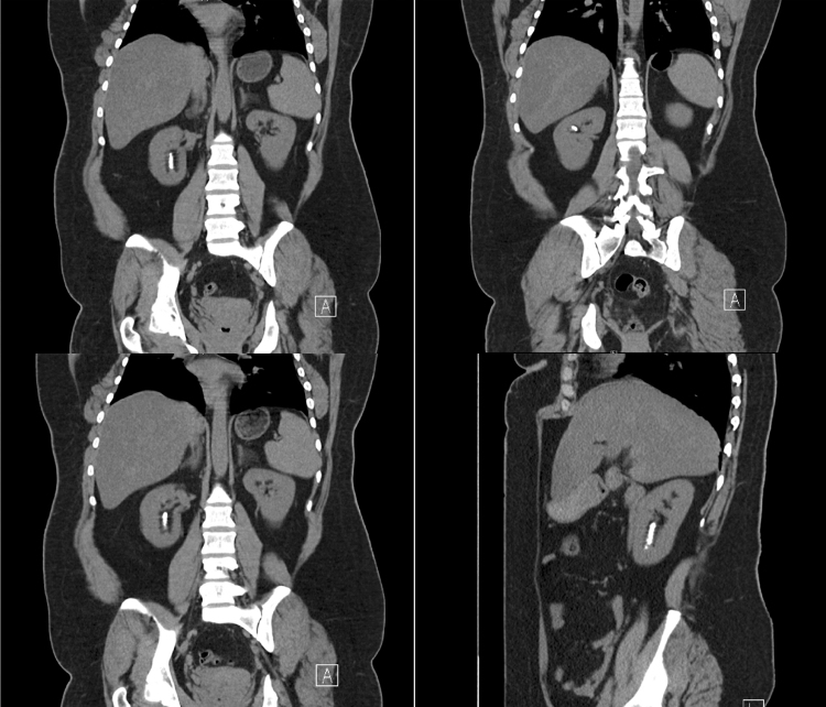
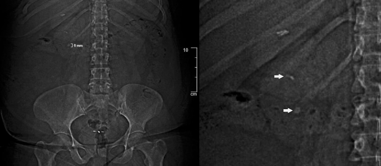
Background: CT has become the gold standard for radiographic evaluation of urolithiasis. CT is highly sensitive for detecting kidney stones and provides valuable information regarding stone size, composition, location, and overall stone burden. Although CT can provide reliable estimations of stone size, we have encountered an instance in which it can be deceiving. Motion artifact in CT images can cause a warping distortion effect that makes renal stones appear larger than they actually are. Case Presentation: We describe a case of a 37-year-old woman with a history of kidney stones and obesity presenting with intermittent flank pain and gross hematuria, found to have a large lower pole renal calculus that appeared deceptively large on CT imaging. Given the apparent size and location of the stone, the patient was counseled and consented for a percutaneous nephrolithotomy (PCNL). Although the stone was initially suspected to be >2 cm based on the preoperative CT scan, intraoperative pyelography revealed a much smaller than expected radio-dense stone. The patient was stone free after PCNL without any immediate postoperative complications. However, her course was later complicated by delayed bleeding causing significant clot hematuria, perinephric hematoma, and reactive pleural effusion. Conclusion: Although CT is especially valuable in preparing for surgery based on its ability to outline collecting system anatomy, it is important to remember that it can be deceiving. Correlation with kidney, ureter, and bladder radiograph and ultrasound is critical to understanding the clinical case and planning the optimal surgical approach.
Keywords: CT imaging; percutaneous nephrolithotomy; urolithiasis.
Copyright 2020, Mary Ann Liebert, Inc., publishers.
Conflict of interest statement
No competing financial interests exist.
Figures
References
-
- Renard-Penna R, Martin A, Conort P, et al. . Kidney stones and imaging: What can your radiologist do for you? World J Urol 2015;33:193. - PubMed
-
- Tisdale BE, Siemens DR, Lysack J, Nolan RL, Wilson JW. Correlation of CT scan versus plain radiography for measuring urinary stone dimensions. Can J Urol 2007;14:3489–3492 - PubMed
-
- Grosjean R, Sauer B, Guerra RM, Daudon M, Blum A, Felblinger J, Hubert J. Characterization of human renal stones with MDCT: Advantage of dual energy and limitations due to respiratory motion. Am J Roentgenol 2008;190:720–728 - PubMed
-
- Albala DM, Assimos DG, Clayman RV, et al. . Lower pole I: A prospective randomized trial of extracorporeal shock wave lithotripsy and percutaneous nephrostolithotomy for lower pole nephrolithiasis-initial results. J Urol 2001;166:2072–2080 - PubMed
LinkOut - more resources
Full Text Sources