Screening and follow-up of chronic liver diseases with understanding their etiology in clinics and hospitals
- PMID: 33102751
- PMCID: PMC7578295
- DOI: 10.1002/jgh3.12406
Screening and follow-up of chronic liver diseases with understanding their etiology in clinics and hospitals
Abstract
Background and aim: Considering the increasing prevalence of non-alcoholic fatty liver disease and non-alcoholic steatohepatitis (NASH), the development of an effective screening and follow-up system that enables the recognition of etiological changes by primary physicians in clinics and specialists in hospitals is required.
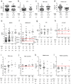
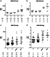
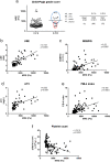
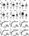
Methods: Chronic hepatitis B (HBV) and C (HCV), NASH, and alcoholic steatohepatitis (ASH) patients who were assayed for Mac-2-binding protein glycosylation isomer (M2BPGi) (n = 272) and underwent magnetic resonance elastography (MRE) (n = 119) were enrolled. Patients who underwent MRE were also tested by ultrasound elastography (USE) (n = 80) and for M2BPGi (n = 97), autotaxin (ATX) (n = 62), and platelet count (n = 119), and their fibrosis-4 (FIB-4) index was calculated (n = 119).
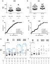
Results: FIB-4 index >2, excluding HBV-infected patients, M2BPGi >0.5, ATX >0.5, and platelet count <20 × 104/μL were the benchmark indices, and we took into consideration other risk factors, such as diabetes mellitus and age, to recommend further examinations, such as USE, based on the local situation to avoid overlooking hepatocellular carcinoma (HCC) in the clinic. During specialty care in the hospital, MRE exhibited high diagnostic ability for fibrosis stages >F3 or F4; it could efficiently predict collateral circulation with high sensitivity, which can replace USE. We also identified etiological features and found that collateral circulation in NASH/ASH patients tended to exceed high-risk levels; moreover, these patients exhibited more variation in HCC-associated liver stiffness than the HBV and HCV patients.
Conclusions: Using appropriate markers and tools, we can establish a stepwise, practical, noninvasive, and etiology-based screening and follow-up system in primary and specialty care.
Keywords: Mac‐2‐binding protein glycosylation isomer; autotaxin; fibrosis‐4 index; hepatocellular carcinoma; magnetic resonance elastography.
© 2020 The Authors. JGH Open published by Journal of Gastroenterology and Hepatology Foundation and John Wiley & Sons Australia, Ltd.
Figures


Similar articles
-
Diagnostic performance of Mac-2 binding protein glycosylation isomer (M2BPGi) in screening liver fibrosis in health checkups.J Clin Lab Anal. 2020 Aug;34(8):e23316. doi: 10.1002/jcla.23316. Epub 2020 Mar 29. J Clin Lab Anal. 2020. PMID: 32227396 Free PMC article.
-
Mac-2 binding protein glycosylation isomer, the FIB-4 index, and a combination of the two as predictors of non-alcoholic steatohepatitis.PLoS One. 2022 Nov 10;17(11):e0277380. doi: 10.1371/journal.pone.0277380. eCollection 2022. PLoS One. 2022. PMID: 36355761 Free PMC article.
-
Liver fibrosis assessments using FibroScan, virtual-touch tissue quantification, the FIB-4 index, and mac-2 binding protein glycosylation isomer levels compared with pathological findings of liver resection specimens in patients with hepatitis C infection.BMC Gastroenterol. 2020 Sep 25;20(1):314. doi: 10.1186/s12876-020-01459-w. BMC Gastroenterol. 2020. PMID: 32977741 Free PMC article.
-
Diagnostic accuracy of elastography and magnetic resonance imaging in patients with NAFLD: A systematic review and meta-analysis.J Hepatol. 2021 Oct;75(4):770-785. doi: 10.1016/j.jhep.2021.04.044. Epub 2021 May 13. J Hepatol. 2021. PMID: 33991635
-
Clinical Utility of Mac-2 Binding Protein Glycosylation Isomer in Chronic Liver Diseases.Ann Lab Med. 2021 Jan;41(1):16-24. doi: 10.3343/alm.2021.41.1.16. Epub 2020 Aug 25. Ann Lab Med. 2021. PMID: 32829576 Free PMC article. Review.
Cited by
-
Transition of clinical and basic studies on liver cirrhosis treatment using cells to seek the best treatment.Inflamm Regen. 2021 Sep 16;41(1):27. doi: 10.1186/s41232-021-00178-3. Inflamm Regen. 2021. PMID: 34530931 Free PMC article. Review.
-
Pruritus in Chronic Cholestatic Liver Diseases, Especially in Primary Biliary Cholangitis: A Narrative Review.Int J Mol Sci. 2025 Feb 22;26(5):1883. doi: 10.3390/ijms26051883. Int J Mol Sci. 2025. PMID: 40076514 Free PMC article. Review.
-
Diagnosis and Therapeutic Management of Liver Fibrosis by MicroRNA.Int J Mol Sci. 2021 Jul 29;22(15):8139. doi: 10.3390/ijms22158139. Int J Mol Sci. 2021. PMID: 34360904 Free PMC article. Review.
-
Fibulin-4 as a potential extracellular vesicle marker of fibrosis in patients with cirrhosis.FEBS Open Bio. 2024 Aug;14(8):1264-1276. doi: 10.1002/2211-5463.13842. Epub 2024 Jun 9. FEBS Open Bio. 2024. PMID: 38853023 Free PMC article.
References
-
- Feld JJ, Jacobson IM, Hezode C et al Sofosbuvir and velpatasvir for HCV genotype 1, 2, 4, 5, and 6 infection. N. Engl. J. Med. 2015; 373: 2599–607. - PubMed
-
- Zeuzem S, Foster GR, Wang S et al Glecaprevir‐pibrentasvir for 8 or 12 weeks in HCV genotype 1 or 3 infection. N. Engl. J. Med. 2018; 378: 354–69. - PubMed
-
- Tang LSY, Covert E, Wilson E, Kottilil S. Chronic hepatitis B infection: a review. JAMA. 2018; 319: 1802–13. - PubMed
LinkOut - more resources
Full Text Sources
Research Materials
Miscellaneous

