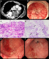
Refractory hemorrhagic esophageal ulcers by Candida esophagitis with advanced systemic sclerosis
- PMID: 33102777
- PMCID: PMC7578314
- DOI: 10.1002/jgh3.12353
Refractory hemorrhagic esophageal ulcers by Candida esophagitis with advanced systemic sclerosis
Abstract
A 64-year-old woman diagnosed with rheumatoid arthritis (RA) and systemic sclerosis (SSc) was admitted to our hospital with chief complaints of uncontrolled bleeding from esophageal ulcers and an inability to consume meals. For RA and SSc, she was treated with prednisolone and abatacept and was taking vonoprazan as prophylaxis for steroid-induced gastric ulcers. She was diagnosed with severe Candida esophagitis, with multiple large and small ulcers with bleeding, based on esophagogastroduodenoscopy and pathological findings. We performed comprehensive treatment; abatacept was discontinued, and total parenteral nutrition was initiated along with antifungal therapy. Improvement in the esophageal ulcers was observed. Although severe Candida esophagitis is a rare condition, we should keep in mind that severe Candida esophagitis can occur in patients with an immunosuppressive compromised host and esophageal movement disorders such as SSc. Regular follow up by endoscopy and prophylactic treatment to prevent severe esophagitis may be necessary.
© 2020 The Authors. JGH Open: An open access journal of gastroenterology and hepatology published by Journal of Gastroenterology and Hepatology Foundation and John Wiley & Sons Australia, Ltd.
Conflict of interest statement
The authors declare no conflict of interest.
Figures

Similar articles
-
Esophageal involvement in scleroderma: gastroesophageal reflux, the common problem.Semin Arthritis Rheum. 2006 Dec;36(3):173-81. doi: 10.1016/j.semarthrit.2006.08.002. Epub 2006 Oct 11. Semin Arthritis Rheum. 2006. PMID: 17045629 Review.
-
Clinical experience of esophageal ulcers and esophagitis in AIDS patients.Kaohsiung J Med Sci. 1996 Nov;12(11):624-9. Kaohsiung J Med Sci. 1996. PMID: 8953856
-
[A case of Behçet's disease with esophageal ulcers complicated with systemic sclerosis, chronic hepatitis C, and pancytopenia].Nihon Rinsho Meneki Gakkai Kaishi. 2004 Jun;27(3):164-70. doi: 10.2177/jsci.27.164. Nihon Rinsho Meneki Gakkai Kaishi. 2004. PMID: 15291253 Review. Japanese.
-
Etiology of esophageal disease in human immunodeficiency virus-infected patients who fail antifungal therapy.Am J Med. 1996 Dec;101(6):599-604. doi: 10.1016/s0002-9343(96)00303-8. Am J Med. 1996. PMID: 9003106
-
[Esophageal pathology in patients with the AIDS virus. Etiology and diagnosis].Acta Gastroenterol Latinoam. 1991;21(2):67-83. Acta Gastroenterol Latinoam. 1991. PMID: 1820692 Spanish.
Cited by
-
Clinical Implications of Proton Pump Inhibitors and Vonoprazan Micro/Nano Drug Delivery Systems for Gastric Acid-Related Disorders and Imaging.Nanotheranostics. 2024 Sep 30;8(4):535-560. doi: 10.7150/ntno.100727. eCollection 2024. Nanotheranostics. 2024. PMID: 39507107 Free PMC article. Review.
-
Genotyping, antifungal susceptibility, enzymatic activity, and phenotypic variation in Candida albicans from esophageal candidiasis.J Clin Lab Anal. 2021 Jul;35(7):e23826. doi: 10.1002/jcla.23826. Epub 2021 May 14. J Clin Lab Anal. 2021. PMID: 33988259 Free PMC article.
-
Dysphagia, reflux and related sequelae due to altered physiology in scleroderma.World J Gastroenterol. 2021 Aug 21;27(31):5201-5218. doi: 10.3748/wjg.v27.i31.5201. World J Gastroenterol. 2021. PMID: 34497445 Free PMC article. Review.
References
-
- Villadsen GE, Storkholm J, Zachariae H, Hendel L, Bendtsen F, Gregersen H. Oesophageal pressure‐cross‐sectional area distributions and secondary peristalsis in relation to subclassification of systemic sclerosis. Neurogastroenterol. Motil. 2001; 13: 199–210. - PubMed
-
- Clements PJ, Becvar R, Drosos AA, Ghattas L, Gabrielli A. Assessment of gastrointestinal involvement. Clin. Exp. Rheumatol. 2003; 21: S15–8. - PubMed
-
- Kirby DF, Chatterjee S. Evaluation and management of gastrointestinal manifestations in scleroderma. Curr. Opin. Rheumatol. 2014; 26: 621–9. - PubMed
Publication types
LinkOut - more resources
Full Text Sources

