

Novel ultrashort self-assembling peptide bioinks for 3D culture of muscle myoblast cells
- PMID: 33102913
- PMCID: PMC7582005
- DOI: 10.18063/IJB.v4i2.129
Novel ultrashort self-assembling peptide bioinks for 3D culture of muscle myoblast cells
Abstract
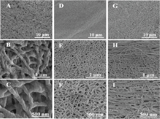
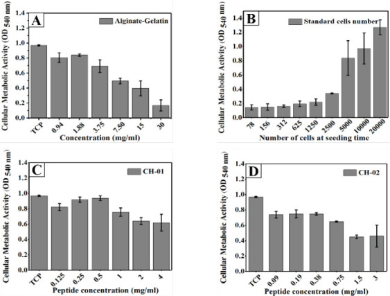
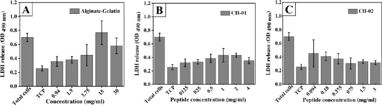
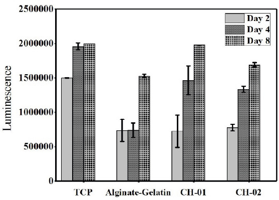
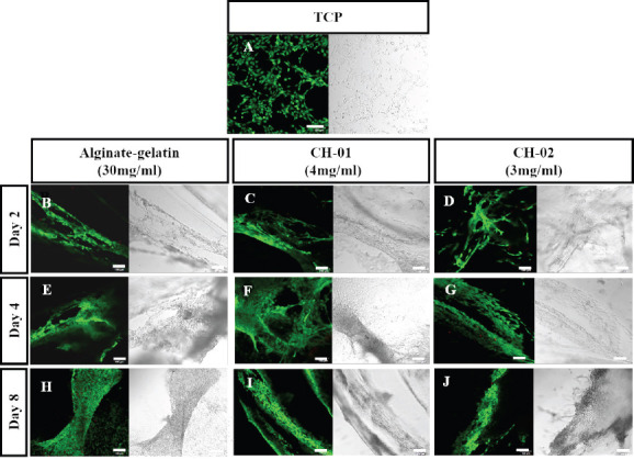
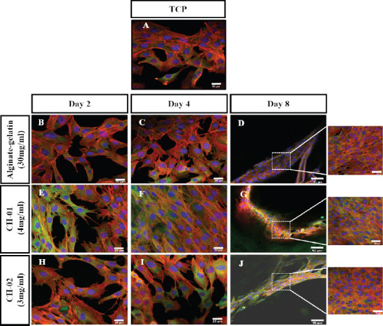
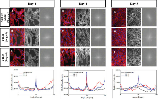
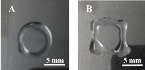
The ability of skeletal muscle to self-repair after a traumatic injury, tumor ablation, or muscular disease is slow and limited, and the capacity of skeletal muscle to self-regenerate declines steeply with age. Tissue engineering of functional skeletal muscle using 3D bioprinting technology is promising for creating tissue constructs that repair and promote regeneration of damaged tissue. Hydrogel scaffolds used as biomaterials for skeletal muscle tissue engineering can provide chemical, physical and mechanical cues to the cells in three dimensions thus promoting regeneration. Herein, we have developed two synthetically designed novel tetramer peptide biomaterials. These peptides are self-assembling into a nanofibrous 3D network, entrapping 99.9% water and mimicking the native collagen of an extracellular matrix. Different biocompatibility assays including MTT, 3D cell viability assay, cytotoxicity assay and live-dead assay confirm the biocompatibility of these peptide hydrogels for mouse myoblast cells (C2C12). Immunofluorescence analysis of cell-laden hydrogels revealed that the proliferation of C2C12 cells was well-aligned in the peptide hydrogels compared to the alginategelatin control. These results indicate that these peptide hydrogels are suitable for skeletal muscle tissue engineering. Finally, we tested the printability of the peptide bioinks using a commercially available 3D bioprinter. The ability to print these hydrogels will enable future development of 3D bioprinted scaffolds containing skeletal muscle myoblasts for tissue engineering applications.
Keywords: 3D cell culture; 3D scaffold; bioinks; biomaterials; skeletal muscle cells; tissue engineering.
Copyright: © 2018 Arab W, et al.
Conflict of interest statement
The authors declare that they do not have any competing interests.
Figures
Similar articles
-
Exploring nanofibrous self-assembling peptide hydrogels using mouse myoblast cells for three-dimensional bioprinting and tissue engineering applications.Int J Bioprint. 2019 Jul 20;5(2):198. doi: 10.18063/ijb.v5i2.198. eCollection 2019. Int J Bioprint. 2019. PMID: 32596536 Free PMC article.
-
Myoblast 3D bioprinting to burst in vitro skeletal muscle differentiation.J Tissue Eng Regen Med. 2022 May;16(5):484-495. doi: 10.1002/term.3293. Epub 2022 Mar 4. J Tissue Eng Regen Med. 2022. PMID: 35246958 Free PMC article.
-
3D bioprinting of ultrashort self-assembling peptides to engineer scaffolds with different matrix stiffness for chondrogenesis.Int J Bioprint. 2023 Mar 24;9(4):719. doi: 10.18063/ijb.719. eCollection 2023. Int J Bioprint. 2023. PMID: 37323500 Free PMC article.
-
Advancing bioinks for 3D bioprinting using reactive fillers: A review.Acta Biomater. 2020 Sep 1;113:1-22. doi: 10.1016/j.actbio.2020.06.040. Epub 2020 Jul 2. Acta Biomater. 2020. PMID: 32622053 Review.
-
Nanocomposite bioinks for 3D bioprinting.Acta Biomater. 2022 Oct 1;151:45-69. doi: 10.1016/j.actbio.2022.08.014. Epub 2022 Aug 13. Acta Biomater. 2022. PMID: 35970479 Review.
Cited by
-
Bioprinting science.Int J Bioprint. 2018 Jul 9;4(2):149. doi: 10.18063/IJB.v4i2.149. eCollection 2018. Int J Bioprint. 2018. PMID: 33102922 Free PMC article. No abstract available.
-
Applications of self-assembling ultrashort peptides in bionanotechnology.RSC Adv. 2019 Jan 8;9(2):844-852. doi: 10.1039/c8ra07533f. eCollection 2019 Jan 2. RSC Adv. 2019. PMID: 35517614 Free PMC article. Review.
-
Three-Dimensional (3D) Printing in Cancer Therapy and Diagnostics: Current Status and Future Perspectives.Pharmaceuticals (Basel). 2022 May 27;15(6):678. doi: 10.3390/ph15060678. Pharmaceuticals (Basel). 2022. PMID: 35745597 Free PMC article. Review.
-
Peptide nanogels as a scaffold for fabricating dermal grafts and 3D vascularized skin models.J Tissue Eng. 2022 Jul 29;13:20417314221111868. doi: 10.1177/20417314221111868. eCollection 2022 Jan-Dec. J Tissue Eng. 2022. PMID: 35923174 Free PMC article.
-
Preparation and printability of ultrashort self-assembling peptide nanoparticles.Int J Bioprint. 2019 Jul 31;5(2):239. doi: 10.18063/ijb.v5i2.239. eCollection 2019. Int J Bioprint. 2019. PMID: 32596541 Free PMC article.
References
-
- Stilhano R S, Madrigal J L, Wong K, et al. Injectable alginate hydrogel for enhanced spatiotemporal control of lentivector delivery in murine skeletal muscle. J Control Release. 2016;237:42–49. http://dx.doi.org/10.1016/j.jconrel.2016.06.047. - PubMed
-
- Chaturvedi V, Dye D E, Kinnear B F, et al. Interactions between skeletal muscle myoblasts and their extracellular matrix revealed by a serum free culture system. PLO S. 2015;10(6):1–27. https://dx.doi.org/10.1371/journal. pone.0127675. - PMC - PubMed
-
- Järvinen T A H, Järvinen T L N, Kääriäinen M, et al. Muscle injuries:Optimising recovery. Best Pract Res Clin Rheumatol. 2007;2(2):317–331. http://dx.doi.org/10.1016/berh.2006.12.004. - PubMed
-
- Grasman J M, Zayas M J, Page R L, et al. Biomimetic scaffolds for regeneration of volumetric muscle loss in skeletal muscle injuries. Acta Biomater. 2015;25:2–15. http://dx.doi.org/10.1016/j.actbio.2015.07.038. - PMC - PubMed
LinkOut - more resources
Full Text Sources