

A multi-scale porous scaffold fabricated by a combined additive manufacturing and chemical etching process for bone tissue engineering
- PMID: 33102914
- PMCID: PMC7582010
- DOI: 10.18063/IJB.v4i2.133
A multi-scale porous scaffold fabricated by a combined additive manufacturing and chemical etching process for bone tissue engineering
Abstract
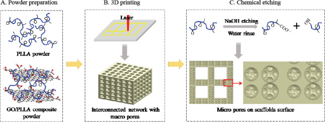
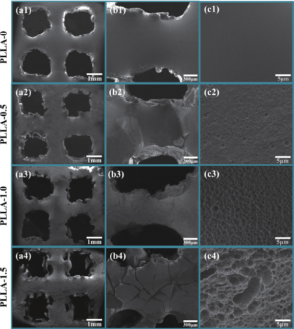
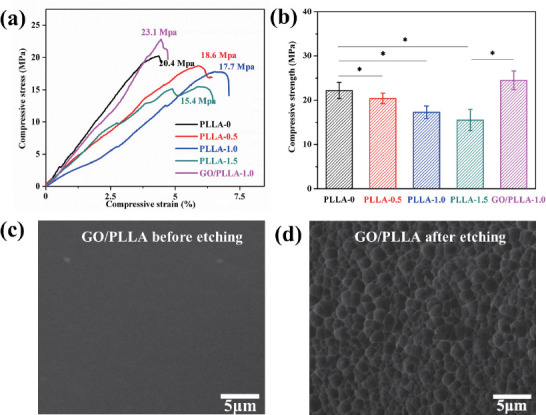
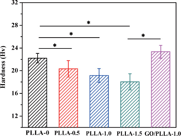
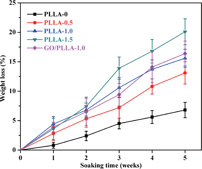
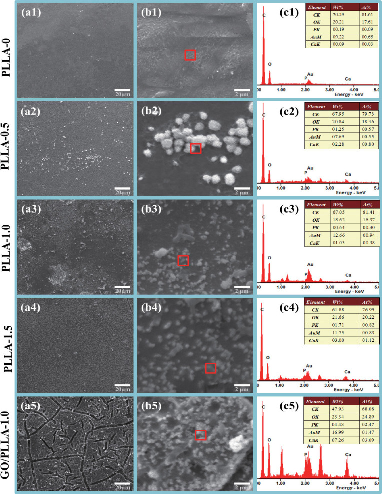
It is critical to develop a fabrication technology for precisely controlling an interconnected porous structure of scaffolds to mimic the native bone microenvironment. In this work, a novel combined process of additive manufacturing (AM) and chemical etching was developed to fabricate graphene oxide/poly(L-lactic acid) (GO/PLLA) scaffolds with multiscale porous structure. Specially, AM was used to fabricate an interconnected porous network with pore sizes of hundreds of microns. And the chemical etching in sodium hydroxide solution constructed pores with several microns or even smaller on scaffolds surface. The degradation period of the scaffolds was adjustable via controlling the size and quantity of pores. Moreover, the scaffolds exhibited surprising bioactivity after chemical etching, which was ascribed to the formed polar groups on scaffolds surfaces. Furthermore, GO improved the mechanical strength of the scaffolds.
Keywords: PLLA; additive manufacturing; chemical etching; multi-scale pores; scaffolds.
Copyright: © 2018 Shuai C et al.
Conflict of interest statement
No conflict of interest was reported by the authors. The authors gratefully acknowledge the following projects and funds for the financial support: (1) The Natural Science Foundation of China (51575537, 81572577, 51705540); (2) Hunan Provincial Natural Science Foundation of China (2016JJ1027); (3) The Project of Innovation-driven Plan of Central South University (2016CX023); (4) The Open-End Fund for the Valuable and Precision Instruments of Central South University; (5) The fund of the State Key Laboratory of Solidification Processing at NWPU (SKLSP201605); (6) The Project of State Key Laboratory of High Performance Complex Manufacturing, Central South University; (7) National Postdoctoral Program for Innovative Talents (BX201700291); (8) The Project of Hunan Provincial Science and Technology Plan (2017RS3008) and (9) The Fundamental Research Funds for the Central Universities of Central South University (2016zzts046).
Figures
References
-
- Tavolaro P, Catalano S, Martino G, et al. Zeolite inorganic scaffolds for novel biomedical application:Effect of physicochemical characteristic of zeolite membranes on cell adhesion and viability. Appi Surf Sci. 2016;380:135–140. http://doi.org/10.1016/j.apsusc.2016.01.279.
-
- Puppi D, Piras A M, Pirosa A, et al. Levofloxacinloaded star poly^-caprolactone) scaffolds by additive manufacturing. J Mater Sci Mater Med. 2016;27(3):44. http://doi.org/10.1007/s10856-015-5658-1. - PubMed
-
- Fradique R, Correia T R, Miguel S P, et al. Production of new 3D scaffolds for bone tissue regeneration by rapid prototyping. J Mater Sci Mater Med. 2016;27(4):1–14. http://doi.org/10.1007/s10856-016-5681-x. - PubMed
-
- Gao C, Deng Y, Feng P, et al. Current progress in bioactive ceramic scaffolds for bone repair and regeneration. Int J Mol Sci. 2014;15(3):4714–4732. http://doi.org/10.3390/ijms15034714. - PMC - PubMed
-
- Yuan H, Zhou Q, Li B, et al. Direct printing of patterned three-dimensional ultrafine fibrous scaffolds by stable jet electrospinning for cellular ingrowth. Biofabrication. 2015;7(4):045004. http://doi.org/10.1088/1758-5090/7/4/045004. - PubMed
LinkOut - more resources
Full Text Sources