

Successful repair of a spontaneous scleral rupture in a patient with type VI Ehlers-Danlos syndrome
- PMID: 33102932
- PMCID: PMC7575778
- DOI: 10.1016/j.ajoc.2020.100961
Successful repair of a spontaneous scleral rupture in a patient with type VI Ehlers-Danlos syndrome
Abstract
Purpose: To describe ocular findings in a patient with Type VI Ehlers-Danlos syndrome (EDS) and make ophthalmologists aware of the potential ophthalmic complications of this particular type of EDS. To briefly report the surgical technique utilized for the repair of spontaneous scleral rupture that may be associated with Type VI Ehlers-Danlos syndrome.
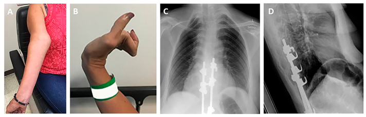
Observations: A 36-year-old female visited the Emergency Room due to sudden vision loss, edema, and redness of the right eye consistent with spontaneous scleral rupture secondary to scleral thinning due to Type VI EDS. Repair with scleral patch graft resulted in improvement in visual acuity, a decrease in hyphema, and discomfort resolution.
Conclusions and importance: Spontaneous scleral perforation may occur in patients with Type VI EDS. A scleral patch graft may serve as a viable surgical repair alternative for such patients.
Keywords: Ehlers danlos syndrome; Scleral patch graft; Scleral rupture.
© 2020 The Authors.
Conflict of interest statement
The following authors have no financial disclosures: RL, CA, SA, NI, AO.
Figures

Similar articles
-
RD repair using 360-degree scleral graft for extensive scleral ectasia in a patient with Ehlers Danlos syndrome.Am J Ophthalmol Case Rep. 2019 Sep 13;17:100554. doi: 10.1016/j.ajoc.2019.100554. eCollection 2020 Mar. Am J Ophthalmol Case Rep. 2019. PMID: 32083221 Free PMC article.
-
[Retinal detachment in Ehlers-Danlos syndrome. Treatment by pars plana vitrectomy].Ophthalmologe. 1997 Sep;94(9):634-7. doi: 10.1007/s003470050173. Ophthalmologe. 1997. PMID: 9410229 German.
-
Homograft of preserved sclera for post-traumatic scleral staphyloma in Ehlers-Danlos syndrome.Graefes Arch Clin Exp Ophthalmol. 1986;224(3):247-50. doi: 10.1007/BF02143064. Graefes Arch Clin Exp Ophthalmol. 1986. PMID: 3710179
-
Vascular type Ehlers-Danlos Syndrome with fatal spontaneous rupture of a right common iliac artery dissection: case report and review of literature.J Radiol Case Rep. 2014 Feb 1;8(2):63-9. doi: 10.3941/jrcr.v8i2.1568. eCollection 2014 Feb. J Radiol Case Rep. 2014. PMID: 24967021 Free PMC article. Review.
-
Management of colonic complications of type IV Ehlers-Danlos syndrome: a systematic review and evidence-based management strategy.Colorectal Dis. 2020 Feb;22(2):129-135. doi: 10.1111/codi.14749. Epub 2019 Jul 31. Colorectal Dis. 2020. PMID: 31260161
Cited by
-
Connective tissue disorders and eye: A review and recent updates.Indian J Ophthalmol. 2023 Jun;71(6):2385-2398. doi: 10.4103/ijo.IJO_286_22. Indian J Ophthalmol. 2023. PMID: 37322648 Free PMC article. Review.
-
Ehlers-Danlos syndromes and their manifestations in the visual system.Front Med (Lausanne). 2022 Sep 27;9:996458. doi: 10.3389/fmed.2022.996458. eCollection 2022. Front Med (Lausanne). 2022. PMID: 36237549 Free PMC article. Review.
References
-
- Giunta C., Baumann M., Fauth C. A cohort of 17 patients with kyphoscoliotic Ehlers-Danlos syndrome caused by biallelic mutations in FKBP14: expansion of the clinical and mutational spectrum and description of the natural history. Genet Med. 2018;20(1):42–54. doi: 10.1038/gim.2017.70. - DOI - PMC - PubMed
Publication types
LinkOut - more resources
Full Text Sources
