Review
. 2020 Oct 10;6(4):998-1011.
doi: 10.1016/j.bioactmat.2020.09.030. eCollection 2021 Apr.

Advanced hydrogels for the repair of cartilage defects and regeneration

Affiliations
Review

Advanced hydrogels for the repair of cartilage defects and regeneration

Wei Wei et al. Bioact Mater. .

Abstract

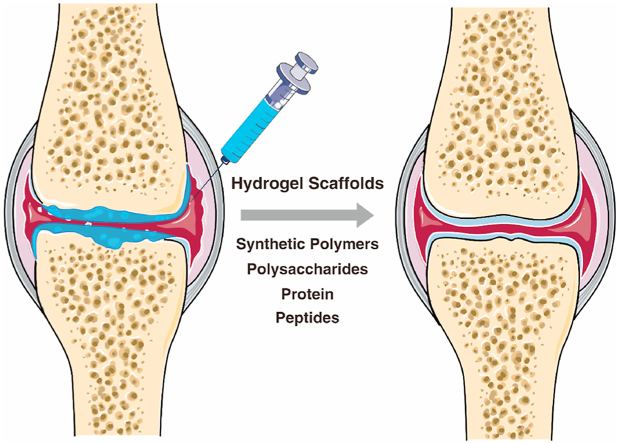
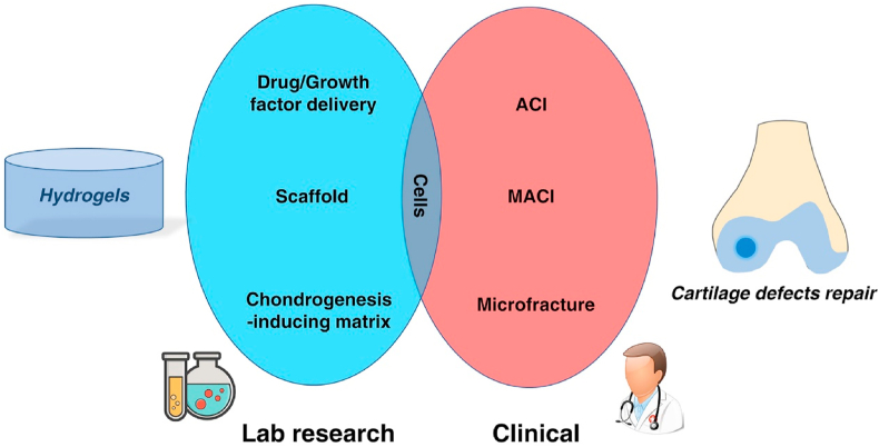
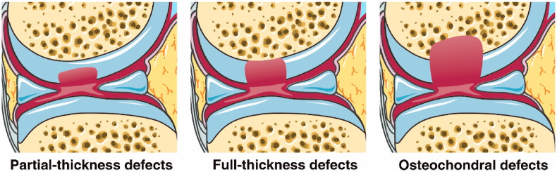
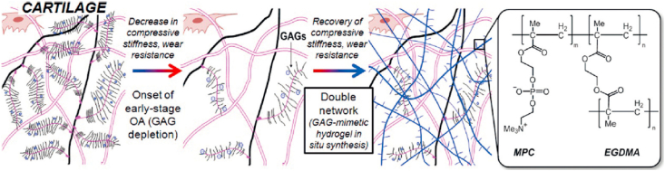
Cartilage defects are one of the most common symptoms of osteoarthritis (OA), a degenerative disease that affects millions of people world-wide and places a significant socio-economic burden on society. Hydrogels, which are a class of biomaterials that are elastic, and display smooth surfaces while exhibiting high water content, are promising candidates for cartilage regeneration. In recent years, various kinds of hydrogels have been developed and applied for the repair of cartilage defects in vitro or in vivo, some of which are hopeful to enter clinical trials. In this review, recent research findings and developments of hydrogels for cartilage defects repair are summarized. We discuss the principle of cartilage regeneration, and outline the requirements that have to be fulfilled for the deployment of hydrogels for medical applications. We also highlight the development of advanced hydrogels with tailored properties for different kinds of cartilage defects to meet the requirements of cartilage tissue engineering and precision medicine.

Keywords: Articular cartilage defects; Clinical translation; Hydrogels; Precision medicine; Tissue engineering.


Conflict of interest statement

The authors declare no conflict of interest.

Figures

Image 1
Graphical abstract
Fig. 1
Fig. 1
Schematic of relationship of hydrogel in lab and cartilage defects repair in clinical. ACI: autologous chondrocyte implantation; MACI: Matrix-induced autologous chondrocyte implantation.
Fig. 2
Fig. 2
Schematic diagram of three kinds of cartilage defects: partial-thickness defects, full-thickness defects, and osteochondral defects. Partial-thickness defects happen in the cartilage surface and do not penetrate the tidemark. No MSC from bone marrow will migrate or be recruited to the defect areas. Full-thickness defects are deeper cartilage defects to the tidemark but do not penetrate the subchondral bone. The environment of subchondral bone matrix is conducive to the migration and adhesion of MSC. Osteochondral defects penetrate the bone marrow and allow MSCs to be recruited to the defect area.
Fig. 3
Fig. 3
Schematic representation of normal cartilage (left), osteoarthritic cartilage (middle), and hydrogel-reinforced cartilage (right). GAG depletion decreases compressive stiffness and wear resistance of cartilage. To recover lost properties, a GAG-mimetic hydrogel was made by polymerizing hydrophilic zwitterionic monomers 2-methacryloyloxyethyl phosphorylcholine (MPC) using ethylene glycol dimethacrylate (EGDMA) as a crosslinker [Reprinted from Refs. [41] with permission from Wiley].
Fig. 4
Fig. 4
Macromer solutions of 4-arm PEG norbornene, MMP fluorescent sensor, TGF-β1, and tethered growth factor were mixed with chondrocytes at 40 million cells/mL. The final hydrogel scaffold networks were UV cross-linked by degradable linker (MMP-degradable peptide sequence) or non-degradable linker (3.5 kDa PEG dithiol) [Reprinted from Refs. [50] with permission from Wiley].
Fig. 5
Fig. 5
Schematic of CS containing hydrogel for cartilage tissue engineering. (a) CS was complexed with polydopamine (PDA). (b) CS was encapsulated in a PAM (polyacrylamide) hydrogel. (c) The CS containing hydrogel was implanted in a cartilage defect. (d) and (e) CS and polydopamine promoted cell adhesion on the hydrogel surfaces [Reprinted from Refs. [57] with permission from ACS Publications © 2018 American Chemical Society].
Fig. 6
Fig. 6
(A) PLGA nanoparticles loaded with KGN, molecule structure of KGN, methacrylated HA (HAMA). (B) The brief operative treatment for cartilage defects repair. (C) The release of KGN from photo-crosslinked HA scaffold and the hyaline cartilage chondrogenesis [Reprinted from Ref. [67] with permission from ACS Publications © 2016 American Chemical Society].
Fig. 7
Fig. 7
Schematic illustration of preparation of oxidized dextran (A) and GelMA(B). (A) Dextran was oxidized by NaIO4 to generate aldehyde groups (red) which can bond to amino groups through Schiff base reaction. (B) GelMA was formed by the reaction between gelatin and methacrylic anhydride. (C) Fabrication process of hydrogel scaffolds: Gelatin, GelMA, and oxidized dextran were firstly mixed with photoinitiator LAP. The mixed solution was pipetted into molds and allowed the Schiff base reaction. After final gelation by UV irradiation, hydrogels were formed and removed from molds. (D) Gross view of different phases of GelMA and M-O-G (Reprinted from Refs. [35] with permission from Elsevier).
Fig. 8
Fig. 8
Agarose/silk fibroin blended hydrogels for cartilage tissue engineering [Reprinted from Refs. [114] with permission from ACS Publications © 2016 American Chemical Society].
Fig. 9
Fig. 9
Summary of the characteristics of some polymers and factors for cartilage tissue engineering applications. Abbreviations: PVA (Polyvinyl alcohol), PEG (Polyethylene glycol), PAM (Polyacrylamide), PMPC (Poly (2-methacryloyloxyethyl phosphorylcholine)), HA (Hyaluronic Acid), CS (Chondroitin sulfate), KGN (Kartogenin), TGF-β1 (Transforming growth factor beta 1), NaCS (Sodium cellulose sulfate), GelMA (Gelatin methacrylamide), PDA (Polydopamine), HA-NB (o-nitrobenzyl alcohol moiety modified hyaluronic acids).
Fig. 10
Fig. 10
Logic diagram of hydrogels designed for different kinds of cartilage defects.

Similar articles

Cited by

References

    1. James S.L., Abate D., Abate K.H., Abay S.M., Abbafati C., Abbasi N. Global, regional, and national incidence, prevalence, and years lived with disability for 354 diseases and injuries for 195 countries and territories, 1990–2017: a systematic analysis for the Global Burden of Disease Study 2017. Lancet. 2018;392:1789–1858. - PMC - PubMed
    1. Hunter D.J., Schofield D., Callander E. The individual and socioeconomic impact of osteoarthritis. Nat. Rev. Rheumatol. 2014;10:437–441. - PubMed
    1. Wu Y., Zhu S.A., Wu C.T., Lu P., Hu C.C., Xiong S. A Bi-lineage conducive scaffold for osteochondral defect regeneration. Adv. Funct. Mater. 2014;24:4473–4483.
    1. Glass K.A., Link J.M., Brunger J.M., Moutos F.T., Gersbach C.A., Guilak F. Tissue-engineered cartilage with inducible and tunable immunomodulatory properties. Biomaterials. 2014;35:5921–5931. - PMC - PubMed
    1. Kang D., Shin J., Cho Y., Kim H.S., Gu Y.R., Kim H. Stress-activated miR-204 governs senescent phenotypes of chondrocytes to promote osteoarthritis development. Sci. Transl. Med. 2019;11 - PubMed

LinkOut - more resources