. 2020 Oct 12;6(4):1040-1050.
doi: 10.1016/j.bioactmat.2020.09.023. eCollection 2021 Apr.

Intelligent H2S release coating for regulating vascular remodeling

Affiliations

Intelligent H2S release coating for regulating vascular remodeling

Bingyang Lu et al. Bioact Mater. .

Abstract

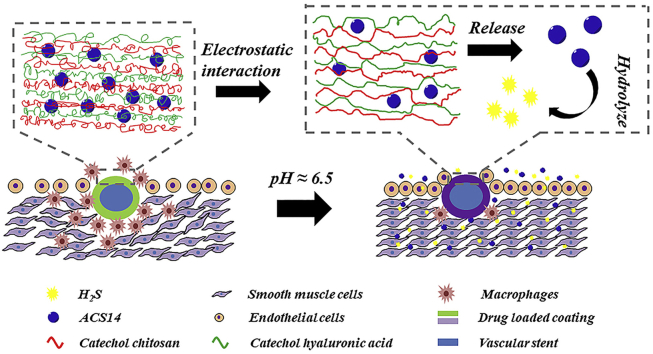
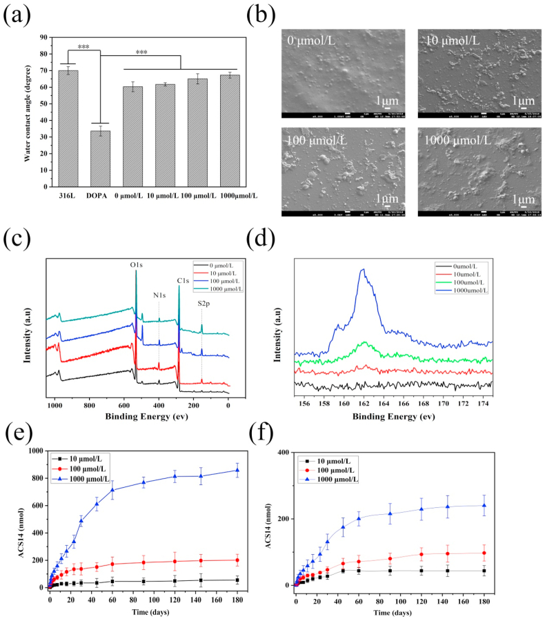
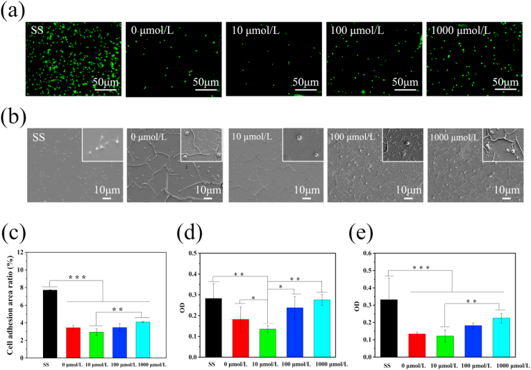
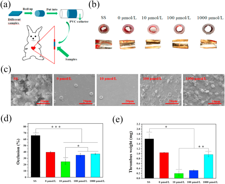
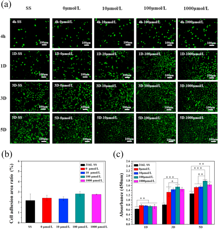
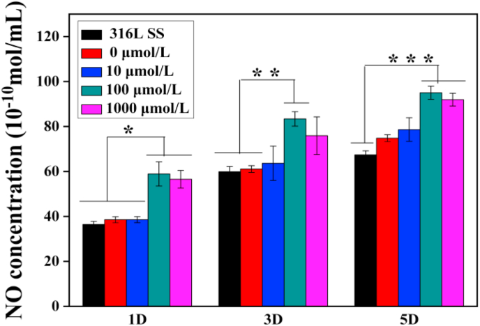
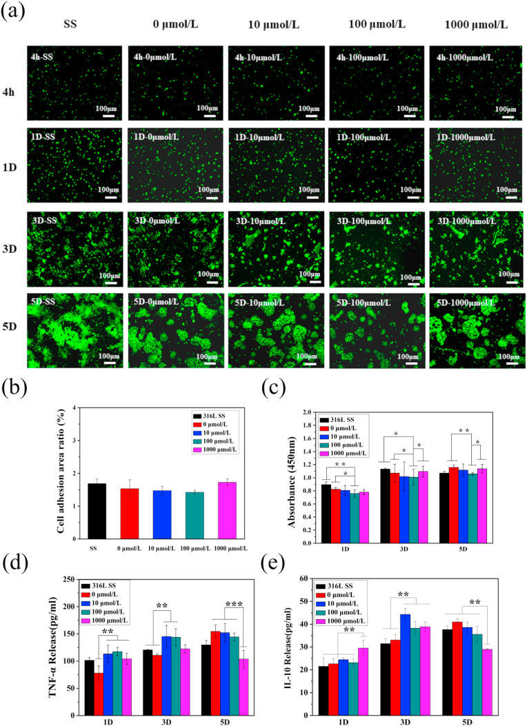
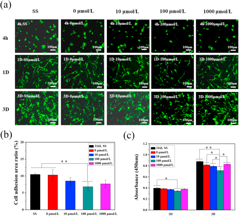
Coronary atherosclerotic lesions exhibit a low-pH chronic inflammatory response. Due to insufficient drug release control, drug-eluting stent intervention can lead to delayed endothelialization, advanced thrombosis, and unprecise treatment. In this study, hyaluronic acid and chitosan were used to prepare pH-responsive self-assembling films. The hydrogen sulfide (H2S) releasing aspirin derivative ACS14 was used as drug in the film. The film regulates the release of the drug adjusted to the microenvironment of the lesion, and the drug balances the vascular function by releasing the regulating gas H2S, which comparably to NO promotes the self-healing capacity of blood vessels. Drug releasing profiles of the films at different pH, and other biological effects on blood vessels were evaluated through blood compatibility, cellular, and implantation experiments. This novel method of self-assembled films which H2S in an amount, which is adjusted to the condition of the lesion provides a new concept for the treatment of cardiovascular diseases.

Keywords: Coronary atherosclerosis; H2S; Layer-by-layer self-assembly film; pH responsive.


Conflict of interest statement

The authors declare that they have no known competing financial interests or personal relationships that could have appeared to influence the work reported in this paper.

Figures

Image 1
Graphical abstract
Fig. 1
Fig. 1
(a) Water contact angle of different ACS14 concentration coatings (n = 5, ***p < 0.001). (b) SEM images of ACS14 coatings at different concentrations. (c) XPS full spectrum of drug-loaded coatings prepared with different concentrations of ACS14, and (d) high-resolution spectra of the S element content. (e) Drug release of the drug-loaded coatings in PBS at pH 6.5. (f) Drug release curve of drug-loaded coating in PBS at pH 7.4.
Fig. 2
Fig. 2
(a) Fluorescence of platelets on the surface of samples prepared with different concentrations of ACS14, and (b) SEM morphology of platelets on the sample surface (n = 3, **p < 0.01, ***p < 0.001). (c) Platelet surface adhesion area of samples prepared with different concentrations of ACS14. (d) Fibrinogen denaturation. (e) Fibrinogen adhesion. (n = 3, *p < 0.05, **p < 0.01, ***p < 0.001).
Fig. 3
Fig. 3
(a) Schematic diagram of the experimental design. (b) The cross-section and inner views of different samples. (c) Coagulation of different samples observed with SEM. (d) Blood flow obstruction rate and (e) thrombus weight.(n = 3, *p < 0.05, **p < 0.01, ***p < 0.001).
Fig. 4
Fig. 4
(a) Fluorescence of endothelial cells growth after 4 h, 1D, 3D, 5D on different concentrations of drug-loaded coatings. (b) Adhesion area after 4 h of implantation on different samples. (c) CCK-8 testing endothelial cells viability map (n = 3, *p < 0.05, **p < 0.01, ***p < 0.001).
Fig. 5
Fig. 5
Quantitative assessment of NO concentration in endothelial cell culture medium on different samples after culturing for 1, 3 and 5 days (n = 3, *p < 0.05, **p < 0.01, ***p < 0.001).
Fig. 6
Fig. 6
(a) Fluorescence of macrophages after 4 h, 1D, 3D, and 5D on different samples. (b) Adhesion area of macrophages after 4 h of implantation on different samples, and (c) CCK-8 cells activity maps tested (n = 3, *p < 0.05, **p < 0.01). (d) ELISA kit detected the secretion of TNF-α from macrophages on different drug-coated surfaces, (e) ELISA kit detected the secretion of interleukin IL-10 on different drug-coated surfaces (n = 3, **p < 0.01, ***p < 0.001).
Fig. 7
Fig. 7
(a) Fluorescence of smooth muscle cells after 4 h, 1D, 3D implantation on different concentrations of drug-loaded coatings. (b) Adhesion area of smooth muscle cells after 4 h of implantation on different samples, and (c) cells detected by CCK-8 activity map. (n = 3, *p < 0.05, **p < 0.01).
Fig. 8
Fig. 8
Cross sections of aortae with bare and coated wires after four weeks implantation in rats. (a–e) HE histological images. (f) Quantitative analysis of tissue proliferation around the different samples, measured as cross-section areas (yellow* is the sample implantation position, n = 6, **p < 0.01,*p < 0.05).

Similar articles

Cited by

References

    1. Rezvan A., Ni C.W., Alberts-Grill N., Jo H. Animal, in vitro , and ex vivo models of flow-dependent atherosclerosis: role of oxidative stress, antioxid. Redox Signaling. 2011;15(5):1433–1448. - PMC - PubMed
    1. Urban P., Benedetti E.D. Thrombosis: the last frontier of coronary stenting? Lancet. 2007;369(9562):619–621. - PubMed
    1. Dangas G., Claessen B.E., Caixeta A., Sanidas E., Mintz G.S., Mehran R. In-stent restenosis in the drug-eluting stent era. J. Am. Coll. Cardiol. 2010;56(23):1897–1907. - PubMed
    1. Lagerqvist B., James S., Stenestrand U., Lindback J., Nilsson T., Wallentin L. Long-Term outcomes with drug-eluting stents versus bare-metal stents in Sweden. N. Engl. J. Med. 2007;356(10):1009–1019. - PubMed
    1. Sheiban I., Villata G., Bollati M., Sillano D., Lotrionte M., Biondizoccai G. Next-generation drug-eluting stents in coronary artery disease: focus on everolimus-eluting stent (Xience V®), Vasc. Healthc. Risk Manag. 2008;4(1):31–38. - PMC - PubMed

LinkOut - more resources