. 2020 Oct 7;3(4):1087-1113.
doi: 10.1016/j.matt.2020.08.002.

Bioinspired Materials for In Vivo Bioelectronic Neural Interfaces

Affiliations

Bioinspired Materials for In Vivo Bioelectronic Neural Interfaces

Grace A Woods et al. Matter. .

Abstract

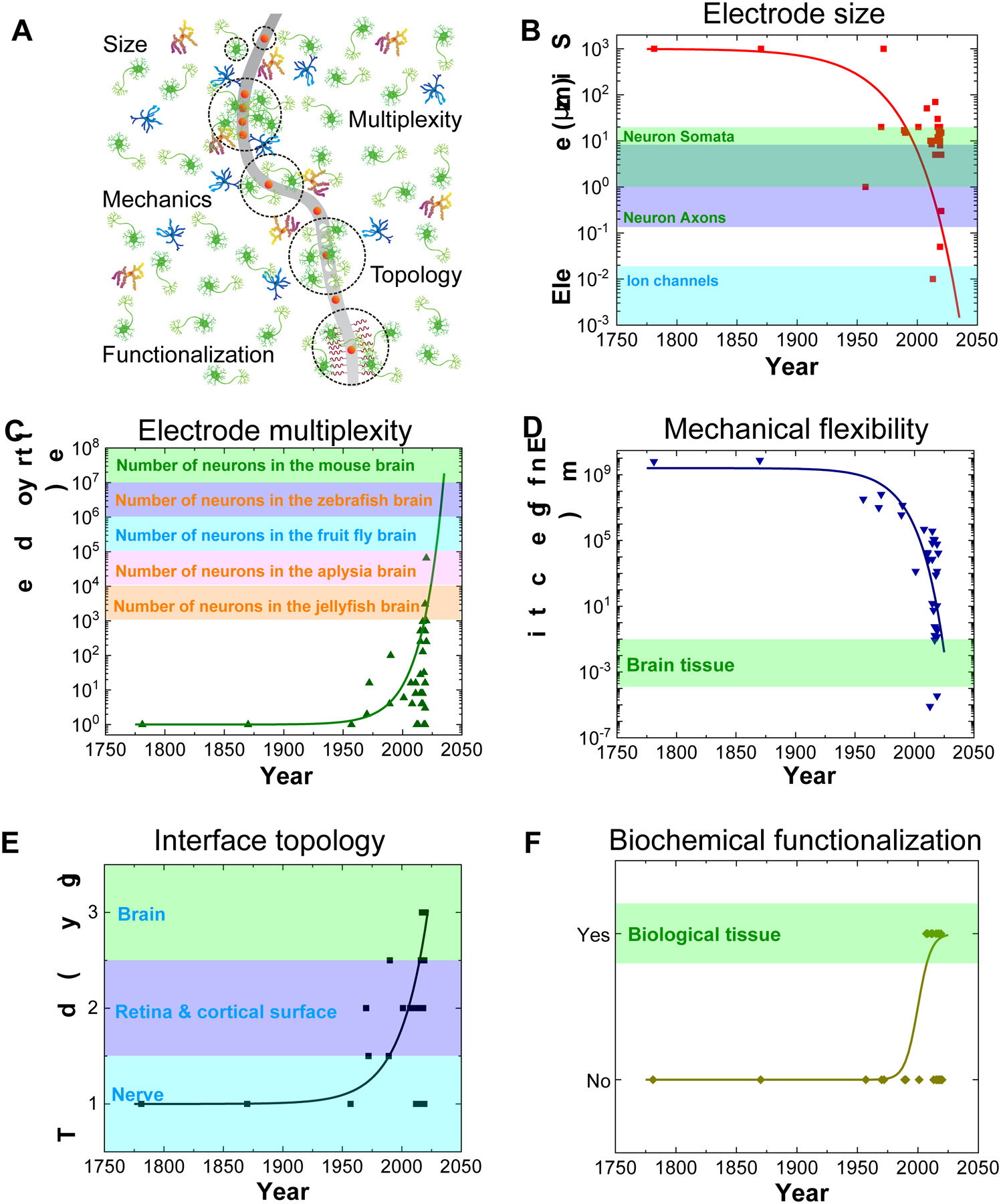
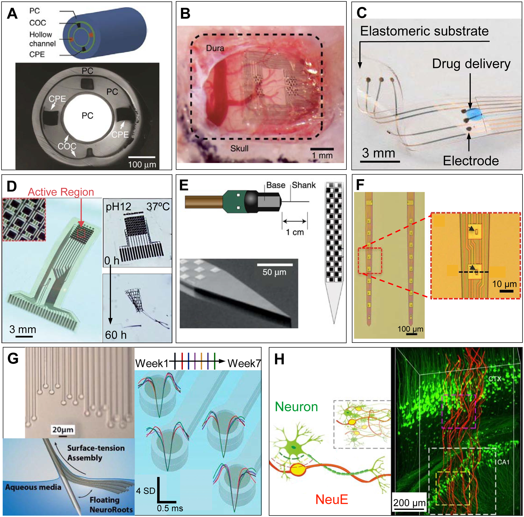
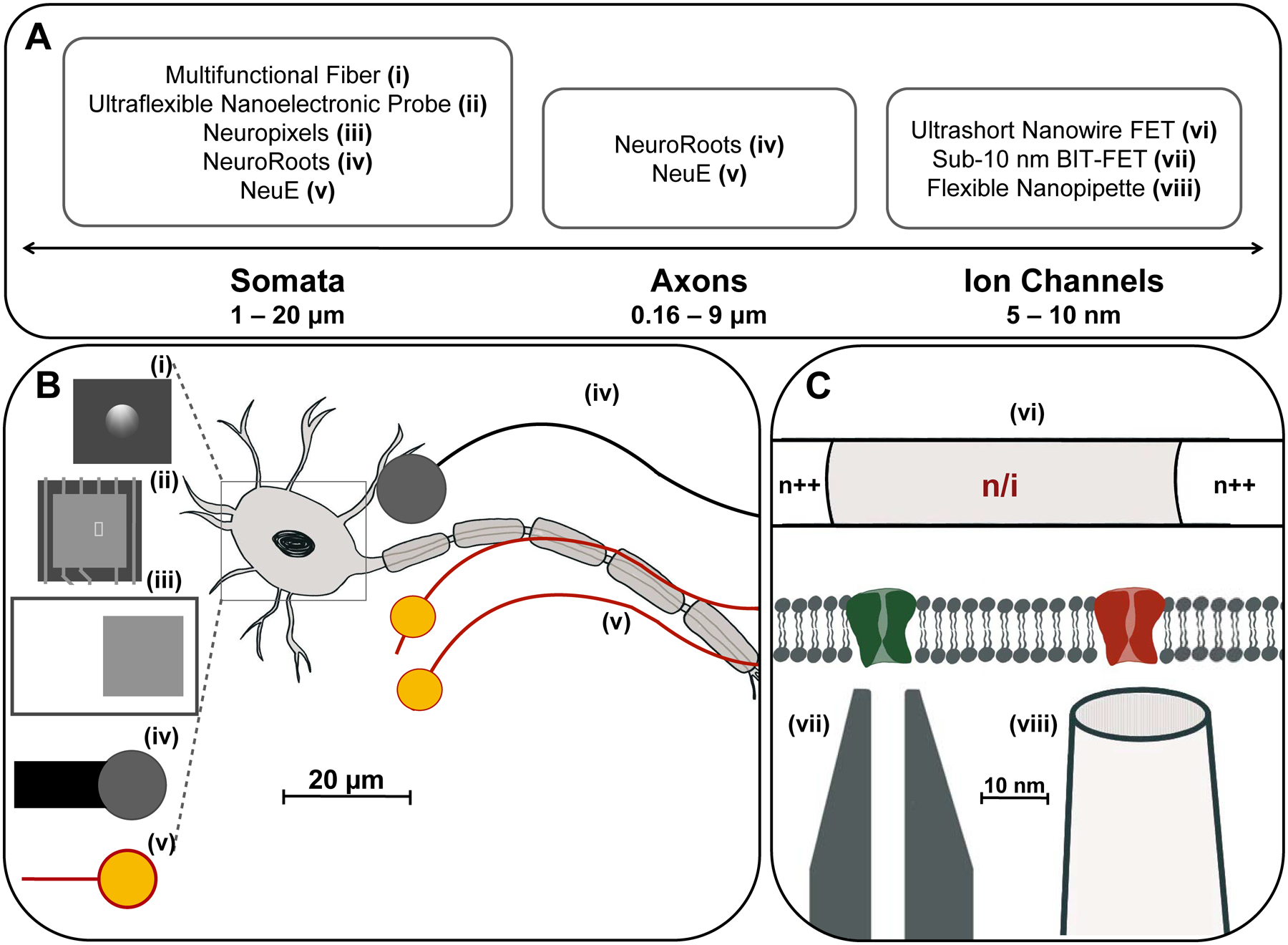
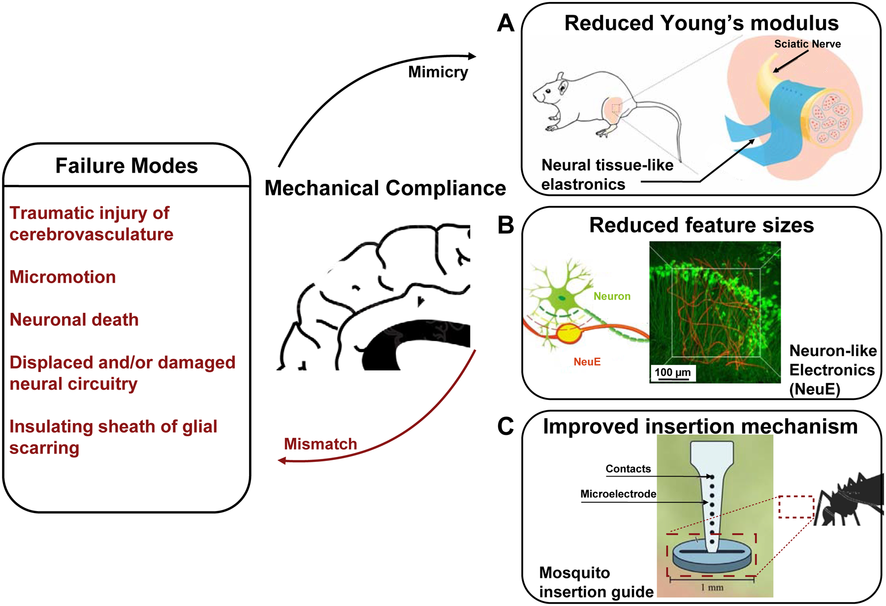
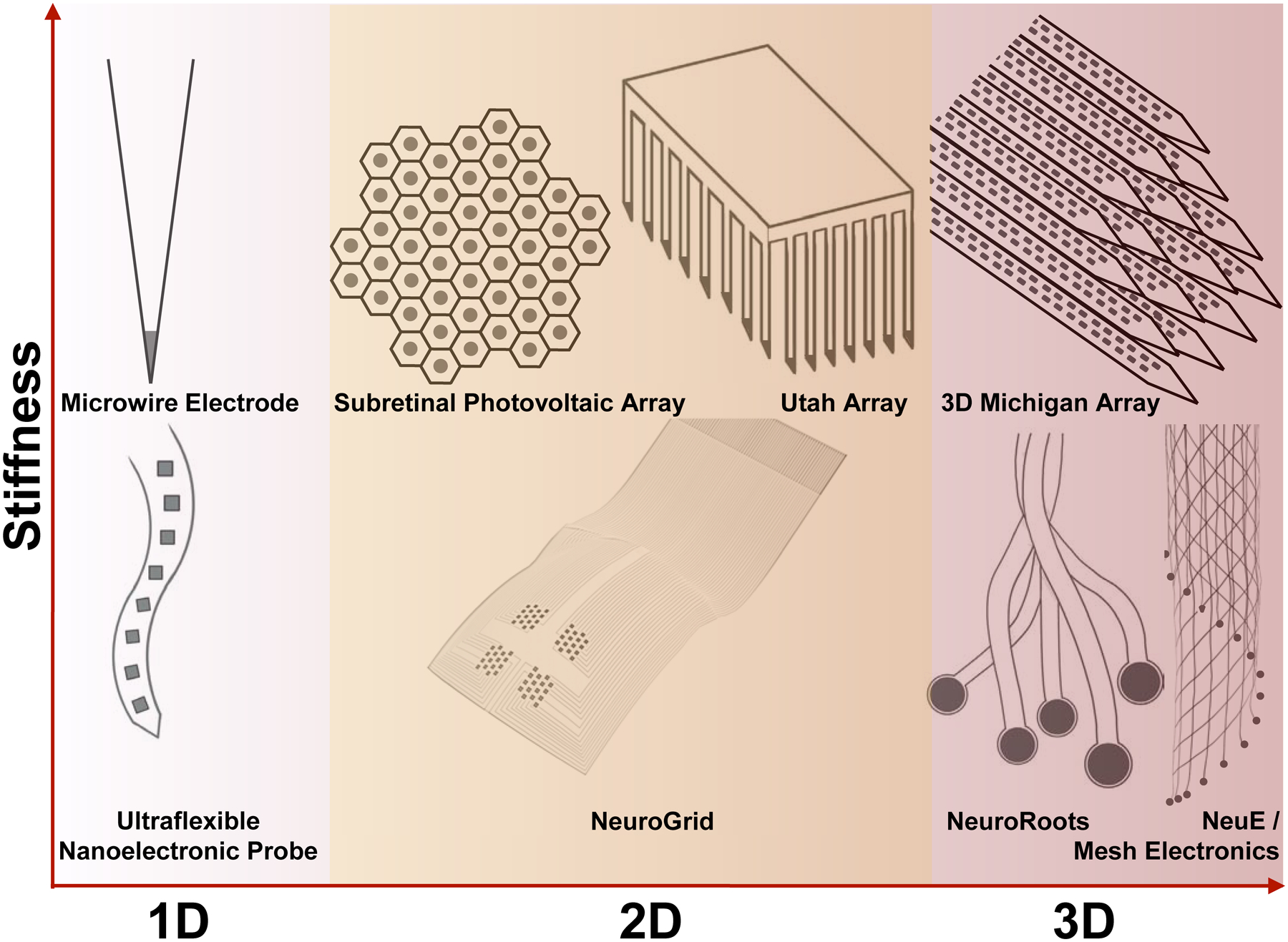
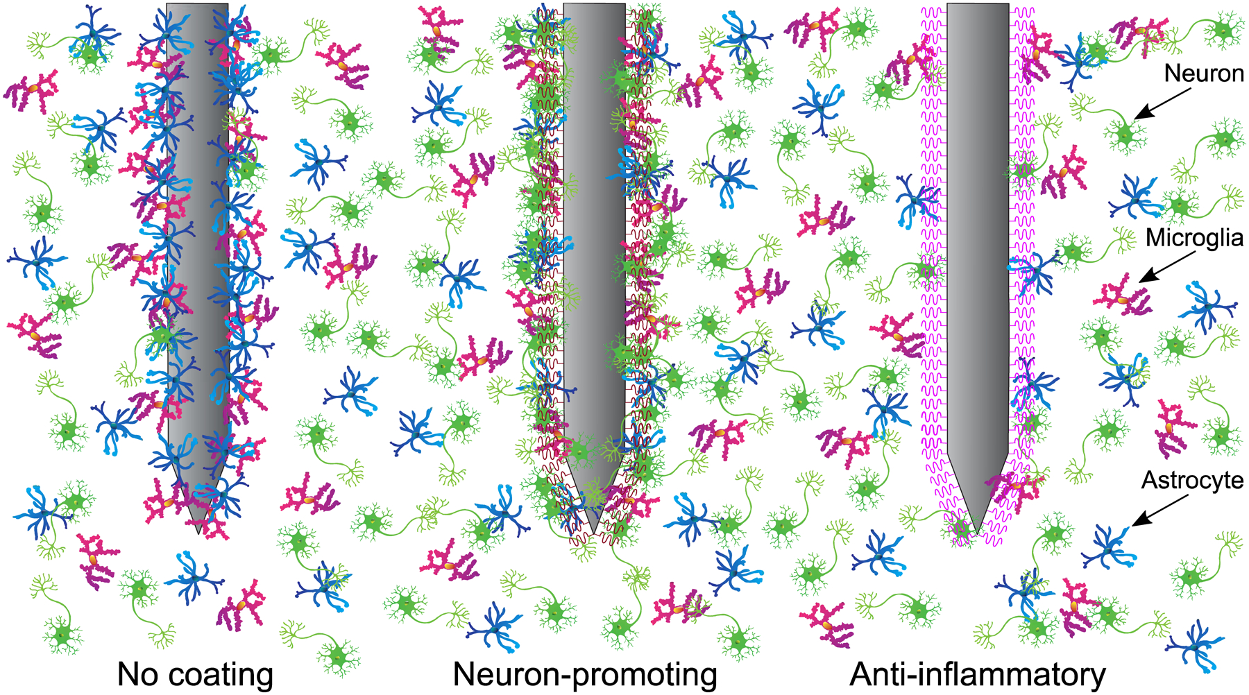
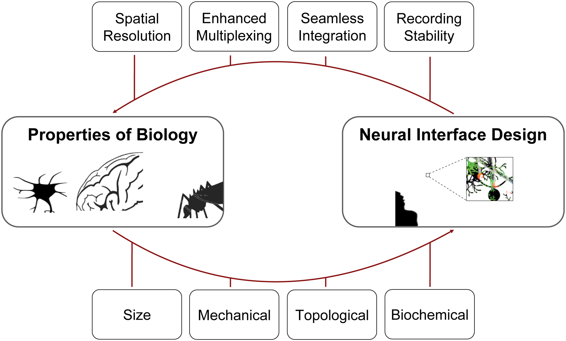
The success of in vivo neural interfaces relies on their long-term stability and large scale in interrogating and manipulating neural activity after implantation. Conventional neural probes, owing to their limited spatiotemporal resolution and scale, face challenges for studying the massive, interconnected neural network in its native state. In this review, we argue that taking inspiration from biology will unlock the next generation of in vivo bioelectronic neural interfaces. Reducing the feature sizes of bioelectronic neural interfaces to mimic those of neurons enables high spatial resolution and multiplexity. Additionally, chronic stability at the device-tissue interface is realized by matching the mechanical properties of bioelectronic neural interfaces to those of the endogenous tissue. Further, modeling the design of neural interfaces after the endogenous topology of the neural circuitry enables new insights into the connectivity and dynamics of the brain. Lastly, functionalization of neural probe surfaces with coatings inspired by biology leads to enhanced tissue acceptance over extended timescales. Bioinspired neural interfaces will facilitate future developments in neuroscience studies and neurological treatments by leveraging bidirectional information transfer and integrating neuromorphic computing elements.

Keywords: Biomimetics; brain implants; brain-machine interfaces; chronic stability; nanoelectronics; neural probes; neural recording; neural stimulation; spatiotemporal resolution; tissue-like electronics.


Conflict of interest statement

Declaration of interests The authors declare no competing interests.

Figures

Figure 1.
Figure 1.
Trends of in vivo bioelectronic neural interfaces with bioinspired size, multiplexity, mechanics, topology, and functionalization. (A) Schematic summarizing the bioinspired design features of in vivo bioelectronic neural interfaces. (B&C) The onset of modern fabrication techniques, such as photolithography, has made a substantial impact on the accessible resolution of device feature sizes, corresponding to a distinct decrease in electrode size (B), and an increase in electrode multiplexity, which is defined as the number of independent channels in a bioelectronic neural interface (C). The size ranges of typical neuronal structures and the ranges of neuron number in different species are shown in the colored shades of B and C, respectively. Note that the range of soma size (green shade) overlaps with that of axon size (blue shade) in B. (D) Engineering devices with soft materials and specific structures allows the effective bending stiffness, which is defined as ratio of bending moment to the product of width and curvature of the neural probe, to decrease by up to ten orders of magnitude, reducing the overall probability of eliciting a chronic immune response. The range of effective bending stiffness of a 20 – 100 μm thick slice of brain tissue is shown as the green shade. (E) A general trend in the topology of neural interfaces is found towards higher dimensions, allowing for more intimate interfacing with the neural tissue due to brain-device conformability. Non-integer topologies, such as radially segmented DBS electrode array (1D – 2D) and Utah slanted electrode array (2D – 3D) are indicated due to variability in design. The dimensionality of different components of the nervous system is shown as colored shades. (F) Progression of the field towards biochemical functionalization represents another clear marker of bioinspired neural interfaces. Biological tissues are intrinsically functionalized with biochemical cues, as labeled in the colored shade. In all graphs, solid curves are intended to guide the visualization of trends of various important features of bioelectronic neural interfaces and are not intended to fit these data to a particular mathematical model, although a similar trend of exponential growth has been found for doubling of simultaneously recorded neurons and Moore’s law. All data points come from technologies listed in Table 1.
Figure 2.
Figure 2.
Examples of novel in vivo bioelectronic neural interfaces. (A) Multifunctional fibers, constructed via thermal drawing processes, offer a multimodal means of interacting with neurons. As demonstrated in the schematic drawing (top) and a cross-sectional image (bottom) of the multifunctional fiber, this design incorporates one cylindrical optical waveguide comprising a polycarbonate (PC) core and a cyclic olefin copolymer (COC) shell, two microfluidic channels (‘hollow channels’) for local drug delivery, and two conductive polyethylene electrodes (CPE) for recording extracellular action potentials. Adapted with permission from Ref. 50. (B) NeuroGrid features poly(3,4-ethylenedioxythiophene) polystyrene sulfonate (PEDOT:PSS) coated electrodes with sizes and interelectrode spacing inspired by the average size of neuron somata and neuronal density within the neocortex, and is capable of resolving both local field potentials (LFPs) and single unit activity when conformally interfacing the curvilinear cortical surface. Adapted with permission from Ref. 52. (C) e-dura is designed to mimic the mechanical properties of the dura mater of the brain and spinal cord. By integrating flexible silicone with thin films of conductive platinum-silicone composites and a microfluidic channel, the e-dura affords seamless integration with the spinal cord and potentially superficial regions of the brain, enabling electrical recording and stimulation in addition to local drug delivery. Adapted with permission from Ref. 51. (D) Bioresorbable silicon electronics are designed to seamlessly interface with cortical tissue for transient monitoring and modulation of brain activity (left). By integrating functional materials that can dissolve under physiological conditions, bioresorbable electronics show rapid dissolution upon immersion in an aqueous buffer solution with pH = 12 at 37 °C (right). Adapted with permission from Ref. 55. (E) Neuropixels facilitates electrophysiological recordings that demand high channel counts (left). Neuropixels incorporates 960 electrodes (layout shown in the right), with 384 active processing units at any one time. Adapted with permission from Ref. 57. (F) Ultraflexible nanoelectronic probes (shown: NET-50, left) combine reduced feature sizes of electrodes (right) and flexible materials to produce a device capable of chronically stable single unit recording. Adapted with permission from Ref. 56. (G) NeuroRoots takes inspiration from axon sizes and distribution within the brain (top left) and can be inserted into the brain through self-assembly, mediated by capillary forces, along the direction of the microwire shuttle (bottom left). Owing to NeuroRoots’ flexibility and small footprint, stable single unit recording is demonstrated for up to 7 weeks (right). Adapted with permission from Ref. 61. (H) Neuron-like electronics (NeuE) combine bioinspired feature sizes, mechanical compliance and topological properties (left) to optimize device-brain interfacing (right) and affords chronic recording stability. Adapted with permission from Ref. 63.
Figure 3.
Figure 3.
Bioelectronic neural interfaces inspired by the size and morphology of neuron somata and neurites. (A) Feature sizes of the subcellular structures of the neuron, ranging from a few nanometers for ion channels, 0.16 – 9 microns for axonal diameter, to 1 – 20 microns for neuron somata. (B) Many modern bioelectronic neural interfaces have recording electrodes of similar sizes to neuron somata. Shown are the electrode regions of (i) Multifunctional fibers (d = 5 μm), (ii) Ultraflexible nanoelectronic probe (d = 10 μm), (iii) Neuropixels (d = 12 μm), (iv) NeuroRoots (d = 10 μm), (v) NeuE (d = 8 – 20 μm), where d is the diameter for round electrodes, or width for square electrodes. Additionally, by mimicking the size characteristics of axons, neural interfaces can encourage acceptance in the endogenous neural tissue via promotion of neural progenitor cell migration and integration with the neuronal network. Shown: (iv) NeuroRoots (w ~ 7 μm), (v) NeuE (w = 1 – 4 μm), where w is the width of interconnect ribbons in the electronics. (C) Devices mimicking the size of ion channels, such as the 50 nm ultrashort-channel FET and sub-10 nm BIT-FET, allow recording from individual ion channels with subcellular resolution. Shown: (vi) Ultrashort nanowire FET, (vii) Sub-10 nm BIT FET, and (viii) Flexible nanopipette.
Figure 4.
Figure 4.
Bioelectronic neural interfaces inspired by the mechanical properties of neural tissue. Bioinspired mechanical compliance may result from the reduction of (A) the material’s Young’s modulus, through engineering thin-film hydrogel elastronics with Young’s moduli on the order of kPa, or (B) feature sizes, with neuron-like electronics (NeuE). A schematic of the intertwined neuron-NeuE interface is shown on the left, and a reconstructed 3D image of neurons (green) interpenetrating the electronic network of NeuE (red) in shown on the right. Finally, improved insertion mechanisms of flexible neural interfaces may be realized through a variety of bioinspired strategies, such as the fascicles of female mosquitos, thereby reducing buckling during implantation (C). Reproduced with permission from Refs. 63, , .
Figure 5.
Figure 5.
Bioelectronic neural interfaces inspired by the topology of neural tissue. This means that, for neurons within the brain (white-red gradient), neural interfaces constantly advance toward 3D topologies. Starting with the microwire electrode, silicon-based fabrication strategies ignited arraying rigid materials for higher-dimensional interfacing, as seen with the Utah array and the 3D Michigan array. In contrast, interrogation of the neurons in the retina (i.e., retinal ganglion cells) and on the cortical surface requires interfacing at two dimensions on a curvilinear surface (yellow highlight in the middle column). Meanwhile, the joint trend towards higher-dimensional topologies is maintained for flexible bioelectronic neural interfaces, evidenced by the ultraflexible nanoelectronic probe (NET-50), NeuroGrid, NeuroRoots, mesh electronics, and NeuE. Dark gray circles and squares indicate recording sites in these examples. Schematics are not to scale in these drawings.
Figure 6.
Figure 6.
Bioelectronic neural interfaces with bioinspired functionalization. At left, a typical immune response to a non-coated neural implant is illustrated. Astrocytes and microglia accumulate at the probe interface, ultimately forming a glial scar, and neuronal loss is observed around the probe. At center, a probe with neuron-promoting coating (such as L1 or Matrigel) prevents local neural loss while reducing immune response, thoroughly incorporating the probe with the endogenous neural tissue. At right, an interface with anti-inflammatory coating (such as α-MSH or IL1-receptor antagonist) significantly reduces the presence of microglia and astrocytes, allowing unhindered access to local neurons.
Figure 7.
Figure 7.
Envisioned synergy between neuroscience, which elucidates the properties of biology in the nervous system, and neuroengineering, via neural interface design. In particular, the knowledge of the size, mechanical, topological, and biochemical properties of the brain affords an unprecedented opportunity for next-generation neural interface design, with features such as high spatiotemporal resolution, enhanced electrode multiplexity (i.e., number of electrodes), the capability for seamless integration, and chronic recording stability.

References

    1. Rivnay J, Wang H, Fenno L, Deisseroth K, and Malliaras GG (2017). Next-generation probes, particles, and proteins for neural interfacing. Sci. Adv 3, e1601649. - PMC - PubMed
    1. Chen R, Canales A, and Anikeeva P (2017). Neural recording and modulation technologies. Nat. Rev. Mater 2, 16093. - PMC - PubMed
    1. Patel SR, and Lieber CM (2019). Precision electronic medicine in the brain. Nat. Biotechnol 37, 1007–1012. - PMC - PubMed
    1. Song E, Li J, Won SM, Bai W, and Rogers JA (2020). Materials for flexible bioelectronic systems as chronic neural interfaces. Nat. Mater 19, 590–603. - PubMed
    1. Galvani L (1791). D viribus electricitatis in motu musculari: Commentarius. Bologna: Tip. Istituto delle Scienze, 1791; 58p.: 4 tavv. ft; in 4.; DCC. f. 70.

LinkOut - more resources