. 2020 Dec 1;319(6):F1003-F1014.
doi: 10.1152/ajprenal.00261.2020. Epub 2020 Oct 26.

Histone deacetylase 6 inhibition mitigates renal fibrosis by suppressing TGF-β and EGFR signaling pathways in obstructive nephropathy

Affiliations

Histone deacetylase 6 inhibition mitigates renal fibrosis by suppressing TGF-β and EGFR signaling pathways in obstructive nephropathy

Xingying Chen et al. Am J Physiol Renal Physiol. .

Abstract

We have recently shown that histone deacetylase 6 (HDAC6) is critically involved in the pathogenesis of acute kidney injury. Its role in renal fibrosis, however, remains unclear. In this study, we examined the effect of ricolinostat (ACY-1215), a selective inhibitor of HDAC6, on the development of renal fibrosis in a murine model induced by unilateral ureteral obstruction (UUO). HDAC6 was highly expressed in the kidney following UUO injury, which was coincident with deposition of collagen fibrils and expression of α-smooth muscle actin, fibronectin, and collagen type III. Administration of ACY-1215 reduced these fibrotic changes and inhibited UUO-induced expression of transforming growth factor-β1 and phosphorylation of Smad3 while increasing expression of Smad7. ACY-1215 treatment also suppressed phosphorylation of epidermal growth factor receptor (EGFR) and several signaling molecules associated with renal fibrogenesis, including AKT, STAT3, and NF-κB in the injured kidney. Furthermore, ACY-1215 was effective in inhibiting dedifferentiation of renal fibroblasts to myofibroblasts and the fibrotic change of renal tubular epithelial cells in culture. Collectively, these results indicate that HDAC6 inhibition can attenuate development of renal fibrosis by suppression of transforming growth factor-β1 and EGFR signaling and suggest that HDAC6 would be a potential therapeutic target for the treatment of renal fibrosis.

Keywords: ACY-1215; epidermal growth factor receptor; histone deacetylase 6; renal fibrosis; transforming growth factor-β1; unilateral ureteral obstruction.


Conflict of interest statement

No conflicts of interest, financial or otherwise, are declared by the authors.

Figures

None
Graphical abstract
Fig. 1.
Fig. 1.
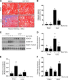
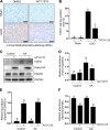
Inhibition of histone deacetylase 6 (HDAC6) with ACY-1215 attenuates renal fibrosis. Mice were subjected to unilateral ureteral obstruction (UUO) and treated daily with ACY-1215 for 7 days before samples were harvested analysis. A: photomicrographs illustrating Masson trichrome staining of kidney tissue. B: the percentage of Masson trichrome-positive tubulointerstitial area (blue) relative to the whole area was quantified (original magnification: ×200). Scale bar = 50 μm. C: kidney tissue lysates were subjected to immunoblot analysis with specific antibodies against HDAC6, acetyl-H3, acetyl-α-tubulin, or GAPDH. DF: protein expression levels of HDAC6 (D), acetyl-H3 (E), or acetyl-α-tubulin (F) were qualified by densitometry and normalized with GAPDH. Values are means ± SD of at least three independent experiments. Bars with different letters (a–c) for each molecule were significantly different from one other (P < 0.05).
Fig. 2.
Fig. 2.
Expression of histone deacetylase 6 (HDAC6) in the kidney. Mice were subjected to unilateral ureteral obstruction (UUO) and treated daily with ACY-1215 for 7 days before samples were harvested for analysis. Photomicrographs show protein expression of HDAC6 (red) and α-smooth muscle actin (α-SMA; green) after immunofluorescent costaining and counterstaining with DAPI (blue) (original magnification: ×400). In the injured kidney, HDAC6 was most abundant in the cytoplasm of renal tubular cells but was also observed in the nucleus of this cell type. Scale bar = 50 μm.
Fig. 3.
Fig. 3.
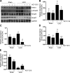
Inhibition of histone deacetylase 6 (HDAC6) with ACY-1215 reduces renal fibroblast activation and extracellular matrix (ECM) protein deposition in the renal interstitium. Mice were subjected to unilateral ureteral obstruction (UUO) and treated daily with ACY-1215 for 7 days before samples were harvested for analysis. A: whole kidney tissue lysates from obstructed (UUO) and contralateral nonobstructed (sham) ureters were processed for immunoblot analysis with antibodies specific to α-smooth muscle actin (α-SMA), fibronectin, collagen type III, and GAPDH. Expression levels of α-SMA (B), fibronectin (C), and collagen type III (D) were qualified by densitometry and normalized with GAPDH. Values are means ± SD of at least three independent experiments. Bars with different letters (a–c) for each molecule were significantly different from one other (P < 0.05).
Fig. 4.
Fig. 4.
Histone deacetylase 6 (HDAC6) blockade inhibits unilateral ureteral obstruction (UUO)-induced activation of transforming growth factor-β (TGF-β)/Smad3 signaling in the kidney. Mice were subjected to UUO and treated daily with ACY-1215 for 7 days before samples were harvested for analysis. A: Whole kidney tissue lysates from obstructed (UUO) and contralateral nonobstructed (sham) were processed for immunoblot analysis with antibodies specific to TGF-β1, phosphorylated (p-)Smad3, Smad3, Smad7, and GAPDH. Expression levels of TGF-β1 (B), p-Smad3 (C), Smda3 (D), and Smad7 (E) were qualified by densitometry and normalized with GAPDH. Values are means ± SD of at least three independent experiments. Bars with different letters (a–d) for each molecule were significantly different from one other (P < 0.05).
Fig. 5.
Fig. 5.
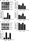
Histone deacetylase 6 (HDAC6) blockade inhibits unilateral ureteral obstruction (UUO)-induced activation of the epidermal growth factor receptor (EGFR)/AKT signaling pathway in the kidney. Mice were subjected to UUO and treated daily with ACY-1215 for 7 days before samples were harvested for analysis. A: whole kidney tissue lysates from obstructed (UUO) and contralateral nonobstructed ureters (sham) were processed for immunoblot analysis with antibodies specific to phosphorylated (p-)EGFR, EGFR, p-AKT, and AKT. B: p-EGFR expression levels were qualified by densitometry and normalized to EGFR. C: p-AKT expression levels were qualified by densitometry and normalized to AKT. Values are means ± SD of at least three independent experiments. Bars with different letters (a–c) for each molecule were significantly different from one other (P < 0.05).
Fig. 6.
Fig. 6.
Histone deacetylase 6 (HDAC6) blockade inhibits unilateral ureteral obstruction (UUO)-induced activation of the STAT3/NF-κB (p65) signaling pathway in the kidney. Mice were subjected to UUO and treated daily with ACY-1215 for 7 days before samples were harvested for analysis. A: whole kidney tissue lysates from obstructed (UUO) and contralateral nonobstructed (sham) groups were processed for immunoblot analysis with antibodies specific to phosphorylated (p-)NF-κB (p65), NF-κB (p65), p-STAT3, STAT3, and GAPDH. B: p-NF-κB (p65) expression levels were qualified by densitometry and normalized to NF-κB (p65). C: p-STAT3 expression levels were qualified by densitometry and normalized to STAT3. Values are means ± SD of at least three independent experiments. Bars with different letters (a–d) for each molecule were significantly different from one another (P < 0.05).
Fig. 7.
Fig. 7.
Inhibition of histone deacetylase 6 (HDAC6) with ACY-1215 reduces activation of renal interstitial fibroblasts in cultured NRK-49F cells. NRK-49F were cultured with 5% FBS and treated with various concentrations of ACY-1215 (0–50 μM) for 36 h. A: Western blot analysis of cell lysates with various antibodies as indicated. Expression levels of α-smooth muscle actin (α-SMA; B), HDAC6 (C), and acetyl-H3 (D) were qualified by densitometry and normalized with GAPDH. Values shown in the graph are means ± SD of at least three independent experiments. Each letter (a–d) indicates that different bars were significantly different from each other (P < 0.05).
Fig. 8.
Fig. 8.
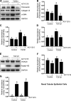
Inhibition of histone deacetylase 6 (HDAC6) with ACY-1215 reduces activation of renal interstitial fibroblasts in cultured NRK-49F cells. Normally cultured NRK-49F was exposed to 5 ng/mL transforming growth factor-β1 (TGF-β1) and then cultured for 36 h in the absence or presence of ACY-1215 (25 μM). A and E: Western blot analysis of cell lysates with various antibodies as indicated. Protein expression levels of fibronectin (B), collagen type III (C), α-smooth muscle actin (α-SMA; D), HDAC6 (F), and acetyl-H3 (G) were qualified by densitometry and normalized with GAPDH. Values shown in the graphs are means ± SD of at least three independent experiments. Each letter (a–c) indicates that different bars were significantly different from each other (P < 0.05).
Fig. 9.
Fig. 9.
ACY-1215 inhibits profibrotic phenotype changes of renal epithelial cells. Serum-starved murine renal tubular epithelial cells (mTECs) were treated with transforming growth factor-β1 (TGF-β1; 5 ng/mL) in the presence or absence of ACY-1215 (25 μM) for 24 h and then harvested. A and E: Western blot analysis of cell lysates with various antibodies as indicated. Protein expression levels of fibronectin (B), collagen type III (C), α-smooth muscle actin (α-SMA; D), histone deacetylase 6 (HDAC6; F), and acetyl-histone H3 (G) were qualified by densitometry and normalized with GAPDH. Values shown in the graphs are means ± SD of at least three independent experiments. Each letter (a–c) indicates that different bars were significantly different from each other (P < 0.05).
Fig. 10.
Fig. 10.
ACY-1215 inhibits expression of transforming growth factor-β1 (TGF-β1) in the kidney after unilateral ureteral obstruction (UUO) and in cultured renal tubular epithelial cells after aristolochic acid (AA) exposure. Mice were subjected to UUO and daily treatment with ACY-1215 for 7 days before samples were harvested for analysis. A: photomicrographs illustrating TGF-β1 staining of kidney tissue. B: the percentage of TGF-β1-positive area (yellow) relative to the whole area was quantified (original magnification: ×200). Scale bar = 100 μm. C: murine renal tubular epithelial cells were treated as indicated in MATERIALS AND METHODS. The prepared cell lysates were subjected to immunoblot analysis using antibodies against TGF-β1, acetyl-histone H3, or histone deacetylase 6 (HDAC6). Protein expression levels of TGF-β1 (D), acetyl histone H3 (E), or HDAC6 (F) were qualified by densitometry and normalized with GAPDH. Values shown in the graphs are means ± SD of at least three independent experiments. Each letter (a–c) indicates that different bars were significantly different from each other (P < 0.05).

Similar articles

Cited by

References

    1. Andrade-Oliveira V, Foresto-Neto O, Watanabe IKM, Zatz R, Câmara NOS. Inflammation in renal diseases: new and old players. Front Pharmacol 10: 1192, 2019. doi:10.3389/fphar.2019.01192. - DOI - PMC - PubMed
    1. Bertos NR, Gilquin B, Chan GK, Yen TJ, Khochbin S, Yang XJ. Role of the tetradecapeptide repeat domain of human histone deacetylase 6 in cytoplasmic retention. J Biol Chem 279: 48246–48254, 2004. doi:10.1074/jbc.M408583200. - DOI - PubMed
    1. Brindisi M, Saraswati AP, Brogi S, Gemma S, Butini S, Campiani G. Old but gold: tracking the new guise of histone deacetylase 6 (HDAC6) enzyme as a biomarker and therapeutic target in rare diseases. J Med Chem 63: 23–39, 2020. doi:10.1021/acs.jmedchem.9b00924. - DOI - PubMed
    1. Butler KV, Kalin J, Brochier C, Vistoli G, Langley B, Kozikowski AP. Rational design and simple chemistry yield a superior, neuroprotective HDAC6 inhibitor, tubastatin A. J Am Chem Soc 132: 10842–10846, 2010. doi:10.1021/ja102758v. - DOI - PMC - PubMed
    1. Chen L, Yang T, Lu DW, Zhao H, Feng YL, Chen H, Chen DQ, Vaziri ND, Zhao YY. Central role of dysregulation of TGF-β/Smad in CKD progression and potential targets of its treatment. Biomed Pharmacother 101: 670–681, 2018. doi:10.1016/j.biopha.2018.02.090. - DOI - PubMed

Publication types

MeSH terms