. 2020 Dec 1;319(6):F1027-F1036.
doi: 10.1152/ajprenal.00322.2020. Epub 2020 Oct 26.

Renal lymphatic vessel dynamics

Affiliations

Renal lymphatic vessel dynamics

Elaine L Shelton et al. Am J Physiol Renal Physiol. .

Abstract

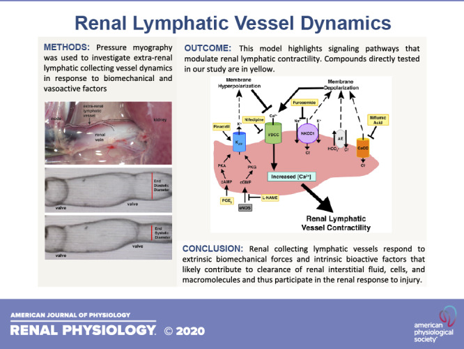
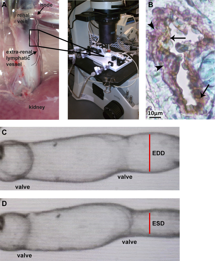
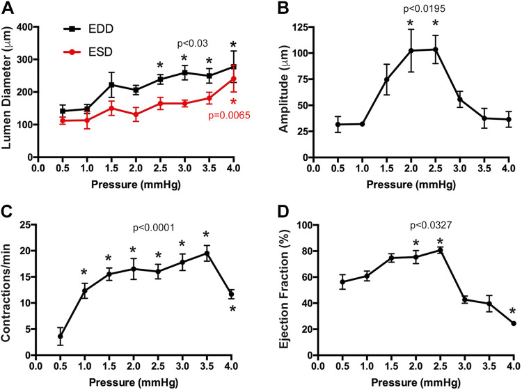
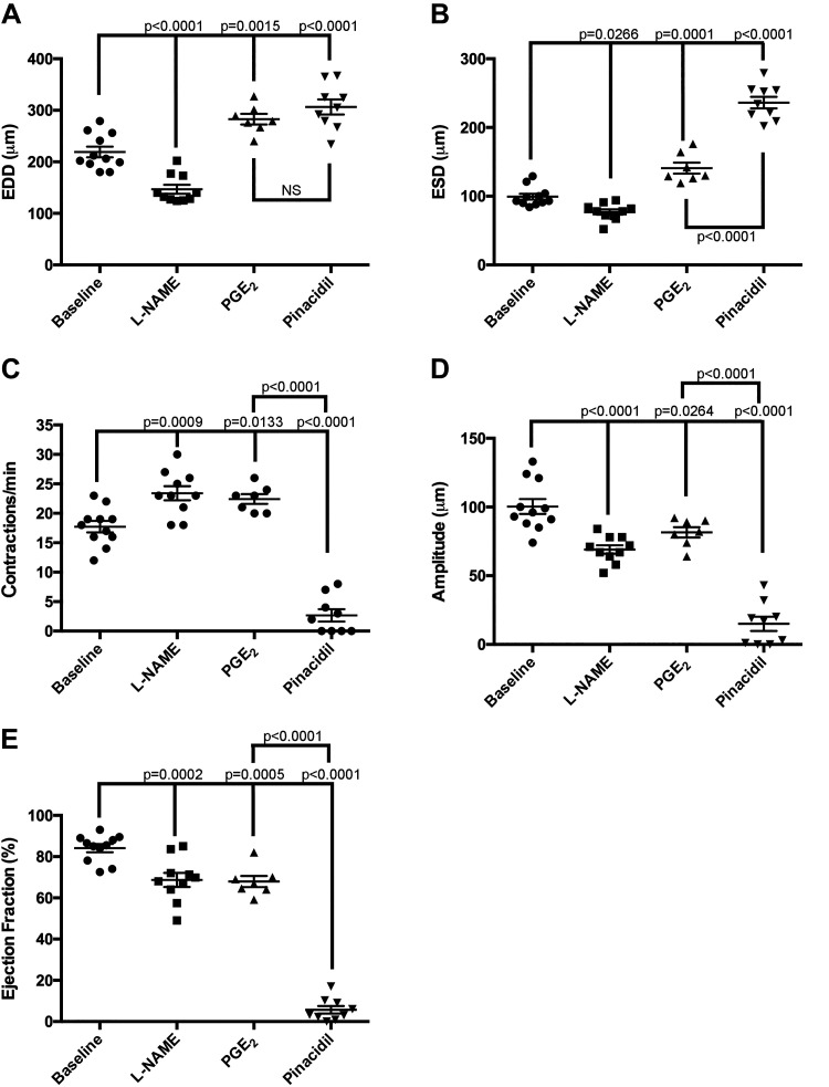
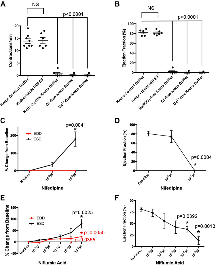
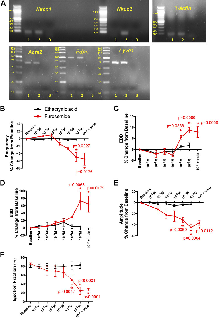
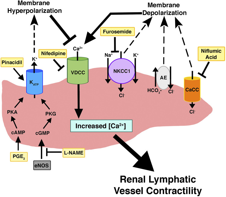
Similar to other organs, renal lymphatics remove excess fluid, solutes, and macromolecules from the renal interstitium. Given the kidney's unique role in maintaining body fluid homeostasis, renal lymphatics may be critical in this process. However, little is known regarding the pathways involved in renal lymphatic vessel function, and there are no studies on the effects of drugs targeting impaired interstitial clearance, such as diuretics. Using pressure myography, we showed that renal lymphatic collecting vessels are sensitive to changes in transmural pressure and have an optimal range of effective pumping. In addition, they are responsive to vasoactive factors known to regulate tone in other lymphatic vessels including prostaglandin E2 and nitric oxide, and their spontaneous contractility requires Ca2+ and Cl-. We also demonstrated that Na+-K+-2Cl- cotransporter Nkcc1, but not Nkcc2, is expressed in extrarenal lymphatic vessels. Furosemide, a loop diuretic that inhibits Na+-K+-2Cl- cotransporters, induced a dose-dependent dilation in lymphatic vessels and decreased the magnitude and frequency of spontaneous contractions, thereby reducing the ability of these vessels to propel lymph. Ethacrynic acid, another loop diuretic, had no effect on vessel tone. These data represent a significant step forward in our understanding of the mechanisms underlying renal lymphatic vessel function and highlight potential off-target effects of furosemide that may exacerbate fluid accumulation in edema-forming conditions.

Keywords: furosemide; loop diuretics; pressure myography; renal lymphatics.


Conflict of interest statement

No conflicts of interest, financial or otherwise, are declared by the authors.

Figures

None
Graphical abstract
Fig. 1.
Fig. 1.
Extrarenal afferent lymphatic vessels were isolated from adult male rats. Lymphangions were mounted in microvessel perfusion chambers for assessment in pressure myography assays (A). Lymphatic vessel identity was confirmed using immunostaining for podoplanin (brown staining, arrows) and α-smooth muscle actin (purple staining, arrowheads). Nuclei were stained with methyl green (B). A digital image capture system was used to measure the frequency of spontaneous contractions, end-diastolic lumen diameter (C), and end-systolic lumen diameter (D). EDD, end-diastolic diameter; ESD, end-systolic diameter.
Fig. 2.
Fig. 2.
Renal lymphatic vessel contractility is regulated by transmural pressure. Isolated vessels were subjected to increasing preload pressure. End-diastolic and end-systolic lumen diameters (A), amplitude of contraction (B), frequency of contractions (C), and calculated ejection fraction (D) were measured at each pressure. Data are expressed as means ± SE. Experimental values were compared with values acquired at 0.5 mmHg to determine statistical significance. n = 6 vessels. EDD, end-diastolic diameter; ESD, end-systolic diameter.
Fig. 3.
Fig. 3.
Renal lymphatic vessels respond to factors known to regulate lymphatic vasoactivity. Vessels were challenged with the nitric oxide synthase inhibitor N-nitro-l-arginine methyl ester (l-NAME; 10−3M), PGE2 (10−9 M), or the ATP-gated K+ (KATP) channel activator pinacidil (10−4 M). The following contractile parameters were measured: end-diastolic diameter (EDD; A), end-systolic diameter (ESD; B), frequency of contraction (C), amplitude of contraction (D), and ejection fraction (E). Baseline values were measured immediately prior to drug addiction. Data are expressed as means ± SE. Experimental values were compared with baseline values to determine statistical significance. PGE2 values were also compared with pinacidil values to determine statistically significant differences. n ≥ 7 vessels. EDD, end-diastolic diameter; ESD, end-systolic diameter.
Fig. 4.
Fig. 4.
Cl and Ca2+ are required for renal lymphatic contractility. Vessels were exposed to standard Krebs, Krebs + HEPES, NaHCO3-free Krebs, Cl-free Krebs, or Ca2+-free Krebs buffer. The frequency of contractions and ejection fraction under each condition were quantified (A and B). Vessels were also challenged with increasing doses of nifedipine, a Ca2+ channel blocker, or niflumic acid, a Ca2+-activated Cl channel inhibitor. End-diastolic diameter (EDD) and end-systolic diameter (ESD; C and E) as well as ejection fraction (D and F) were quantified and expressed as a percentage of baseline values. Data are expressed as means ± SE. In A and B, experimental values were compared with baseline values (control Krebs buffer) to determine statistical significance. In CF, experimental values were compared with baseline values to determine statistical significance. n ≥ 6 vessels.
Fig. 5.
Fig. 5.
Furosemide regulates renal lymphatic contractility via Na+-K+-2Cl cotransporter NKCC1. Images of conventional PCR gels show amplification products for Na+-K+-2Cl cotransporters Nkcc1 and Nkcc2. Lane 1 shows extrarenal lymphatic vessel samples, lane 2 shows whole kidney samples, and lane 3 shows the water-negative control. α2-Smooth muscle actin (Acta2), podoplanin (Pdpn), and lymphatic vessel endothelial receptor 1 (Lyve1) were used to confirm the lymphatic vessel identity of samples in lane 1. The housekeeping gene β-actin was used as a loading control (A). Renal lymphatic vessels were exposed to increasing concentrations of the loop diuretics furosemide or ethacrynic acid. The following contractile parameters were quantified: frequency of contraction (B), end-diastolic diameter (EDD; C), end-systolic diameter (ESD; D) amplitude of contraction (E), and ejection fraction (F). Baseline values were measured immediately prior to drug addiction. Vessels that received the highest dose of furosemide were then challenged with indomethacin. Data points represent means ± SE. Experimental values were compared with baseline values to determine statistical significance. n ≥ 6 vessels.
Fig. 6.
Fig. 6.
Biochemical regulation of renal lymphatic vessels. This model highlights the signaling pathways that modulate renal lymphatic contractility. Compounds that were directly tested in our study are in yellow. CaCC, Ca2+-activated Cl channels; NKCC1, Na+-K+-2Cl cotransporter; AE, anion exchanger; eNOS, endothelial nitric oxide synthase.

References

    1. Alexander JS, Chaitanya GV, Grisham MB, Boktor M. Emerging roles of lymphatics in inflammatory bowel disease. Ann N Y Acad Sci 1207, Suppl 1: E75–E85, 2010. doi: 10.1111/j.1749-6632.2010.05757.x. - DOI - PubMed
    1. Alguire PC, Bailie MD, Weaver WJ, Taylor DG, Hook JB. Differential effects of furosemide and ethacrynic acid on electrolyte excretion in anesthetized dogs. J Pharmacol Exp Ther 190: 515–522, 1974. - PubMed
    1. Baluk P, Fuxe J, Hashizume H, Romano T, Lashnits E, Butz S, Vestweber D, Corada M, Molendini C, Dejana E, McDonald DM. Functionally specialized junctions between endothelial cells of lymphatic vessels. J Exp Med 204: 2349–2362, 2007. doi: 10.1084/jem.20062596. - DOI - PMC - PubMed
    1. Basile DP, Anderson MD, Sutton TA. Pathophysiology of acute kidney injury. Compr Physiol 2: 1303–1353, 2012. - PMC - PubMed
    1. Beutler JJ, Boer WH, Koomans HA, Dorhout Mees EJ. Comparative study of the effects of furosemide, ethacrynic acid and bumetanide on the lithium clearance and diluting segment reabsorption in humans. J Pharmacol Exp Ther 260: 768–772, 1992. - PubMed

Publication types

LinkOut - more resources