. 2020 Jan-Dec;12(1):1829335.
doi: 10.1080/19420862.2020.1829335.

Beyond affinity: selection of antibody variants with optimal biophysical properties and reduced immunogenicity from mammalian display libraries

Affiliations

Beyond affinity: selection of antibody variants with optimal biophysical properties and reduced immunogenicity from mammalian display libraries

Michael R Dyson et al. MAbs. 2020 Jan-Dec.

Abstract

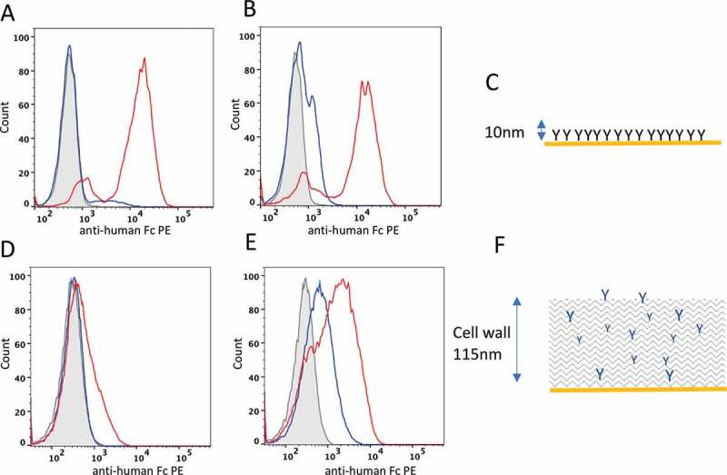
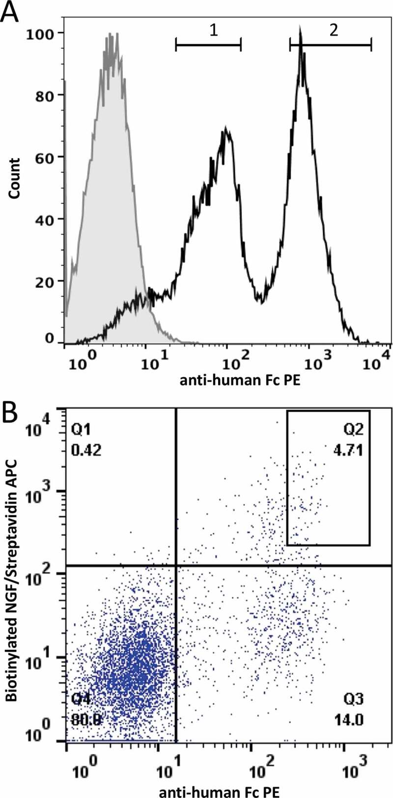
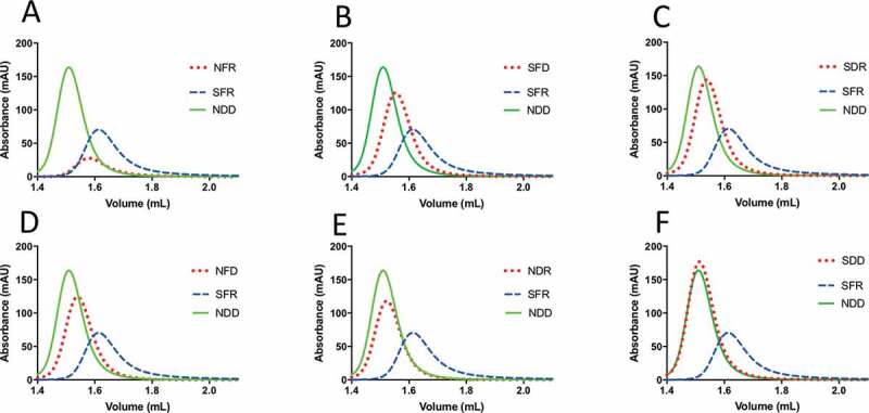
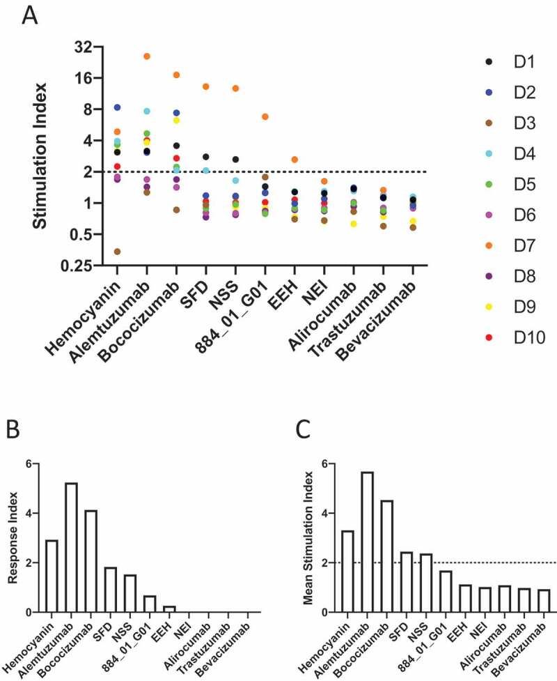
The early phase of protein drug development has traditionally focused on target binding properties leading to a desired mode of therapeutic action. As more protein therapeutics pass through the development pipeline; however, it is clear that non-optimal biophysical properties can emerge, particularly as proteins are formulated at high concentrations, causing aggregation or polyreactivity. Such late-stage "developability" problems can lead to delay or failure in traversing the development process. Aggregation propensity is also correlated with increased immunogenicity, resulting in expensive, late-stage clinical failures. Using nucleases-directed integration, we have constructed large mammalian display libraries where each cell contains a single antibody gene/cell inserted at a single locus, thereby achieving transcriptional normalization. We show a strong correlation between poor biophysical properties and display level achieved in mammalian cells, which is not replicated by yeast display. Using two well-documented examples of antibodies with poor biophysical characteristics (MEDI-1912 and bococizumab), a library of variants was created based on surface hydrophobic and positive charge patches. Mammalian display was used to select for antibodies that retained target binding and permitted increased display level. The resultant variants exhibited reduced polyreactivity and reduced aggregation propensity. Furthermore, we show in the case of bococizumab that biophysically improved variants are less immunogenic than the parental molecule. Thus, mammalian display helps to address multiple developability issues during the earliest stages of lead discovery, thereby significantly de-risking the future development of protein drugs.

Keywords: Mammalian display; antibody aggregation; antibody developability; antibody discovery; antibody half-life; biophysical antibody screening; gene editing; pharmacokinetics; polyreactivity; polyspecificity.


Figures

Figure 1.
Figure 1.
Distinguishing developable antibody by mammalian cell display. Flow cytometry plots of HEK293 (A and B) and yeast cells (D and E) displaying the Ang2 pair (A and D, respectively) or MEDI-1912 pair (B and E, respectively) on the cell surface. Histogram plots represent the cell count (x-axis) plotted against fluorescence intensity of anti-Fc-PE stain) for non-transfected cells (gray, filled), parental antibody (blue) and improved antibody (red). C and F show a scale representations of antibody presentation by mammalian cell display and yeast display, respectively
Figure 2.
Figure 2.
FACS separation of the MEDI-1912 variants based on antibody presentation level. A. Histogram plot of HEK293 population targeted by nuclease-directed integration with an equal mix of MEDI-1912 parent and MEDI-1912 STT variants. Gates 1 and 2 represent the sorting gates used to sort the 2 populations. B. MEDI-1912 mammalian display library was constructed based on mutagenesis of amino-acid position 30, 31 and 56. Cells were stained with anti-Fc-PE (x axis) and NGF-biotin/streptavidin-APC (y axis). The gate chosen for sorting is represented as a box on the dot-plot
Figure 3.
Figure 3.
Engineering bococizumab by random mutagenesis of selected paratope residues. A. Structure of bococizumab bound to PCSK9 (PDB: 3SQO). Upper panel depicts PCSK9 as a yellow ribbon structure and bococizumab as a molecular surface with its hydrophobic, positive, and negative patches shaded green, blue and red, respectively. The lower panel depicts a zoomed in image of the VH region that was mutated. The left panels show wild-type bococizumab and the right panels show a model of the bococizumab VH variant S52N, F54S R57S. Dot plot of HEK293 cells displaying either B. the mutagenized VH library of bococizumab, or C. the mutagenized VH and VL bococizumab library stained with biotinylated PCSK9 (y-axis) and anti-human Fc PE (x-axis)
Figure 4.
Figure 4.
HPLC-SEC analysis of bococizumab variants. Purified antibodies were loaded on a Superdex 200 column integrated in an Agilent 1100 HPLC instrument. Absorbance at 215 nm is plotted against elution volume for each antibody. In each plot samples are compared against the problematic bococizumab (labeled SFR) and the NDD variant, which is a well-behaved positive control. Samples are named according to amino acid substitution at positions 52, 54 and 57 within the VH CDR2. A. NFR, B. SFD, C. SDR, D. NFD E. NDR, F. SDD
Figure 5.
Figure 5.
Induction of proliferation of CD4 + T cells by bococizumab and bococizumab variants selected by mammalian display. A. PBMCs from 10 individual haplotyped human donors (labeled D1 to D10) were labeled with CFDA-SE. After 8 days the cells were analyzed with an iQue screener flow cytometer (Intellicyt) where the viable singlet CD3+ CD4+ gated cells (see Supplementary Figure 7 for gating strategy) were analyzed for CFSE mean fluorescence intensity (MFI). The dot plot (A) shows the stimulation index (SI) for each donor. The SI was calculated by the average percentage of divided cells (% of parent) plus antibody or KLH divided by the average percentage of divided cells in the media only control samples. B. The average response index (RI) is calculated by averaging SI values across donors and multiplying by the percentage frequency of donors responding (p < .05 SI≥2). RI could not be calculated for Trastuzumab, Bevacizumab, Alirocumab and NEI due to a lack of samples meeting positive hit cut off criteria. C. Mean SI for all 10 donors for each antibody or antigen used for stimulation
Figure 6.
Figure 6.
Up-regulation activation markers on CD3+ CD4 + T lymphocytes by bococizumab and bococizumab variants selected by mammalian display. PBMCs from 10 individual haplotyped human donors (labeled D1 to D10) were incubated with different antibodies or KLH. After 8 days, the cells were stained with anti-CD3, anti-CD4, TO-PRO™ −3 and either A. anti-CD69, B. anti-CD25, C. anti-CD71 or D. anti-CD40L and analyzed by flow cytometry. SI was calculated by the percentage of marker positive cells plus antibody or KLH divided by the percentage of marker positive cells in the media only control sample
Figure 7.
Figure 7.
Screening for polyreactivity by mammalian display. Dual staining with (a) Heparin-FITC, (b) Hsp70 or (c) Hsp90 (x-axis) and anti-human Fc APC (y-axis). Dot plots showing overlay of ustekinumab (gray) and briakinumab (black). Gate within the overlay plots indicates the cells to be high expressers and non-binders to heparin or Hsp proteins

References

    1. Kaplon H, Muralidharan M, Schneider Z, Reichert JM.. Antibodies to watch in 2020. mAbs. 2020;12:1703531. - PMC - PubMed
    1. Wörn A, Plückthun A. Stability engineering of antibody single-chain Fv fragments1. J Mol Biol. 2001;305(5):989–18. doi:10.1006/jmbi.2000.4265. - DOI - PubMed
    1. Dudgeon K, Rouet R, Kokmeijer I, Schofield P, Stolp J, Langley D, Stock D, Christ D. General strategy for the generation of human antibody variable domains with increased aggregation resistance. Proc Natl Acad Sci U S A. 2012;109(27):10879–84. doi:10.1073/pnas.1202866109. - DOI - PMC - PubMed
    1. Famm K, Hansen L, Christ D, Winter G. Thermodynamically stable aggregation-resistant antibody domains through directed evolution. J Mol Biol. 2008;376(4):926–31. doi:10.1016/j.jmb.2007.10.075. - DOI - PubMed
    1. Jain T, Sun T, Durand S, Hall A, Houston NR, Nett JH, Sharkey B, Bobrowicz B, Caffry I, Yu Y. Biophysical properties of the clinical-stage antibody landscape. Proc Natl Acad Sci U S A. 2017;114(5):944–49. doi:10.1073/pnas.1616408114. - DOI - PMC - PubMed

Publication types

MeSH terms

Substances