. 2020 Oct;77(10):365-378.
doi: 10.1002/cm.21637. Epub 2020 Nov 18.

Cilium axoneme internalization and degradation in chytrid fungi

Affiliations

Cilium axoneme internalization and degradation in chytrid fungi

Claire M Venard et al. Cytoskeleton (Hoboken). 2020 Oct.

Abstract

Loss of the cilium is important for cell cycle progression and certain developmental transitions. Chytrid fungi are a group of basal fungi that have retained centrioles and cilia, and they can disassemble their cilia via axoneme internalization as part of the transition from free-swimming spores to sessile sporangia. While this type of cilium disassembly has been observed in many single-celled eukaryotes, it has not been well characterized because it is not observed in common model organisms. To better characterize cilium disassembly via axoneme internalization, we focused on chytrids Rhizoclosmatium globosum and Spizellomyces punctatus to represent two lineages of chytrids with different motility characteristics. Our results show that each chytrid species can reel in its axoneme into the cell body along its cortex on the order of minutes, while S. punctatus has additional faster ciliary compartment loss and lash-around mechanisms. S. punctatus retraction can also occur away from the cell cortex and is partially actin dependent. Post-internalization, the tubulin of the axoneme is degraded in both chytrids over the course of about 2 hr. Axoneme disassembly and axonemal tubulin degradation are both partially proteasome dependent. Overall, chytrid cilium disassembly is a fast process that separates axoneme internalization and degradation.

Keywords: Chytridiomycota; RRID:AB_2535778; RRID:AB_2535809; RRID:AB_2650560; RRID:AB_2715541; RRID:AB_477583; RRID:AB_609894; RRID:AB_621847; RRID:AB_621848; RRID:SCR_000432; RRID:SCR_002285; RRID:SCR_003070; RRID:SCR_016865; axoneme; cilia; proteolysis; tubulin.


Conflict of interest statement

CONFLICT OF INTEREST

The authors report no conflict of interest.

Figures

FIGURE 1
FIGURE 1
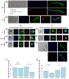
Characterizing cilium disassembly mechanisms in R. globosum and S. punctatus. R. globosum and S. punctatus zoospores were concentrated and placed between an agar pad and glass coverslip for live cell imaging. Still images were taken from individual movies for each species. (a) Severing and (b) reeling in retraction occurred for R. globosum. (c) Severing, (d) reeling in, (e) lash-around, and (f) ciliary compartment loss mechanisms were observed in S. punctatus. (g) Box plots with each individual retraction event show the distribution of time to retraction for both R. globosum (n = 23 cells) and S. punctatus (n = 32 cells). Black dots indicate reeling in retraction (R. globosum, n = 23 cells; S. punctatus, n = 9 cells), red dots indicate ciliary compartment loss retraction (S. punctatus, n = 19 cells), and blue dots indicate lash-around retraction (S. punctatus, n = 4 cells). White arrow heads indicate proximal ends of cilia, and white arrows indicate distal ends of cilia. All scale bars are 5 μm
FIGURE 2
FIGURE 2
Characterizing axoneme localization post-retraction in R. globosum and S. punctatus. Zoospores were fixed and stained for acetylated tubulin (green) and DNA (blue) for (a) R. globosum and (b) S. punctatus. After zoospores were plated for 10 min, fixed, and stained, partial retractions at the cortex were seen for (c) R. globosum and (d) S. punctatus. (e) A partial retraction with axoneme not associated with the cortex was only seen for S. punctatus. Hoop shaped axonemes were observed for (f) R. globosum and (g) S. punctatus. (h) Off-cortex retraction was also seen in S. punctatus. Cells without acetylated tubulin staining for (i) R. globosum and (j) S. punctatus were also observed. TEM 10 min post-retraction show intact axonemes at the cortex for (k) R. globosum and (l) S. punctatus. Off-cortex axonemes were only observed in (m) S. punctatus. (k-m) Axonemes are shown longitudinally (top) and in cross-section (bottom). Red arrows indicate axonemes. Unlabeled scale bars are 3 μm
FIGURE 3
FIGURE 3
S. punctatus cilium retraction is partially actin dependent. Cells were fixed and stained for (a) R. globosum and (b) S. punctatus zoospores. (c) R. globosum and (d) S. punctatus zoospores were plated for 10 min with no treatment, fixed, and stained. (e) R. globosum and (f) S. punctatus zoospores were plated for 10 min with 0.5% ethanol, fixed, and stained. (g) R. globosum and (h) S. punctatus zoospores were plated for 10 min with 1.2 μM latrunculin A, fixed, and stained. All staining is acetylated tubulin (green), rhodamine-phalloidin (red), and DNA (blue). (i) Quantification of the percent of cells with a retracted axoneme after 10 min are shown for R. globosum and S. punctatus under ethanol (EtOH) and latrunculin A (LatA) treatment (R. globosum p = .586; S. punctatus p = .02673). (j) Quantification of the percent of cells with a remaining exterior cilium after 10 min are shown for R. globosum and S. punctatus under ethanol (EtOH) and latrunculin A (LatA) treatment (R. globosum p = .2909; S. punctatus p = .03787). The quantifications are for four independent experiments for R. globosum and six for S. punctatus. Each condition per experiment quantified 162–757 cells for R. globosum and 53–202 cells for S. punctatus. Error bars are SEM. * indicates p < .05, and n.s. indicates no significant difference. Scale bars are 3 μm
FIGURE 4
FIGURE 4
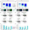
Axonemal tubulin is degraded after internalization. Cells were plated for 10 min, 1hr, and 2 hr, fixed, and stained for acetylated tubulin (green) and DNA (blue). Timepoints were quantified for (a) R. globosum and (b) S. punctatus by the percentage of cells in each of the following categories: cilia, cells that have an exterior cilium; hoop, cells that have a hoop shaped internalized axoneme; pretzel, cells that have an off-cortex internalized axoneme; partial, cells that have one or more fragments of axoneme remaining; and empty, cells that have no axoneme internalized due to axonemal severing or degradation. The quantification reflects four independent experiments for each species. Each timepoint per experiment quantified 47–286 cells for R. globosum and 153–408 cells for S. punctatus. Examples are shown for R. globosum of (c) arcs of axoneme and (d) pieces of axoneme. Examples are also shown for S. punctatus of (e) arcs of axoneme and (f) pieces of axoneme. Western blots for α-tubulin, acetylated tubulin, β-tubulin, and histone H3 using lysates for 0 min,10 min, 1 hr, and 2 hr are shown for (g) R. globosum and (h) S. punctatus. Below each blot is the quantified value for each band normalized to total protein and 0 min. Results were quantified from 3 to 4 independent experiments for each antibody for (i) R. globosum and (j) S. punctatus. Error bars are SEM. Scale bars are 3 μm
FIGURE 5
FIGURE 5
Axoneme depolymerization and tubulin degradation are partially proteasome dependent. Lysates were made for cells treated with 250 μM MG132 or 1% DMSO vehicle control for 10 min, 1hr, and 2 hr. Western blots for acetylated tubulin are shown for (a) R. globosum and (b) S. punctatus. Below each blot is the quantified value for each band normalized to total protein and DMSO 10 min. The results were quantified for (c) R. globosum (2 hr p = .03075) and (d) S. punctatus (1 hr p = .04013; 2 hr p = .03465) for four independent experiments for each species. Cells were treated with 250 μM MG132 or 1% DMSO for 10 min, 1 hr, and 2 hr, fixed, and stained for acetylated tubulin and DNA. The percentage of completely intact axonemes were quantified for (e) R. globosum (2 hr p = .02813) and (f) S. punctatus (1 hr p = .02259). The quantification reflects four independent experiments for R. globosum and three for S. punctatus. Each condition per experiment quantified 73–444 cells for R. globosum and 90–292 cells for S. punctatus. Error bars are SEM. * indicates p < .05. All other pairs are not significantly different

References

    1. Antikajian G. (1949). A developmental, morphological, and cytological study of Asterophlyctis with special reference to its sexuality, taxonomy, and relationships. American Journal of Botany, 36(2), 245–262. 10.1002/j.1537-2197.1949.tb05254.x - DOI
    1. Barr DJS (1986). Allochytridium expandens rediscovered: Morphology, physiology and zoospore ultrastructure. Mycologia, 78(3), 439–448. 10.1080/00275514.1986.12025267 - DOI
    1. Barr DJS, & Hartmann VE (1976). Zoospore ultrastructure of three chytridium species and Rhizoclosmatium globosum. Canadian Journal of Botany, 54(17), 2000–2013. 10.1139/b76-214 - DOI
    1. Bedford JM (1972). An electron microscopic study of sperm penetration into the rabbit egg after natural mating. American Journal of Anatomy, 133(2), 213–254. 10.1002/aja.1001330206 - DOI - PubMed
    1. Berger L, Hyatt AD, Speare R, & Longcore JE (2005). Life cycle stages of the amphibian chytrid Batrachochytrium dendrobatidis. Diseases of Aquatic Organisms, 68, 51–63. 10.3354/dao068051 - DOI - PubMed

Publication types