Observational Study
. 2020 Nov 6;19(11):4455-4469.
doi: 10.1021/acs.jproteome.0c00606. Epub 2020 Oct 26.

Evidence of Structural Protein Damage and Membrane Lipid Remodeling in Red Blood Cells from COVID-19 Patients

Affiliations
Observational Study

Evidence of Structural Protein Damage and Membrane Lipid Remodeling in Red Blood Cells from COVID-19 Patients

Tiffany Thomas et al. J Proteome Res. .

Abstract

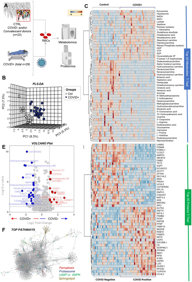
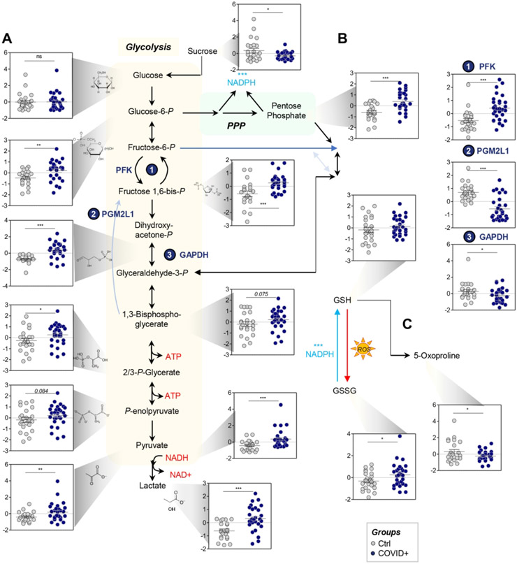
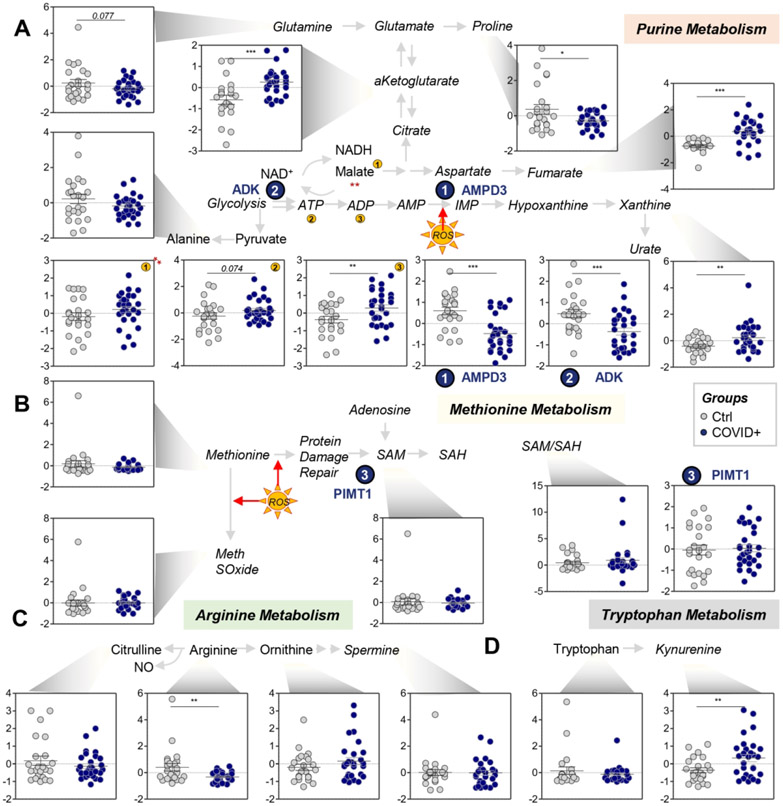
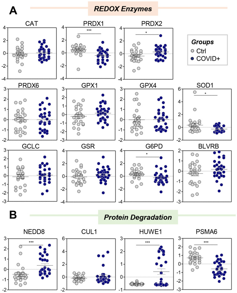
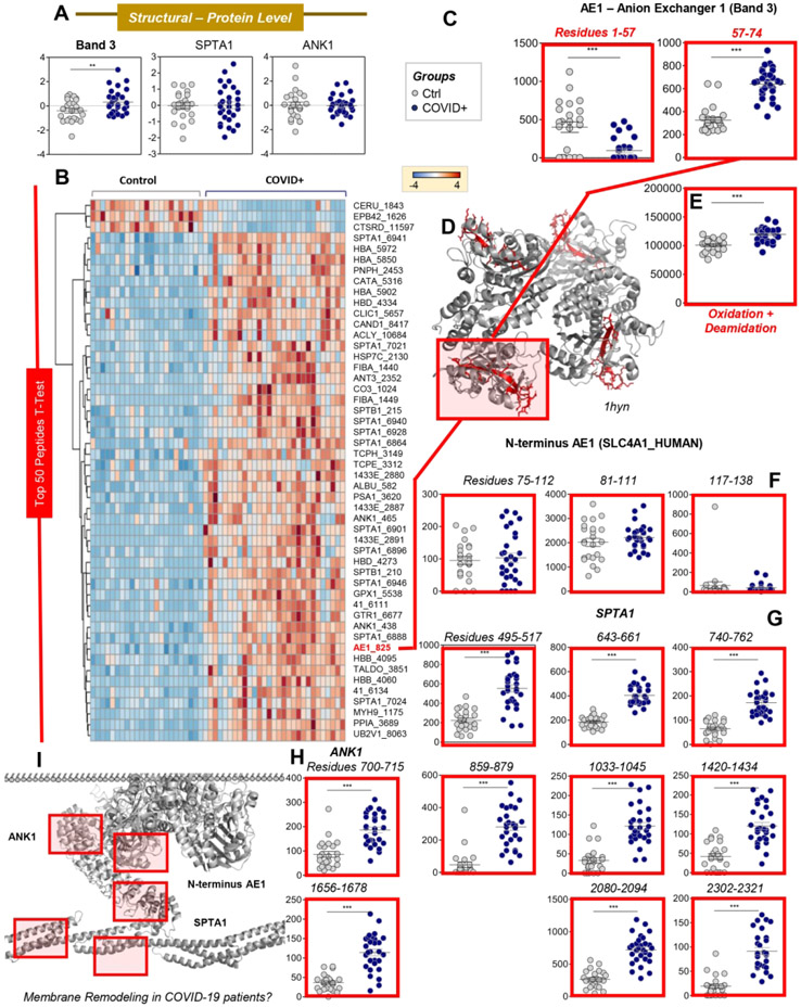
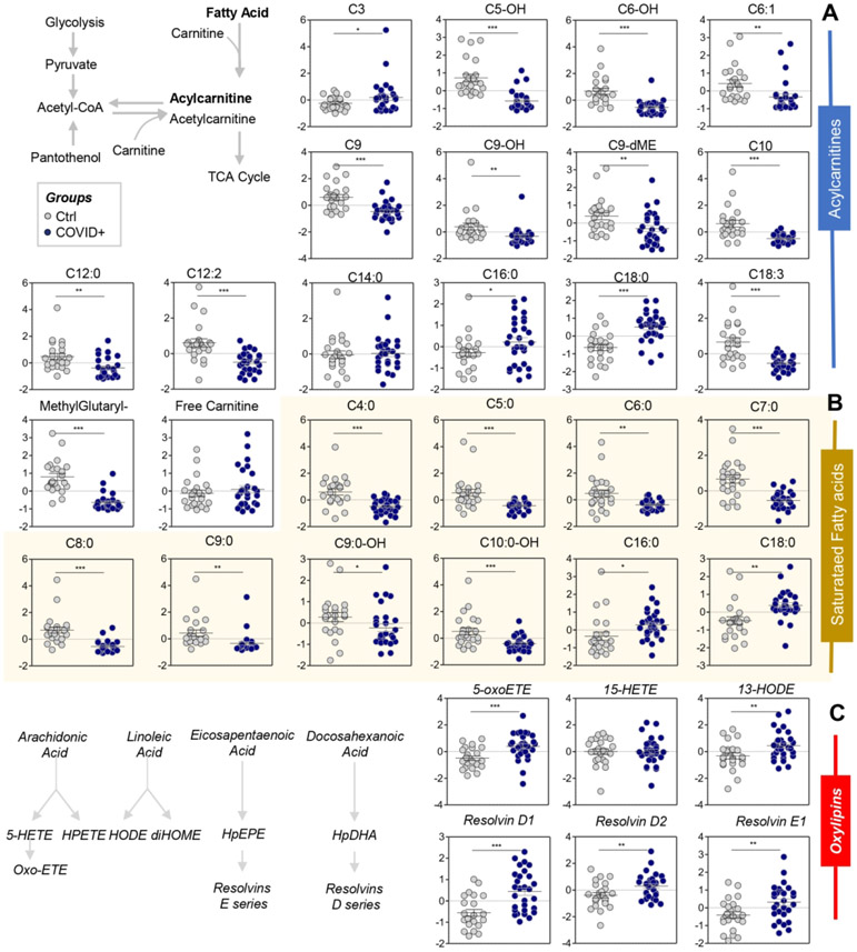
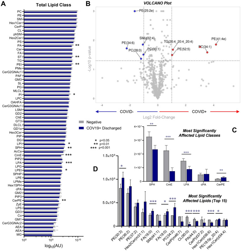
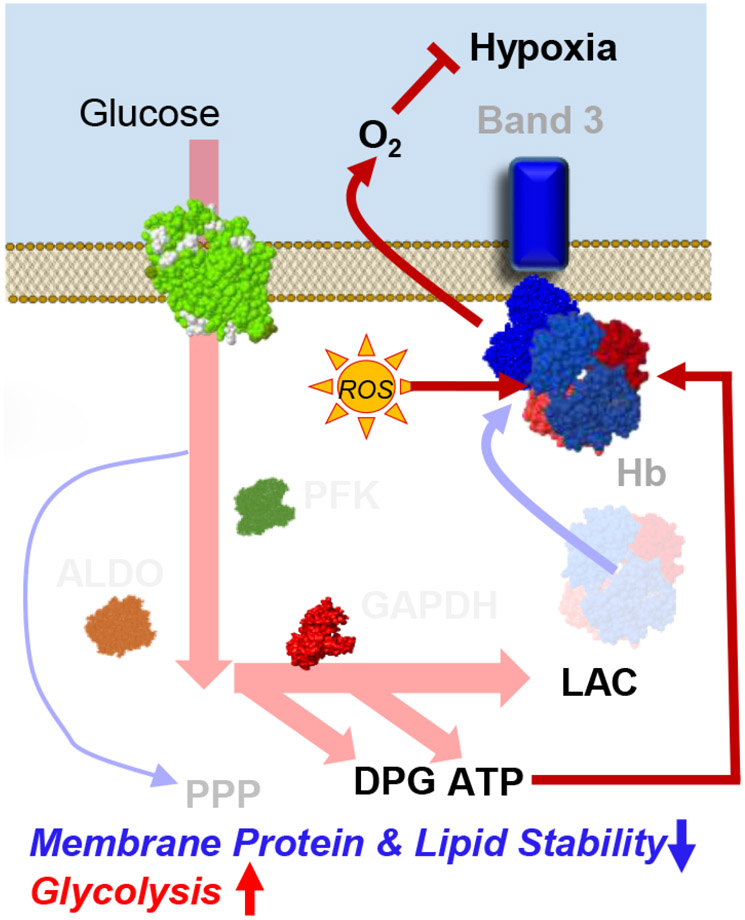
The SARS-CoV-2 beta coronavirus is the etiological driver of COVID-19 disease, which is primarily characterized by shortness of breath, persistent dry cough, and fever. Because they transport oxygen, red blood cells (RBCs) may play a role in the severity of hypoxemia in COVID-19 patients. The present study combines state-of-the-art metabolomics, proteomics, and lipidomics approaches to investigate the impact of COVID-19 on RBCs from 23 healthy subjects and 29 molecularly diagnosed COVID-19 patients. RBCs from COVID-19 patients had increased levels of glycolytic intermediates, accompanied by oxidation and fragmentation of ankyrin, spectrin beta, and the N-terminal cytosolic domain of band 3 (AE1). Significantly altered lipid metabolism was also observed, in particular, short- and medium-chain saturated fatty acids, acyl-carnitines, and sphingolipids. Nonetheless, there were no alterations of clinical hematological parameters, such as RBC count, hematocrit, or mean corpuscular hemoglobin concentration, with only minor increases in mean corpuscular volume. Taken together, these results suggest a significant impact of SARS-CoV-2 infection on RBC structural membrane homeostasis at the protein and lipid levels. Increases in RBC glycolytic metabolites are consistent with a theoretically improved capacity of hemoglobin to off-load oxygen as a function of allosteric modulation by high-energy phosphate compounds, perhaps to counteract COVID-19-induced hypoxia. Conversely, because the N-terminus of AE1 stabilizes deoxyhemoglobin and finely tunes oxygen off-loading and metabolic rewiring toward the hexose monophosphate shunt, RBCs from COVID-19 patients may be less capable of responding to environmental variations in hemoglobin oxygen saturation/oxidant stress when traveling from the lungs to peripheral capillaries and vice versa.

Keywords: AE1; SARS-CoV-2; band 3; erythrocyte; lipidomics; metabolomics; proteomics.


Conflict of interest statement

Disclosure of Conflict of interest Though unrelated to the contents of this manuscript, the authors declare that AD, KCH, and TN are founders of Omix Technologies Inc and Altis Biosciences LLC. AD and SLS are consultants for Hemanext Inc. SLS is also a consultant for Tioma, Inc. JCZ is a consultant for Rubius Therapeutics. All the other authors disclose no conflicts of interest relevant to this study.

Figures

Figure 1 –
Figure 1 –. RBC metabolism and proteome are influenced by COVID-19.
Metabolomics and proteomics analyses were performed on RBCs from COVID-19-negative (n=23) and -positive (n=29) subjects, as determined by molecular testing of nasopharyngeal swabs (A). The effects of COVID-19 on RBCs, as gleaned by PLS-DA (B) and hierarchical clustering analysis of the top 50 metabolites (C) and proteins (D) by t-test. In E, the volcano plot highlights the significant metabolites and proteins increasing (red) or decreasing (blue) in RBCs from COVID-19 patients, as compared to non-infected controls. In F, pathway analyses were performed on the significant features from the analyses in B-E.
Figure 2 –
Figure 2 –. COVID-19 significantly affects RBC glycolysis (A) and the pentose phosphate pathway (PPP - B), with no significant effect on glutathione homeostasis (C).
Metabolomics of RBCs from COVID-19 subjects identified significant increase in several glycolytic intermediates, as compared to controls, including glucose 6-phosphate, fructose bisphosphate, glyceraldehyde 3-phosphate, 2,3-diphosphoglycerate, lactate, and NADH. This phenomenon was at least in part explained by the higher protein levels of PFK, the rate limiting enzyme of glycolysis, in RBCs from COVID-19 subjects, as compared to controls. These subjects also had significant decreases in the levels of PGM2L1, which catalyzes the synthesis of hexose bisphosphate and, thus, slows down glycolysis, and GAPDH, a redox-sensitive enzyme. On the other hand, ribose phosphate (isobars), the end product of the PPP, significantly accumulated in RBCs from COVID-19 patients, suggesting a higher degree of oxidant stress in these RBCs; this was confirmed, in part, by significantly higher levels of GSSG and lower levels of 5-oxoproline (C). Asterisks indicate significance by t-test (*p<0.05; ** p<0.01; *** p<0.001). Groups are color coded according to the legend in the bottom right corner of the figure.
Figure 3 –
Figure 3 –. COVID-19 significantly affects transamination and carboxylic acid metabolism in RBCs (A), but not purine deamination (B), with only limited effects on arginine (C) and tryptophan (D) metabolism.
Asterisks indicate significance by t-test (* p<0.05; ** p<0.01; *** p<0.001). Groups are color coded according to the legend in the center of the figure.
Figure 4 –
Figure 4 –. RBCs from COVID-19 patients have limited alterations in antioxidant enzyme levels (A), but increased levels of components of the ubiquitination/NEDDylation system (B).
Asterisks indicate significance by t-test (* p<0.05; ** p<0.01; *** p<0.001). Groups are color coded according to the legend in the bottom right corner of the figure.
Figure 5 –
Figure 5 –. COVID-19 promotes oxidation and alteration of key structural RBC proteins.
Despite no significant changes in the total levels of key structural proteins (e.g., band 3: AE1; spectrin alpha: SPTA1; ankyrin: ANK1; A), peptidomics analyses showed significant increases and decreases in specific peptides from these proteins (heat map in B shows the top 50 significant changes by t-test). Further analysis of AE1 identified significant increases in levels of the peptide spanning amino acid residues 57-74, in contrast to decreased levels of the N-terminal 1-57 peptide (C), as mapped (red) against the pdb:1hyn spanning residues 56-346 of AE2 (grey; D). In addition, in COVID-19 patients, RBC AE1 was significantly more oxidized (M oxidation + N/Q deamidation) than in control RBCs (E), in the absence of detectable changes in the levels of peptides beyond residue 75 (F). Similar increases in the levels and oxidation of peptides for SPTA1 (G) and ANK1 (H) were observed, consistent with an effect of COVID-19 on the integrity of RBC structural membrane proteins (I).
Figure 6 –
Figure 6 –. RBC acyl-carnitines (A), saturated fatty acids (B), and oxylipins and resolvins (C) were significantly affected by COVID-19.
Asterisks indicate significance by t-test (* p<0.05; ** p<0.01; *** p<0.001). Groups are color coded according to the legend in the top left corner of the figure.
Figure 7 –
Figure 7 –. Lipidomics analyses of RBCs from COVID-19-positive and control patients.
(A) An overview of all the lipid classes investigated in this study (log10 normalized areas) and their variation across groups (symbols indicating significance by t-test are reported in the legend in the center of the panel). (B) Volcano plot showing the most significantly affected lipids, comparing COVID-19-positive subjects and controls. (C and D) Expanded view of the top lipid classes and lipids, respectively, affected by COVID-19. Asterisks indicate significance by t-test (* p<0.05; ** p<0.01; *** p<0.001). Groups are color coded according to the legend in the center of the figure.
Figure 8.
Figure 8.
Model summarizing the proposed findings. Increases in glycolytic metabolites in COVID-19 RBCs are consistent with a theoretically improved capacity of hemoglobin to off-load oxygen as a function of allosteric modulation by high-energy phosphate compounds, perhaps to counteract COVID-19-induced hypoxia. Conversely, because the N-terminus of AE1 stabilizes deoxyhemoglobin and finely tunes oxygen off-loading, RBCs from COVID-19 patients may be incapable of responding to environmental variations in hemoglobin oxygen saturation when traveling from the lungs to peripheral capillaries and, as such, may have a compromised capacity to transport and deliver oxygen. However, this interpretation of the data seems to be confuted by recent reassuring evidence of the lack of alteration of gas exchange and oxygen affinity properties in COVID patients.31,32 On the contrary, damage to the N-term of AE1 may compromise the RBC capacity to inhibit glycolysis and activate the PPP in response to oxidant stress, making the RBCs from COVID patients more susceptible to oxidant stress. Because the damage to AE1 is irreversible, RBCs circulate for up to 120 days without de novo protein synthesis capacity, and this damage may contribute to explaining some of the long-lasting sequelae to COVID-19.

Update of

Similar articles

Cited by

References

    1. Wu F; Zhao S; Yu B; Chen Y-M; Wang W; Song Z-G; Hu Y; Tao Z-W; Tian J-H; Pei Y-Y; Yuan M-L; Zhang Y-L; Dai F-H; Liu Y; Wang Q-M; Zheng J-J; Xu L; Holmes EC; Zhang Y-Z A New Coronavirus Associated with Human Respiratory Disease in China. Nature 2020, 579 (7798), 265–269. 10.1038/s41586-020-2008-3. - DOI - PMC - PubMed
    1. Andersen KG; Rambaut A; Lipkin WI; Holmes EC; Garry RF The Proximal Origin of SARS-CoV-2. Nat. Med 2020, 26 (4), 450–452. 10.1038/s41591-020-0820-9. - DOI - PMC - PubMed
    1. Gordon DE; Jang GM; Bouhaddou M; Xu J; Obernier K; O’Meara MJ; Guo JZ; Swaney DL; Tummino TA; Huettenhain R; Kaake RM; Richards AL; Tutuncuoglu B; Foussard H; Batra J; Haas K; Modak M; Kim M; Haas P; Polacco BJ; Braberg H; Fabius JM; Eckhardt M; Soucheray M; Bennett MJ; Cakir M; McGregor MJ; Li Q; Naing ZZC; Zhou Y; Peng S; Kirby IT; Melnyk JE; Chorba JS; Lou K; Dai SA; Shen W; Shi Y; Zhang Z; Barrio-Hernandez I; Memon D; Hernandez-Armenta C; Mathy CJP; Perica T; Pilla KB; Ganesan SJ; Saltzberg DJ; Ramachandran R; Liu X; Rosenthal SB; Calviello L; Venkataramanan S; Liboy-Lugo J; Lin Y; Wankowicz SA; Bohn M; Sharp PP; Trenker R; Young JM; Cavero DA; Hiatt J; Roth TL; Rathore U; Subramanian A; Noack J; Hubert M; Roesch F; Vallet T; Meyer B; White KM; Miorin L; Rosenberg OS; Verba KA; Agard D; Ott M; Emerman M; Ruggero D; García-Sastre A; Jura N; von Zastrow M; Taunton J; Ashworth A; Schwartz O; Vignuzzi M; d’Enfert C; Mukherjee S; Jacobson M; Malik HS; Fujimori DG; Ideker T; Craik CS; Floor S; Fraser JS; Gross J; Sali A; Kortemme T; Beltrao P; Shokat K; Shoichet BK; Krogan NJ A SARS-CoV-2-Human Protein-Protein Interaction Map Reveals Drug Targets and Potential Drug-Repurposing. bioRxiv 2020, 2020.03.22.002386. 10.1101/2020.03.22.002386. - DOI
    1. Lan J; Ge J; Yu J; Shan S; Zhou H; Fan S; Zhang Q; Shi X; Wang Q; Zhang L; Wang X Structure of the SARS-CoV-2 Spike Receptor-Binding Domain Bound to the ACE2 Receptor. Nature 2020, 1–6. 10.1038/s41586-020-2180-5. - DOI - PubMed
    1. Fantini J; Di Scala C; Chahinian H; Yahi N Structural and Molecular Modelling Studies Reveal a New Mechanism of Action of Chloroquine and Hydroxychloroquine against SARS-CoV-2 Infection. Int. J. Antimicrob. Agents 2020, 55 (5), 105960. 10.1016/jijantimicag.2020.105960. - DOI - PMC - PubMed

Publication types

MeSH terms