. 2022 May 4;225(9):1653-1662.
doi: 10.1093/infdis/jiaa672.

Influence of Strongyloides stercoralis Coinfection on the Presentation, Pathogenesis, and Outcome of Tuberculous Meningitis

Affiliations

Influence of Strongyloides stercoralis Coinfection on the Presentation, Pathogenesis, and Outcome of Tuberculous Meningitis

Joseph Donovan et al. J Infect Dis. .

Abstract

Background: Helminth infections may modulate the inflammatory response to Mycobacterium tuberculosis and influence disease presentation and outcome. Strongyloides stercoralis is common among populations with high tuberculosis prevalence. Our aim was to determine whether S. stercoralis coinfection influenced clinical presentation, cerebrospinal fluid (CSF) inflammation, and outcome from tuberculous meningitis (TBM).

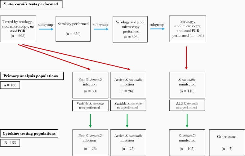
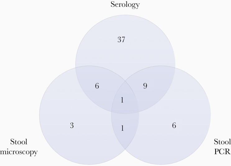
Methods: From June 2017 to December 2019, 668 Vietnamese adults with TBM, enrolled in the ACT HIV or LAST ACT trials (NCT03092817 and NCT03100786), underwent pretreatment S. stercoralis testing by serology, stool microscopy, and/or stool polymerase chain reaction. Comparisons of pretreatment TBM severity, CSF inflammation (including cytokines), and 3-month clinical end points were performed in groups with or without active S. stercoralis infection.

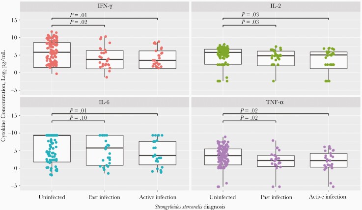
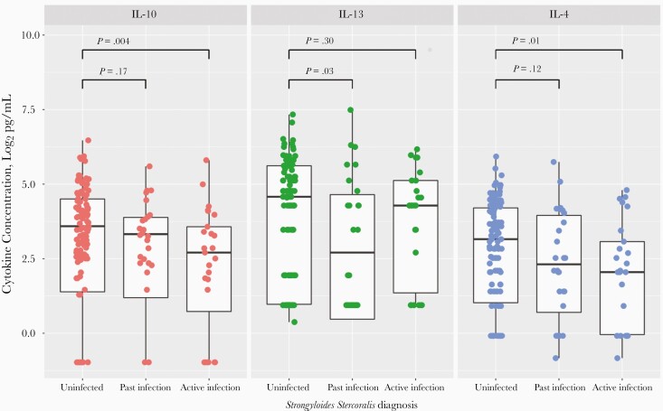
Results: Overall, 9.4% participants (63 of 668) tested positive for S. stercoralis. Active S. stercoralis infection was significantly associated with reduced pretreatment CSF neutrophil counts (median [interquartile range], 3/μL [0-25/μL] vs 14 /μL [1-83/μL]; P = .04), and with reduced CSF interferon ɣ, interleukin 2, and tumor necrosis factor α concentrations (11.4 vs 56.0 pg/mL [P = .01], 33.1 vs 54.5 pg/mL [P = .03], and 4.5 vs 11.9 pg/mL [P = .02], respectively), compared with uninfected participants. Neurological complications by 3 months were significantly reduced in participants with active S. stercoralis infection compared with uninfected participants (3.8% [1 of 26] vs 30.0% [33 of 110], respectively; P = .01).

Conclusions: S. stercoralis coinfection may modulate the intracerebral inflammatory response to M. tuberculosis and improve TBM clinical outcomes.

Keywords: Strongyloides stercoralis; cytokines; immunomodulation; inflammation; outcome; tuberculous meningitis.


Figures

Figure 1.
Figure 1.
Strongyloides stercoralis testing populations. A total of 668 participants underwent ≥1 S. stercoralis test, serology in 659, serology and stool microscopy in 523, and serology, stool microscopy, and stool polymerase chain reaction (PCR) in 141. Following the light gray arrows, each group is a subgroup of the previous group. Dark gray arrows show how primary analysis populations were developed. All S. stercoralis–uninfected participants had serology, stool microscopy, and stool PCR performed. Past S. stercoralis infection and active S. stercoralis infection groups were selected independently of the number of S. stercoralis tests performed; therefore, these are taken from the population in which ≥1 S. stercoralis test was performed (N = 668). Black arrows show how cytokine testing populations were formed. From a total of 173 patients initially eligible for cytokine testing, 10 samples were omitted; 4 were excluded from testing when no stored cerebrospinal fluid (CSF) sample was available, 5 were not tested because S. stercoralis tests returned a positive result after cytokine testing had been arranged and set up, and 1 sample result was lost owing to a computer error during cytokine analysis. Therefore, 163 CSF samples underwent cytokine testing, of which 156 fit into primary analysis population definitions (uninfected, past infection, or active infection). For the uninfected group, all 3 testing methods were used, all with negative results. For the past infection group, results of S. stercoralis serology were positive with no positive stool testing results (but with stool microscopy and/or stool PCR performed). In the active infection group, results of stool microscopy or stool PCR were positive for S. stercoralis, regardless of other testing performed. The “Other status” group includes participants who underwent cytokine testing but did not meet criteria for any of the 3 primary analysis population groups.
Figure 2.
Figure 2.
Venn diagram of 81 Strongyloides stercoralis tests with positive results, including serology (n = 53), stool microscopy (n = 11), and stool polymerase chain reaction (PCR) (n = 17). The tests were performed in 63 participants testing positive for S. stercoralis with serology, stool microscopy, and/or stool PCR; these participants include the past infection (n = 30) and active infection (n = 26) primary analysis populations, as well as 7 participants with a positive S. stercoralis test result not meeting the criteria for those populations.
Figure 3.
Figure 3.
Log2 cerebrospinal fluid interferon (IFN) ɣ, interleukin 2 (IL-2), interleukin 6 (IL-6), and tumor necrosis factor (TNF) α concentrations in participants uninfected with Strongyloides stercoralis, with past S. stercoralis infection, or with active infection. The log2 cytokine concentrations 15, 10, 5, 0, and −5 correspond to the following measured cytokine concentrations: 32 768, 1024, 32, 1, and 0.03 pg/mL, respectively. For each individual box plot, the central horizontal bar represents the median value, and the box contains data between the third and first quartiles (upper and lower ends of box, respectively); vertical lines above and below each box extend to the most extreme data point within 1.5 times the height of the box; and dots represent individual data points. For the uninfected group, all 3 testing methods were used, all with negative results. For the past infection group, results of S. stercoralis serology were positive with no positive stool testing results (but with stool microscopy and/or stool polymerase chain reaction [PCR] performed). In the active infection group, results of stool microscopy or stool PCR were positive for S. stercoralis, regardless of other testing performed. Statistical comparisons of cytokine concentrations were performed using Wilcoxon rank sum tests.
Figure 4.
Figure 4.
Log2 cerebrospinal fluid interleukin 10, 13, and 4 (IL-10, IL-13, and IL-4) concentrations in participants uninfected with Strongyloides stercoralis, with past S. stercoralis infection, or with active infection. For each individual box plot, the central horizontal bar represents the median value, and the box contains data between the third and first quartiles (upper and lower ends of box, respectively); vertical lines above and below each box extend to the most extreme data point within 1.5 times the height of the box; and dots represent individual data points. For the uninfected group, all 3 testing methods were used, all with negative results. For the past infection group, results of S. stercoralis serology were positive with no positive stool testing results (but with stool microscopy and/or stool polymerase chain reaction [PCR] performed). In the active infection group, results of stool microscopy or stool PCR were positive for S. stercoralis, regardless of other testing performed. Statistical comparison of cytokine concentrations was performed using Wilcoxon rank sum tests.

References

    1. World Health Organization. Strongyloidiasis. World Health Organization; 2016. http://www.who.int/intestinal_worms/epidemiology/strongyloidiasis/en/. Accessed 26 June 2020.
    1. Christensen ASH, Roed C, Omland LH, Andersen PH, Obel N, Andersen ÅB. Long-term mortality in patients with tuberculous meningitis: a Danish nationwide cohort study. PLoS One 2011; 6:e27900. - PMC - PubMed
    1. Vinnard C, King L, Munsiff S, et al. . Long-term mortality of patients with tuberculous meningitis in New York City: a cohort study. Clin Infect Dis 2016; 64:ciw763. - PMC - PubMed
    1. Thwaites GE, Bang ND, Dung NH, et al. . Dexamethasone for the treatment of tuberculous meningitis in adolescents and adults. N Engl J Med 2004; 351:1741–51. - PubMed
    1. Ruslami R, Ganiem AR, Dian S, et al. . Intensified regimen containing rifampicin and moxifloxacin for tuberculous meningitis: an open-label, randomised controlled phase 2 trial. Lancet Infect Dis 2013; 13:27–35. - PubMed

Publication types