. 2020 Oct 26;16(10):e1009009.
doi: 10.1371/journal.ppat.1009009. eCollection 2020 Oct.

MAVS regulates the quality of the antibody response to West-Nile Virus

Affiliations

MAVS regulates the quality of the antibody response to West-Nile Virus

Marvin O'Ketch et al. PLoS Pathog. .

Abstract

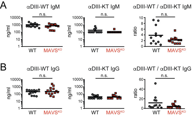
A key difference that distinguishes viral infections from protein immunizations is the recognition of viral nucleic acids by cytosolic pattern recognition receptors (PRRs). Insights into the functions of cytosolic PRRs such as the RNA-sensing Rig-I-like receptors (RLRs) in the instruction of adaptive immunity are therefore critical to understand protective immunity to infections. West Nile virus (WNV) infection of mice deficent of RLR-signaling adaptor MAVS results in a defective adaptive immune response. While this finding suggests a role for RLRs in the instruction of adaptive immunity to WNV, it is difficult to interpret due to the high WNV viremia, associated exessive antigen loads, and pathology in the absence of a MAVS-dependent innate immune response. To overcome these limitations, we have infected MAVS-deficient (MAVSKO) mice with a single-round-of-infection mutant of West Nile virus. We show that MAVSKO mice failed to produce an effective neutralizing antibody response to WNV despite normal antibody titers against the viral WNV-E protein. This defect occurred independently of antigen loads or overt pathology. The specificity of the antibody response in infected MAVSKO mice remained unchanged and was still dominated by antibodies that bound the neutralizing lateral ridge (LR) epitope in the DIII domain of WNV-E. Instead, MAVSKO mice produced IgM antibodies, the dominant isotype controlling primary WNV infection, with lower affinity for the DIII domain. Our findings suggest that RLR-dependent signals are important for the quality of the humoral immune response to WNV.


Conflict of interest statement

The authors have declared that no competing interests exist.

Figures

Fig 1
Fig 1. Impaired neutralizing antibody response in RWN-infected MAVSKO mice.
(A, B) WNV-E-specific IgM (A) and IgG (B) on day 8 after infection with RWN (105 pfu/footpad) as measured by ELISA. (C) Virus neutralization by sera of infected MAVSKO and MAVSWT mice. RWN was incubated with serial dilutions of sera prior to infection of Vero cells in vitro. The number of infected cells was determined 30–48 hours later by staining with an anti-WNV-E antibody. The reduction of infected cells by 90% was scored (PRNT90). (D) Neutralization index on day 8 post infection. The index normalizes virus neutralization to the total amount of WNV-E-specific antibodies in MAVSKO and MAVSWT mice. The index was calculated by dividing the dilution factor (PRNT90) of each mouse by the total amount of WNV-E-specific IgM and IgG of the same mouse. The data were normalized across multiple experiments to MAVSWT mice. Each dot represents one mouse, the lines represent the median. **, p <0.005; ***; p < 0.0005; n.s., not significant; Mann-Whitney test.
Fig 2
Fig 2. Similar levels of WNV-E protein in RWN-infected MAVSKO and MAVSWT mice.
(A) Viral RNA levels in the dLNs on day 1 post infection with RWN (105 pfu/footpad) as measured by qPCR using primer pairs located in the WNV-E or NS4b genes of the viral genome. Data were normalized to the RNA level of RWN-infected MAVSWT mice. (B, C) Production of WNV-E protein in cells from the dLNs of RWN-infected MAVSKO and MAVSWT mice. Cells from the dLNs were isolated 24 hours after infection and cultured for an additional 24 hours in vitro. The amount of WNV-E protein in the combined cell lysates and supernatants was quantified by flow cytometry using an anti-WNV-E bead assay. Samples from naïve mice or assay buffer were used as controls. (B) A representative experiment is shown. Shaded area represents the background staining of samples from uninfected animals, red lines represent RWN-infected animals. (C) Statistical summary of geometric means of multiple independent experiments. The data were normalized to the background staining of uninfected mice in each experiment. Each dot represents one mouse; the line is the median; n.s., not significant, ***, p < 0.0005, Mann-Whitney test.
Fig 3
Fig 3. Increased antigen production does not impair virus neutralization.
(A) Comparison of the viral load in the dLNs of MAVSKO mice infected with 105 Pfu RWN and MAVSWT mice infected with an increased dose of 106 Pfu RWN. Cells from the dLNs were isolated 24 hours after infection and cultured for an additional 24 hours in vitro. The amount of WNV-E protein in the combined cell lysates and supernatants was quantified by flow cytometry using an anti-WNV-E bead assay. (B) Increased doses of RWN do not impair virus neutralization in MAVSWT mice. Mice were infected with indicated doses of RWN. The amount of WNV-E-specific IgM and IgG as well as PRNT90 were determined in order to calculate the neutralization index. Shown are the combined data of 3 experiments. Each dot represents one mouse, the line represents the median. ns, not significant; Mann-Whitney test.
Fig 4
Fig 4. Enhanced GC B and CD4+ T cell response to RWN in MAVSKO mice.
The cellularity of the dLNs from MAVSKO and MAVSWT mice was analyzed 8 days post RWN infection (105 pfu/footpad) by flow cytometry. (A) Total numbers of CD19+ B cells. (B) Left panels: Frequency of Germinal Center (GC) B cells. Right panel: Total GC B cell numbers of 5 experiments normalized to the average of MAVSWT mice in each experiment. (C) Absolute number of CD4+ T cells. (D) Left panels: Frequency of E641:I-Ab class II tetramer+ CD4+ T cells specific for the immunodominant E641 epitope derived from WNV-E. Right panel: Total E641:I-Ab+ CD4+ T cells numbers of 3 experiments normalized to the average of MAVSWT mice in each experiment. (E) Left panels: Frequency of non-GC CXCR5+ PD-1- T follicular helper (Tfh) cells and CXCR5+ PD-1+ GC Tfh cells. Right panel: Total Tfh cell numbers of 5 experiments normalized to the average of MAVSWT mice in each experiment. Frequencies are shown as mean ± SEM. Cell numbers: Each dot is one mouse, lines are the medians. *, p < 0.05; ***; p < 0.0005; n.s., not significant; Mann-Whitney test.
Fig 5
Fig 5. Production of cytokines and interferon-stimulated genes (ISGs) in the dLNs of MAVSKO mice.
(A) Expression of IL-1β, IL-6, and TNF-α mRNA RWN-infected MAVSKO and MAVSWT mice. (B) Expression of type I and type III IFNs mRNA in RWN-infected MAVSKO and MAVSWT mice. (C) Expression of representative ISGs in RWN-infected MAVSKO and MAVSWT mice. (A-C) mRNA was isolated from whole dLNs cells of mice 24 hours after infection with 105 Pfu RWN per footpad and measured by qPCR. Shown is the expression over that of dLNs from naïve WT mice. Expression of GAPDH was used to normalize the samples. Shown are the combined data of at least 4 experiments using 8–20 mice/genotype **, p < 0.005; ***, p < 0.0005; Mann-Whitney test.
Fig 6
Fig 6. Normal specificity of the antibody response of MAVSKO mice to the neutralizing lateral ridge (LR) epitope in the WNV-E DIII domain.
(A, B) The antibody response of RWN-infected MAVSKO and MAVSWT mice was measured 8 days post infection. Shown are the titers of IgM (A) and IgG (B) specific for the WT DIII domain or the mutant form DIII-KT containing loss-of-function mutations in the neutralizing LR epitope (DIII-K307E/T330I). Ratios represent the excess of antibodies directed at the neutralizing epitope over the amount of non-neutralizing antibodies directed at the DIII domain. Combined data of 4 experiments. Each data point is one mouse, the line is the median; n.s., not significant; Mann-Whitney test.
Fig 7
Fig 7. Impaired avidity of the IgM response to the DIII domain of WNV-E protein in RWN-infected MAVSKO mice.
(A, B) DIII-specific IgM titers from RWN-infected mice were measured by ELISA in the presence of BSA to reduce binding avidity and increasing amounts of NaSCN to enhance stringency of binding. Recombinant DIII protein was used as antigen. Shown are the absolute titers (A) and the titers as fraction of the total amount of DIII-specific antibodies in the absence of BSA and NaSCN for each mouse (B). (C, D) Same as before for DIII-specific IgG. Shown are the combined data of 4 experiments. Each dot represents one mouse, the line is the median. *, p < 0.05; ***, p < 0.0005; n.s., not significant; Mann-Whitney test.

References

    1. Schenten D, Medzhitov R. The control of adaptive immune responses by the innate immune system. Adv Immunol. 2011;109:87–124. 10.1016/B978-0-12-387664-5.00003-0 - DOI - PubMed
    1. Iwasaki A, Medzhitov R. Control of adaptive immunity by the innate immune system. Nat Immunol. 2015;16:343–353. 10.1038/ni.3123 - DOI - PMC - PubMed
    1. Moyer TJ, Zmolek AC, Irvine DJ. Beyond antigens and adjuvants: Formulating future vaccines. Journal of Clinical Investigation. 2016. 10.1172/JCI81083 - DOI - PMC - PubMed
    1. Georg P, Sander LE. Innate sensors that regulate vaccine responses. Current Opinion in Immunology. 2019. 10.1016/j.coi.2019.02.006 - DOI - PubMed
    1. Pulendran B, Oh JZ, Nakaya HI, Ravindran R, Kazmin DA. Immunity to viruses: Learning from successful human vaccines. Immunol Rev. 2013. 10.1111/imr.12099 - DOI - PMC - PubMed

Publication types

MeSH terms