. 2021 Mar;78(6):2929-2948.
doi: 10.1007/s00018-020-03683-9. Epub 2020 Oct 26.

Macrophage ubiquitin-specific protease 2 contributes to motility, hyperactivation, capacitation, and in vitro fertilization activity of mouse sperm

Affiliations

Macrophage ubiquitin-specific protease 2 contributes to motility, hyperactivation, capacitation, and in vitro fertilization activity of mouse sperm

Mayuko Hashimoto et al. Cell Mol Life Sci. 2021 Mar.

Abstract

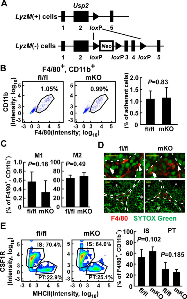
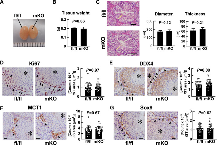
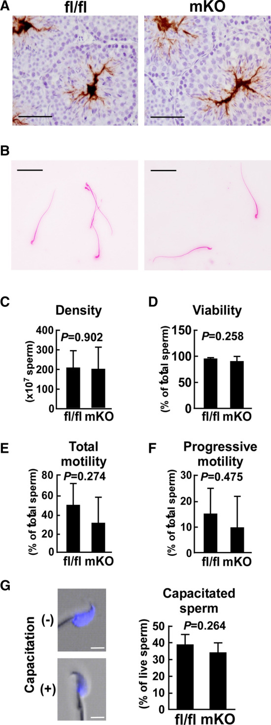
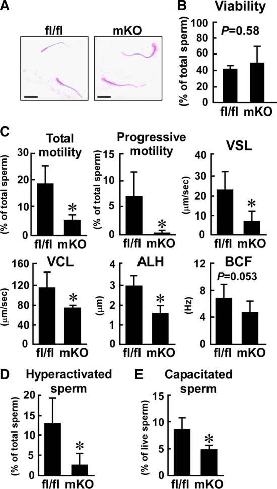
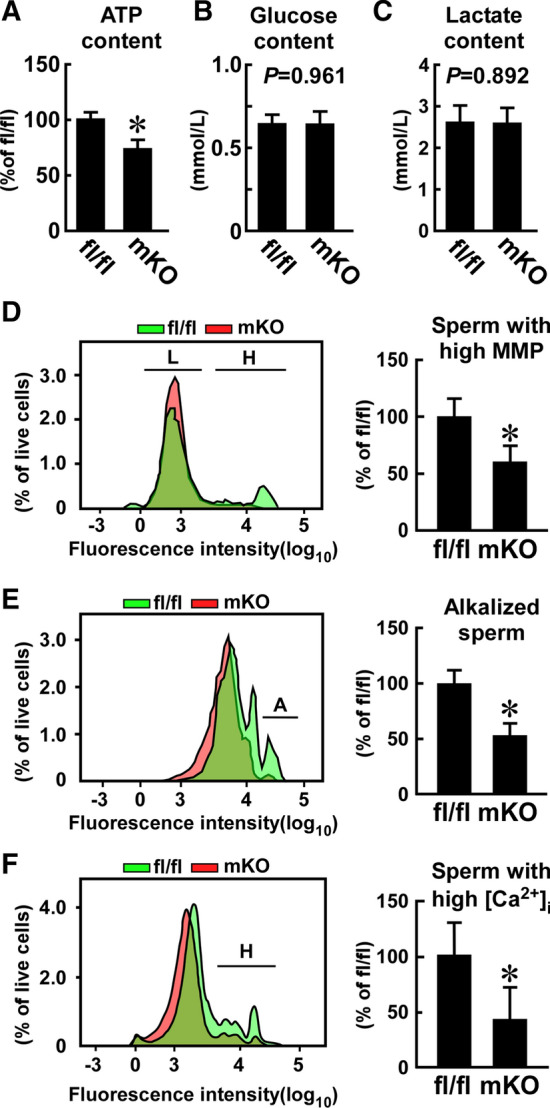
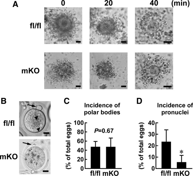
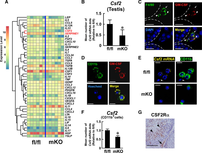
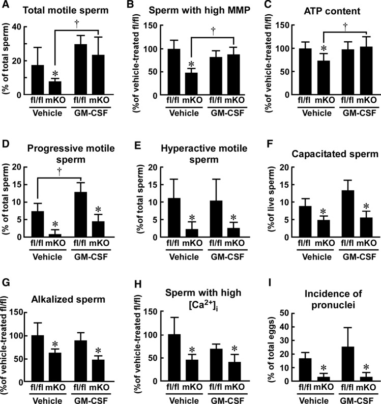
Macrophages are innate immune cells that contribute to classical immune functions and tissue homeostasis. Ubiquitin-specific protease 2 (USP2) controls cytokine production in macrophages, but its organ-specific roles are still unknown. In this study, we generated myeloid-selective Usp2 knockout (msUsp2KO) mice and specifically explored the roles of testicular macrophage-derived USP2 in reproduction. The msUsp2KO mice exhibited normal macrophage characteristics in various tissues. In the testis, macrophage Usp2 deficiency negligibly affected testicular macrophage subpopulations, spermatogenesis, and testicular organogenesis. However, frozen-thawed sperm derived from msUsp2KO mice exhibited reduced motility, capacitation, and hyperactivation. In addition, macrophage Usp2 ablation led to a decrease in the sperm population exhibiting high intracellular pH, calcium influx, and mitochondrial membrane potential. Interrupted pronuclei formation in eggs was observed when using frozen-thawed sperm from msUsp2KO mice for in vitro fertilization. Administration of granulocyte macrophage-colony stimulating factor (GM-CSF), whose expression was decreased in testicular macrophages derived from msUsp2KO mice, restored mitochondrial membrane potential and total sperm motility. Our observations demonstrate a distinct role of the deubiquitinating enzyme in organ-specific macrophages that directly affect sperm function.

Keywords: Capacitation; Granulocyte macrophage-colony stimulating factor; Male sterility; Myeloid cells; USP.


Conflict of interest statement

The authors have no conflicts of interest/competing interests to declare.

Figures

Fig. 1
Fig. 1
Effect of myeloid-selective Usp2 knockout on the proportion of the macrophage subpopulation in the testis. a Schematic presentation of the Usp2 locus of LyzM ( +) and (−) cells. Exon numbers and loxP sites are shown. Neo represents the neomycin-resistant gene. b-e: Myeloid-selective Usp2 knockout mice (mKO) and Usp2fl/fl mice (fl/fl) were subjected to FACS analysis (b, c, e) and histological analysis (d). b The proportion of F4/80+, CD11b+ macrophages in the testicular adherent cell fraction. Representative flow cytometry scatter plots (left panels) and quantification of the macrophages (right panel) are shown. c Proportion of Ly6chigh, CD206 M1 macrophages (left) and Ly6cint, CD206+ M2 macrophages (right) in F4/80+, CD11b+ cells. d Representative images of interstitial (arrows) and peritubular (arrowheads) macrophages in the testis that were stained with an anti-F4/30 antibody. The top panels show an enlarged view of the bottom panels. Seminiferous tubules are shown by dashed lines and asterisks. Scale bars represent 20 µm. Data were reproducibly obtained from three individual mice. e The proportion of CSF1R+, MHCII interstitial (IS) and CSF1R, MHCII+ peritubular (PT) macrophages in F4/80+, CD11b+ cells. Representative flow cytometry scatter plots (left panels) and quantification of IS and PT macrophages (right panel) are shown. Data are shown as the means ± SD of five mice (b, c, e). P values are shown in each graph (b, c, e)
Fig. 2
Fig. 2
Effect of myeloid-selective Usp2 knockout on testicular organogenesis. a, b Morphology (a) and weight (b) of the testis in myeloid-selective Usp2 knockout mice (mKO) and Usp2fl/fl mice (fl/fl). c The outer diameter and thickness of seminiferous tubules. Representative microscopic images (left panels) and quantification of their diameter and thickness (right panels). Yellow bars indicate the wall of the seminiferous tubules. The average thickness of the seminiferous tubule wall at four respective ends of the major and minor axes was calculated. d–g Immunohistochemical staining with antibodies against Ki67 (d), DDX4 (e), MCT1 (f), and Sox9 (g). Representative microscopic images and the density of immune-positive cells are shown in the left and right panels (d–g), respectively. Arrowheads indicate immunopositive cells, and asterisks show seminiferous tubules. Scale bars represent 20 µm. Immunopositive cells were counted in the seminiferous tubules (ST; d, e, g) or in the interstitial regions (IS; f) in five randomly selected microscopic fields for each individual. Data are shown as means ± SD of six (b, c) or five (d–g) mice, with counts of immunopositive cells in each area shown as dots. P values are shown in the graphs (b–g)
Fig. 3
Fig. 3
Effect of myeloid-selective Usp2 deficiency on the characteristics of freshly isolated sperm. a Representative histochemical images of mature sperm in the testis of myeloid-selective Usp2 knockout mice (mKO) and Usp2fl/fl mice (fl/fl). The sperm were stained with an anti-MCT2 antibody. b The morphology of eosin-stained sperm from the epididymis. c, d The density (c) and viability (d) of the epididymal sperm. e, f Total (e) and progressive (f) sperm motility were evaluated by computer-assisted sperm motility analysis (CASA). g Representative images of chlortetracycline (CTC)-stained sperm (left) and capacitation rate in total sperm (right). Capacitated ( +) and non-capacitated (-) sperm are shown. Scale bars represent 50 µm (a, b) or 5 µm (g). Reproducible histological data were obtained from three (a) or five (b, g) mice for each genotype. Data are shown as the means ± SD of 9–13 (c), 4–5 (d), 6 (e, f) or 5 (g) mice. Approximately, 300 (e, f) or 200 (c, d, g) sperm per mouse was used for the analysis. The P values are shown in graphs (c–g)
Fig. 4
Fig. 4
Effect of myeloid-selective Usp2 knockout on the motility, hyperactivation, and capacitation of frozen–thawed sperm. Sperm of myeloid-selective Usp2 knockout mice (mKO) and Usp2fl/fl mice (fl/fl) were subjected to the freeze–thaw process. a The morphology of the epididymal sperm. Scale bars represent 50 µm. Representative microscopic images were obtained from five mice. b The viability of the epididymal sperm. c Sperm motility assessed by CASA. Total motility, progressive motility, straight-line velocity (VSL), curvilinear velocity (VCL), amplitude of lateral head displacement (ALH), and beat-cross frequency (BCF) of sperm were evaluated. d The proportion of hyperactivated sperm. K-means clustering was used to analyze the CASA data. Hyperactive sperm belonged to the cluster with high VCL and low linearity, as shown in Supplementary Fig. 8. e The proportion of capacitated sperm. Capacitation was determined by CTC staining. Approximately 200 (b, e), 300 (c), 340 (d, mKO), or 420 (d, fl/fl) sperm from each mouse was used for analysis. Data are shown as means ± SD of five (b, e) or six (c, d) mice. *P < 0.05 vs. Usp2fl/fl mice (ce). The P values are also shown in the graphs (b, c)
Fig. 5
Fig. 5
Effect of myeloid-selective Usp2 knockout on the biochemical and physiological responses of frozen–thawed sperm. a, b, c Intracellular ATP (a), glucose (b), and lactate (c) contents. Approximately 10,000 (a) or 25,000 (b, c) sperm per mouse was used for the analyses. d, f FACS analysis of mitochondrial membrane potential (MMP, d), intracellular pH (e), and calcium ion content (f) assessed with TMRM, BCECF-AM, and Calbryte-520AM, respectively. FACS images indicate sperm with high (H, d) and low (L, d) MMP, high alkalization (A, e), and high calcium influx (H, f) (left). The proportion of sperm with high MMP (d), high alkalization (e), and high calcium influx (f) in approximately 25,000 live sperm are also indicated (right panel). Data are shown as the means ± SD of six mice (a–f). *P < 0.05 vs. Usp2fl/fl mice. P values are also shown in graphs (b, c)
Fig. 6
Fig. 6
Effect of myeloid-selective Usp2 knockout on the efficiency of in vitro fertilization. Frozen–thawed 4 × 104 live sperm, which were isolated from myeloid-selective Usp2 knockout mice (mKO) and Usp2fl/fl mice (fl/fl), were subjected to in vitro fertilization (IVF) using the ova of C57BL/6 mice. Approximately, 60 ova were used for an IVF series. a Dispersion of cumulus cells. Ova surrounded with cumulus cells were incubated with sperm from the mice for 0, 20, and 40 min. b Ova after 8 h incubation with sperm. Arrows and arrowheads represent polar bodies and pronuclei, respectively. c, d Incidence of polar bodies (c) or pronuclei (d) in ova after incubation with sperm. Representative images of seven IVF series are shown (a, b). Scale bars represent 50 (a), or 20 (b) μm. Data are shown as means ± SD of seven male mice (c, d). *P < 0.05 vs Usp2fl/fl mice (d). P value is also shown (c)
Fig. 7
Fig. 7
Effect of myeloid-selective Usp2 knockout on Csf2 expression in the testis. a Comprehensive qRT-PCR for cytokine expression in the testis of myeloid-selective Usp2 knockout mice (mKO) and Usp2fl/fl mice (fl/fl). The clustering tree is drawn on the left of the heatmap. The Csf2 and Serpine 1 rows are highlighted. b qRT-PCR analysis of Csf2 expression in the testis. Data were normalized to the level of Hprt1 expression. c, d Immunofluorescent detection of GM-CSF (red) and F4/80 (green, c) or CD11b (green, d) in the testis (c) or isolated testicular adherent cells (d) of C57BL/6 mice. Dashed lines show seminiferous tubules (c). Arrows and arrowheads indicate GM-CSF, F4/80+ and GM-CSF+, F4/80+ cells, respectively (c). e, f Q-FISH analysis of Csf2 expression in the testicular adherent fraction of myeloid-selective Usp2 knockout mice (mKO) and Usp2fl/fl mice (fl/fl). Representative images for Q-FISH for Csf2 mRNA (e) and the mean abundance of Csf2 mRNA dots in CD11b+ cells (f) are shown. An arrow indicates sperm (e). The number of Csf2 mRNA dots were counted in 100 randomly selected CD11b+ cells in 10 microscopic fields for each mouse (f). g Representative images of CSF2 receptor α chain (CSF2R α)-immunostaining in the testis of C57BL/6 mice. An arrow and arrowheads indicate a CSF2R α+ spermatid and testicular interstitial cells, respectively. Nuclei were stained with DAPI (c), Hoechst33342 (d, e), and hematoxylin (g). Scale bars represent 20 µm (c, g) and 15 µm (d, e). Data are shown as means ± SD of six (b) or five (f) mice. Microscope analyses were reproducibly obtained from three (c, g), four (d), or seven (e) mice. *P < 0.05 vs Usp2fl/fl mice (b, f)
Fig. 8
Fig. 8
Effect of GM-CSF on frozen–thawed sperm isolated from myeloid-selective Usp2 knockout mice. Sperm from myeloid-selective Usp2 knockout mice (mKO) and Usp2fl/fl mice (fl/fl) were treated with.either GM-CSF (5 nM) or vehicle, before and during the freeze–thaw process. a The proportion of total motile sperm. b The proportion of sperm with a high mitochondrial membrane potential (MMP). c The intracellular ATP content in total sperm. d–f The proportion of progressive motile sperm (d), hyperactive motile sperm (e), or capacitated sperm (f). g, h The proportion of sperm with high alkalization (g) and high intracellular calcium (h). i The incidence of pronuclei in eggs after in vitro fertilization. For each experiment, the presence of pronuclei in ~ 60 ova was evaluated after an 8 h incubation with sperm (4 × 105 sperm/mL). Approximately, 300 (a, d), 10,000 (c) 630 (e, mKO) or 460 (e, fl/fl) total sperm, or 25,000 (b, g, h) or 200 (f) live sperm from each mouse were used in these assays. Data are shown as the means ± SD of four (d, f), five (a, b, g, h, i), or six (c, e) mice. *P < 0.05 vs Usp2fl/fl mice. P < 0.05 vs. vehicle-treated groups (a–d)

References

    1. Kharraz Y, Guerra J, Mann CJ, et al. Macrophage plasticity and the role of inflammation in skeletal muscle repair. Mediat Inflamm. 2013;2013:491497. doi: 10.1155/2013/491497. - DOI - PMC - PubMed
    1. Lavine KJ, Pinto AR, Epelman S, et al. The Macrophage in cardiac homeostasis and disease: JACC macrophage in CVD series (part 4) J Am Coll Cardiol. 2018;72:2213–2230. doi: 10.1016/j.jacc.2018.08.2149. - DOI - PMC - PubMed
    1. Rosin JM, Kurrasch DM. Emerging roles for hypothalamic microglia as regulators of physiological homeostasis. Front Neuroendocrinol. 2019;54:100748. doi: 10.1016/j.yfrne.2019.100748. - DOI - PubMed
    1. Benoit M, Desnues B, Mege J-L. Macrophage polarization in bacterial infections. J Immunol. 2008;181:3733–3739. doi: 10.4049/jimmunol.181.6.3733. - DOI - PubMed
    1. Kitamura H, Naoe Y, Kimura S, et al. Beneficial effects of Brazilian propolis on type 2 diabetes in ob/ob mice. Adipocyte. 2013;2:227–236. doi: 10.4161/adip.25608. - DOI - PMC - PubMed

MeSH terms

LinkOut - more resources