. 2021 Jan;40(1):57-74.
doi: 10.14366/usg.20088. Epub 2020 Aug 25.

Obstetric ultrasound: where are we and where are we going?

Affiliations

Obstetric ultrasound: where are we and where are we going?

Jacques S Abramowicz. Ultrasonography. 2021 Jan.

Abstract

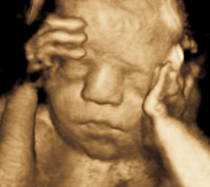
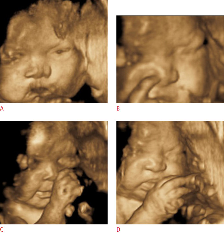
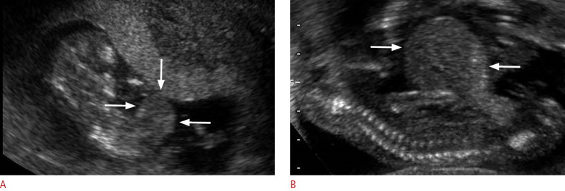
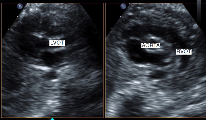
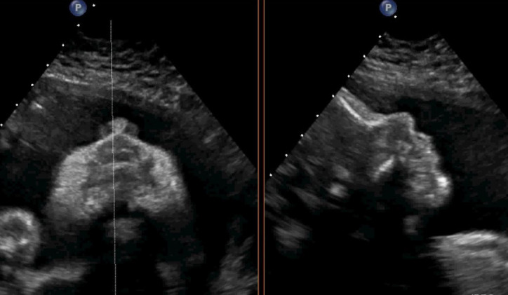
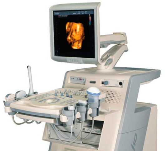
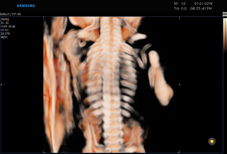
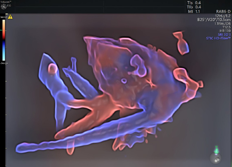
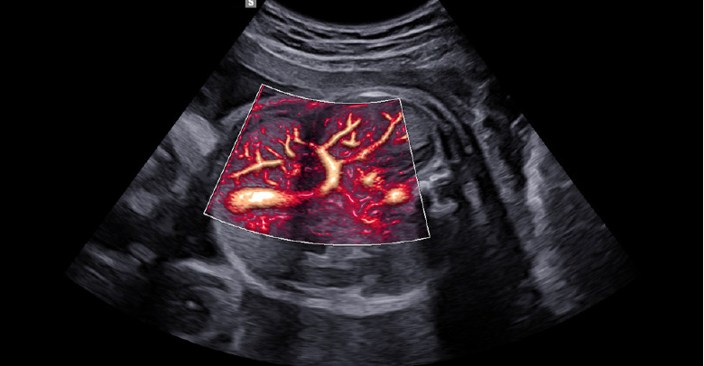
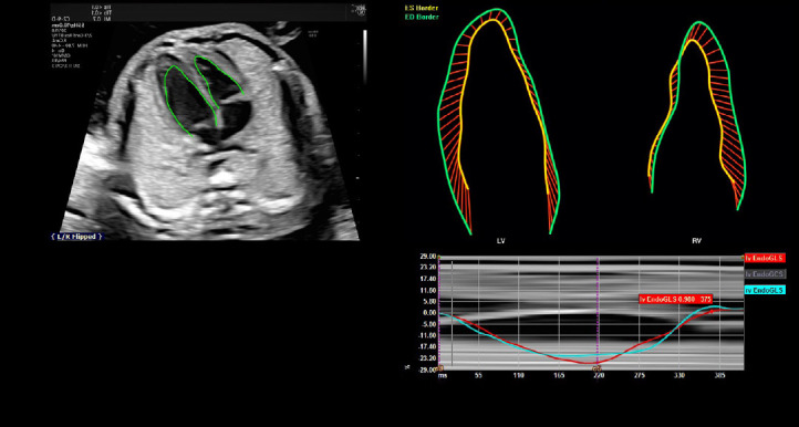
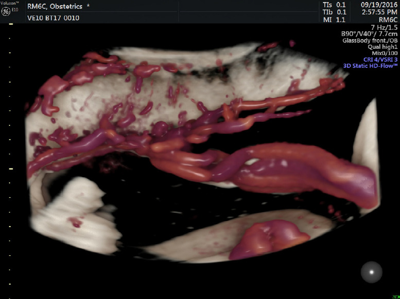
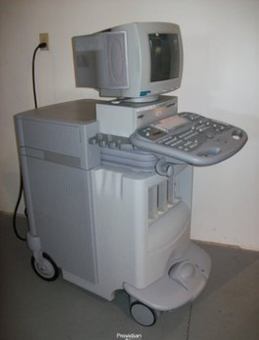
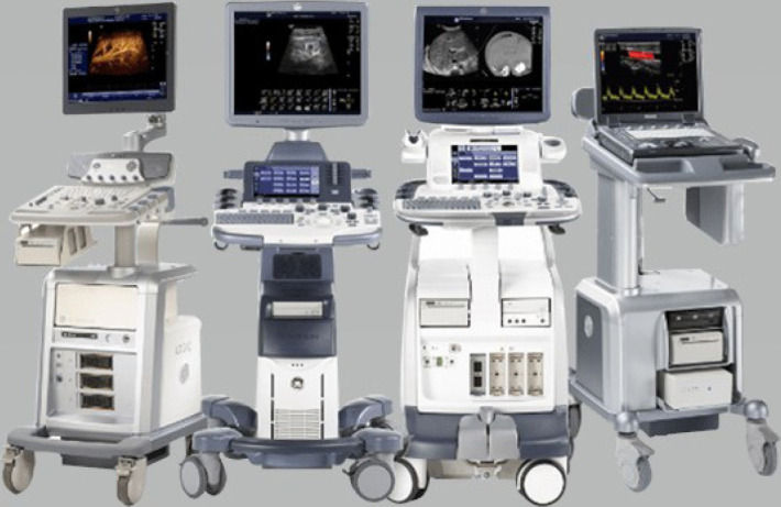
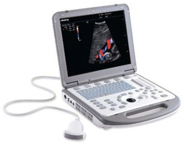
Diagnostic ultrasound (DUS) is, arguably, the most common technique used in obstetrical practice. From A mode, first described by Ian Donald for gynecology in the late 1950s, to B mode in the 1970s, real-time and gray-scale in the early 1980s, Doppler a little later, sophisticated color Doppler in the 1990s and three dimensional/four-dimensional ultrasound in the 2000s, DUS has not ceased to be closely associated with the practice of obstetrics. The latest innovation is the use of artificial intelligence which will, undoubtedly, take an increasing role in all aspects of our lives, including medicine and, specifically, obstetric ultrasound. In addition, in the future, new visualization methods may be developed, training methods expanded, and workflow and ergonomics improved.

Keywords: 3-D; 4-D; Artificial intelligence; Doppler; Obstetrics; training; ultrasound.


Conflict of interest statement

I am on the advisory board of Samsung and an author for UpToDate.

Figures

Fig. 1.
Fig. 1.. B-scan image of the fetus (circa 1980).
The white "smudge" in the center of the picture is the fetal head (within the yellow circle).
Fig. 2.
Fig. 2.. 3D reconstruction of fetal face.
Facial features are very clear in this surface reconstruction.
Fig. 3.
Fig. 3.. Composite picture of various fetal expressions.
These are various expressions: open eyes (A), nose scratching (B), smile (C), and "disgust" (D).
Fig. 4.
Fig. 4.. Omphalocele at 12 weeks gestational age in a fetus with trisomy 13 (A) and 20 weeks gestational age (B).
In both images the fetus on his/her back with the head on left and the fetal spine oriented longitudinally on the bottom of the images. Omphalocele is the bulge on the right side of each image indicated by arrows.
Fig. 5.
Fig. 5.. Use of matrix probe.
Image of the left ventricular outflow (LVOT) on the left and the right ventricular outflow (RVOT) or short axis on the right are obtained simultaneously. Cross section of the LVOT/aorta is seen on the right since this is a perpendicular view to the RVOT view.
Fig. 6.
Fig. 6.. Matrix probe image.
Simultaneous viewing of the fetal face in coronal view, face up on the left and the fetal profile on the right.
Fig. 7.
Fig. 7.. Monitor on articulated arm.
This allows the monitor to be moved in all directions to allow easier visualization for the patient and the clinician.
Fig. 8.
Fig. 8.. "Deconstructed" and "reconstructed" ultrasound system.
This is an example of a "revolutionary" concept of separating various elements of the ultrasound system. The probes are on one side (right) of the examination chair with a probe holder with recoiling cables while the monitor and keyboard are on the other side (left) of the chair and the actual CPU under the chair.
Fig. 9.
Fig. 9.. Crystal view of the fetal spine (used with permission of Samsung Healthcare).
This is one of the new advanced 3D volume post-rendering technology.
Fig. 10.
Fig. 10.. HDlive Silhouette of fetal circulation (used with permission of GE Healthcare).
This is a rendering method that produces realistic images by using an advanced illumination model with vitreous-like clarity.
Fig. 11.
Fig. 11.. Fetal abdomen vasculature, with MV-Flow with Lumi Flow (used with permission of Samsung Healthcare).
MV-Flow, for microvascular, is an advanced Doppler technology that provides detailed documentation of microvascular perfusion into tissues and organs and LumiFlow displays a "3D-like" appearance to 2D color Doppler, enhancing spatial comprehension of blood vessels and aiding in the understanding of vessel boundaries as can be seen in this detailed view of fetal abdominal vasculature.
Fig. 12.
Fig. 12.. Analysis of fetal heart function with Fetal HQ (used with permission of GE Healthcare).
This tool, previously utilized in adults only, allows automatically tracking and measuring strain while performing a fetal echocardiogram by segmenting the heart in many small elements and analyzing directions and intensities of the various movement vectors. This furnishes simultaneous information on the size, shape and function of the fetal heart.
Fig. 13.
Fig. 13.. Umbilical cord insertion into the placenta and placental surface vessels.
This is imaged with special software (Radiant flow). This gives the impression of a real-life picture.
Fig. 14.
Fig. 14.. "Old" ultrasound system.
Note the massive size, rendering transport relatively difficult and the "old style" monitor that can be only minimally moved.
Fig. 15.
Fig. 15.. Various "new" portable ultrasound systems (modified from Enterprise Ultrasound, https://enterpriseultrasound.com).
They have common characteristics: much smaller than older systems, LCD monitors than can move virtually in every direction, movable keyboard, often touch screen, larger wheels that allow easy transportation to various parts of the medical facility, and, in general, upgradability.
Fig. 16.
Fig. 16.. A laptop model of ultrasound system.
Many vendors offer these machines that, with time, have become of excellent quality, albeit, not quite as good as the "stars" from the major vendors.
Fig. 17.
Fig. 17.. Handheld "miniature" system.
This is becoming more and more common because of the ease of transport in the practician’s pocket and ease of use.
Fig. 18.
Fig. 18.. Two systems using smart phones as monitors.
This is a further advancement with the probe attaching directly to a cell phone. One only need to download the application.
Fig. 19.
Fig. 19.. Elastography of a myomatous uterus.
A. On the left panel, the regular 2D gray-scale image, the myoma is visible as the round structure on the right (arrows). The myoma is visible with elastography on the right panel, as it appears surrounded by a layer of tissue (arrows) with different elasticity ("capsule"). B. A different elastography color scheme is used which optimally demonstrates the myoma (arrows).
Fig. 20.
Fig. 20.. Automatic display of fetal brain planes (used with permission of GE Healthcare).
This was obtained after a single sweep. Measurements of the biparietal diameter (BPD), head circumference (HC), cerebellum, cisterna magna (CM), and posterior horn of the lateral ventricle (Vp) are automatic. GA, gestational age; OFD, occipitofrontal diameter.
Fig. 21.
Fig. 21.. Nine views of the fetal heart (used with permission of Samsung Healthcare).
These are displayed automatically after a single sweep. Labels are added automatically by the machine and move with the structures, for instance if zoom is applied.

References

    1. Langevin P. Les ondes ultrasonores. Rev Gen Electr. 1928;23:626–634.
    1. Harvey EN, Loomis AL. The destruction of luminous bacteria by high frequency sound waves. J Bacteriol. 1929;17:373–376. - PMC - PubMed
    1. Gohe H, Wedekind T. Der Ultraschall in der Medizin. Klin Wochenschr. 1940;19:25–29.
    1. Aroso A, Ferreira E. Ultrasonic therapy in microcystic disease of the ovary. C R Soc Fr Gyncol. 1951;21:351–353. - PubMed
    1. Donald I, Macvicar J, Brown TG. Investigation of abdominal masses by pulsed ultrasound. Lancet. 1958;1:1188–1195. - PubMed

LinkOut - more resources