Review
. 2020 Oct 22;21(21):7830.
doi: 10.3390/ijms21217830.

Hereditary Hypofibrinogenemia with Hepatic Storage

Affiliations
Review

Hereditary Hypofibrinogenemia with Hepatic Storage

Rosanna Asselta et al. Int J Mol Sci. .

Abstract

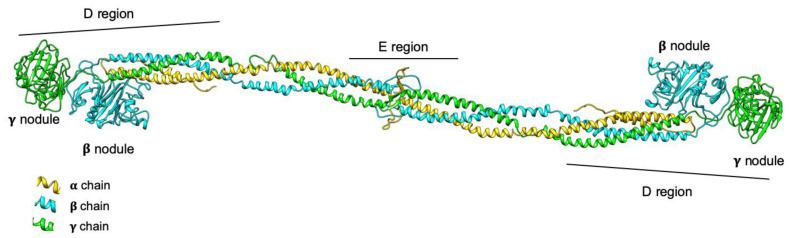
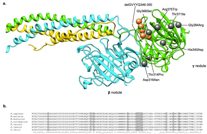
Fibrinogen is a 340-kDa plasma glycoprotein constituted by two sets of symmetrical trimers, each formed by the Aα, Bβ, and γ chains (respectively coded by the FGA, FGB, and FGG genes). Quantitative fibrinogen deficiencies (hypofibrinogenemia, afibrinogenemia) are rare congenital disorders characterized by low or unmeasurable plasma fibrinogen antigen levels. Their genetic basis is represented by mutations within the fibrinogen genes. To date, only eight mutations, all affecting a small region of the fibrinogen γ chain, have been reported to cause hereditary hypofibrinogenemia with hepatic storage (HHHS), a disorder characterized by protein aggregation in the endoplasmic reticulum, hypofibrinogenemia, and liver disease of variable severity. Here, we will briefly review the clinic characteristics of HHHS patients and the histological feature of their hepatic inclusions, and we will focus on the molecular genetic basis of this peculiar type of coagulopathy.

Keywords: FGG gene; fibrinogen; hepatic inclusion; hereditary hypofibrinogenemia with hepatic storage; hypofibrinogenemia; mutation; prevalence; storage disease.


Conflict of interest statement

The authors declare no conflict of interest.

Figures

Figure 1
Figure 1
3D model representation of the fibrinogen protein. The three fibrinogen chains (Aα, Bβ, and γ) are represented in yellow, cyan, and green, respectively; the domains composing the molecule are also shown. The D regions correspond to lateral globular parts containing the C-terminus of Bβ and γ chains; the E region is the central nodule, containing the N-terminus of all chains. The model was obtained using the pdb structure 3GHG and the UCSF Chimera package.
Figure 2
Figure 2
Localization and conservation of HHHS -causing mutations in the fibrinogen γ nodule. (a) The localization of all the mutations responsible for the HHHS phenotype, described in the literature, are indicated as grey spheres (in case of missense mutations), and orange spheres (in case of deletion). Mutations are numbered on the mature protein. The fibrinogen chains (Aα, Bβ, γ) are represented in yellow, cyan, and green, respectively. The model was obtained using the pdb structure 3GHG, and the UCSF Chimera package. (b) Multiple alignments of the fibrinogen γ chain terminal regions of different species. Sequences were retrieved from NCBI, and aligned using clustalW. The last line of the alignment shows in a schematic way the conservation among species. The * symbol indicates a conserved residue, the : symbol indicates conservation between groups of strongly similar properties, the . symbol indicates conservation between groups of weakly similar properties, the space indicates lack of conservation. Amino acids shaded in grey corresponds to those involved in HHHS mutations.

References

    1. Mosesson M.W. Fibrinogen and fibrin structure and functions. J. Thromb. Haemost. 2005;3:1894–1904. doi: 10.1111/j.1538-7836.2005.01365.x. - DOI - PubMed
    1. Genotype-Tissue Expression (GTex) Database, v8. [(accessed on 3 August 2020)]; Available online: https://www.gtexportal.org/home/
    1. Spraggon G., Everse S.J., Doolittle R.F. Crystal structures of fragment D from human fibrinogen and its crosslinked counterpart from fibrin. Nature. 1997;389:455–462. doi: 10.1038/38947. - DOI - PubMed
    1. Yee V.C., Pratt K.P., Côté H.C., Trong I.L., Chung D.W., Davie E.W., Stenkamp R.E., Teller D.C. Crystal structure of a 30 kDa C-terminal fragment from the gamma chain of human fibrinogen. Structure. 1997;5:125–138. doi: 10.1016/S0969-2126(97)00171-8. - DOI - PubMed
    1. Madrazo J., Brown J.H., Litvinovich S., Dominguez R., Yakovlev S., Medved L., Cohen C. Crystal structure of the central region of bovine fibrinogen (E5 fragment) at 1.4-A resolution. Proc. Natl. Acad. Sci. USA. 2001;98:11967–11972. doi: 10.1073/pnas.211439798. - DOI - PMC - PubMed

LinkOut - more resources