Review
. 2020 Oct 22;9(11):2343.
doi: 10.3390/cells9112343.

The SARS-CoV-2 Spike Glycoprotein as a Drug and Vaccine Target: Structural Insights into Its Complexes with ACE2 and Antibodies

Affiliations
Review

The SARS-CoV-2 Spike Glycoprotein as a Drug and Vaccine Target: Structural Insights into Its Complexes with ACE2 and Antibodies

Anastassios C Papageorgiou et al. Cells. .

Abstract

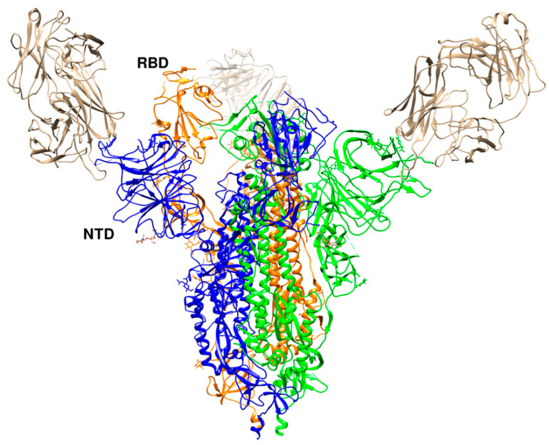
Severe Acute Respiratory Syndrome Coronavirus 2 (SARS-CoV-2), the causative agent of the Coronavirus disease (COVID-19) pandemic, has so far resulted in more than 1.1 M deaths and 40 M cases worldwide with no confirmed remedy yet available. Since the first outbreak in Wuhan, China in December 2019, researchers across the globe have been in a race to develop therapies and vaccines against the disease. SARS-CoV-2, similar to other previously identified Coronaviridae family members, encodes several structural proteins, such as spike, envelope, membrane, and nucleocapsid, that are responsible for host penetration, binding, recycling, and pathogenesis. Structural biology has been a key player in understanding the viral infection mechanism and in developing intervention strategies against the new coronavirus. The spike glycoprotein has drawn considerable attention as a means to block viral entry owing to its interactions with the human angiotensin-converting enzyme 2 (ACE2), which acts as a receptor. Here, we review the current knowledge of SARS-CoV-2 and its interactions with ACE2 and antibodies. Structural information of SARS-CoV-2 spike glycoprotein and its complexes with ACE2 and antibodies can provide key input for the development of therapies and vaccines against the new coronavirus.

Keywords: COVID-19; SARS; antibodies; glycoprotein; receptor binding; spike; structure.


Conflict of interest statement

The authors declare no conflict of interest.

Figures

Figure 1
Figure 1
Genome organization of SARS-CoV-2. The polypeptides expressed by open reading frame (ORF)-1a and ORF-1b are cleaved-off to constituent components by the viral proteases papain-like protease (nsp3) and the main protease (nsp5), respectively.
Figure 2
Figure 2
The overall topology of the SARS-CoV-2 S monomer. FP, fusion peptide; HR1, heptad repeat 1; HR2, heptad repeat 2; IC, intracellular domain; NTD, N-terminal domain; SD1, subdomain 1; SD2, subdomain 2; TM, transmembrane region [15].
Figure 3
Figure 3
The spike protein trimer in the two conformations: “down” (left) and “up” (right). The moving receptor-binding domain (RBD) in monomer B is shown in red. Each monomer is colored differently with A, B, and C in yellow, orange, and cyan, respectively (PDB ids 6vxx and 6vyb).
Figure 4
Figure 4
Angiotensin-converting enzyme 2 (ACE2) conformations. (Left) Open conformation (PDB id 6m1d). (Right) Close conformation (PDB id 6m18). RBD is shown in orange color. ACE2 chains are shown in cyan and blue colors, while B0AT1 chains are shown in pink and grey colors.
Figure 5
Figure 5
SARS-CoV-2 spike receptor-binding domain (in orange) bound to the ACE2 receptor (in cornflower blue) (PDB id 6m0j) [15].
Figure 6
Figure 6
SARS-CoV-2 S protein HR1 and HR2 domains (a) and their parallel-antiparallel view (b). HR1 helices are colored in yellow, hot pink, and sky blue and those of HR2 in coral, purple, and chartreuse.
Figure 7
Figure 7
Three different binding modes of neutralizing antibodies. (a) Human neutralizing CB6 antibody in complex with SARS-CoV-2 RBD (PDB id 7c01). RBD, H, and L chains are shown in orange, cyan, and cornflower blue colors, respectively. (b) REGN antibodies bound to RBD (PDB id 6xdg, (c) CR3022 and CC12.1 in complex with ACE2 RBD (PDB id 6xc3). RBD is colored in orange, heavy chains in cyan and green, and L chains in cornflower blue and magenta. The RBD (in orange) is shown in the same orientation in all three complexes.
Figure 8
Figure 8
Cryo-Electron Microscopy (Cryo-EM) structure of S309 antibody with the S glycoprotein trimer (PDB id 6wpt). The antibody chains are colored in cyan (L chain) and cornflower blue (H chain). The chains of the S trimer are shown in blue, red, and yellow.
Figure 9
Figure 9
Crystal structure of SARS-CoV-2 P2B-2F6 antibody with RBD (PDB id 7bwj [59] superimposed onto the RBD-ACE2 complex structure (PDB id 6m0j). The RBD is shown in orange color and ACE2 in medium purple. The antibody chains are colored in cyan (L chain) and cornflower blue (H chain).
Figure 10
Figure 10
Crystal structure of SARS-CoV-2 RBD with 4A8 (PDB id 7c2l). The RBD in the “up” conformation is labelled. The trimer is colored in blue (B), green (C), and orange (A). The three 4A8 antibodies, each bound to an NTD, are shown in brown.

References

    1. Zhu N., Zhang D., Wang W., Li X., Yang B., Song J., Zhao X., Huang B., Shi W., Lu R., et al. China Novel Coronavirus Investigating and Research Team A Novel Coronavirus from Patients with Pneumonia in China, 2019. N. Engl. J. Med. 2020;382:727–733. doi: 10.1056/NEJMoa2001017. - DOI - PMC - PubMed
    1. Tang D., Comish P., Kang R. The hallmarks of COVID-19 disease. PLoS Pathog. 2020;16:e1008536. doi: 10.1371/journal.ppat.1008536. - DOI - PMC - PubMed
    1. Cui J., Li F., Shi Z.-L. Origin and evolution of pathogenic coronaviruses. Nat. Rev. Microbiol. 2019;17:181–192. doi: 10.1038/s41579-018-0118-9. - DOI - PMC - PubMed
    1. Zhang T., Wu Q., Zhang Z. Probable Pangolin Origin of SARS-CoV-2 Associated with the COVID-19 Outbreak. Curr. Biol. 2020;30:1346.e2–1351.e2. doi: 10.1016/j.cub.2020.03.022. - DOI - PMC - PubMed
    1. Bedford J., Enria D., Giesecke J., Heymann D.L., Ihekweazu C., Kobinger G., Lane H.C., Memish Z., Oh M.-D., Sall A.A., et al. WHO Strategic and Technical Advisory Group for Infectious Hazards COVID-19: Towards controlling of a pandemic. Lancet. 2020;395:1015–1018. doi: 10.1016/S0140-6736(20)30673-5. - DOI - PMC - PubMed

MeSH terms

LinkOut - more resources