Review
. 2021 Feb 10:83:183-203.
doi: 10.1146/annurev-physiol-031620-091043. Epub 2020 Oct 26.

Voltage-Gated Calcium Channels in Nonexcitable Tissues

Affiliations
Review

Voltage-Gated Calcium Channels in Nonexcitable Tissues

Geoffrey S Pitt et al. Annu Rev Physiol. .

Abstract

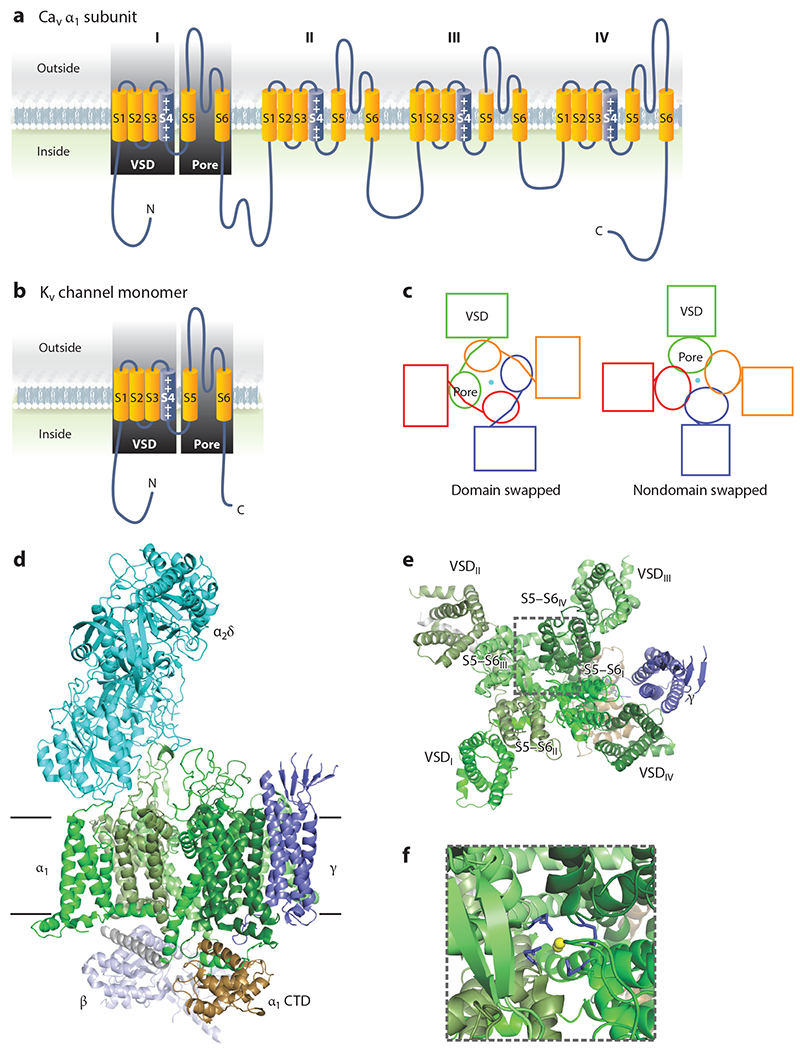
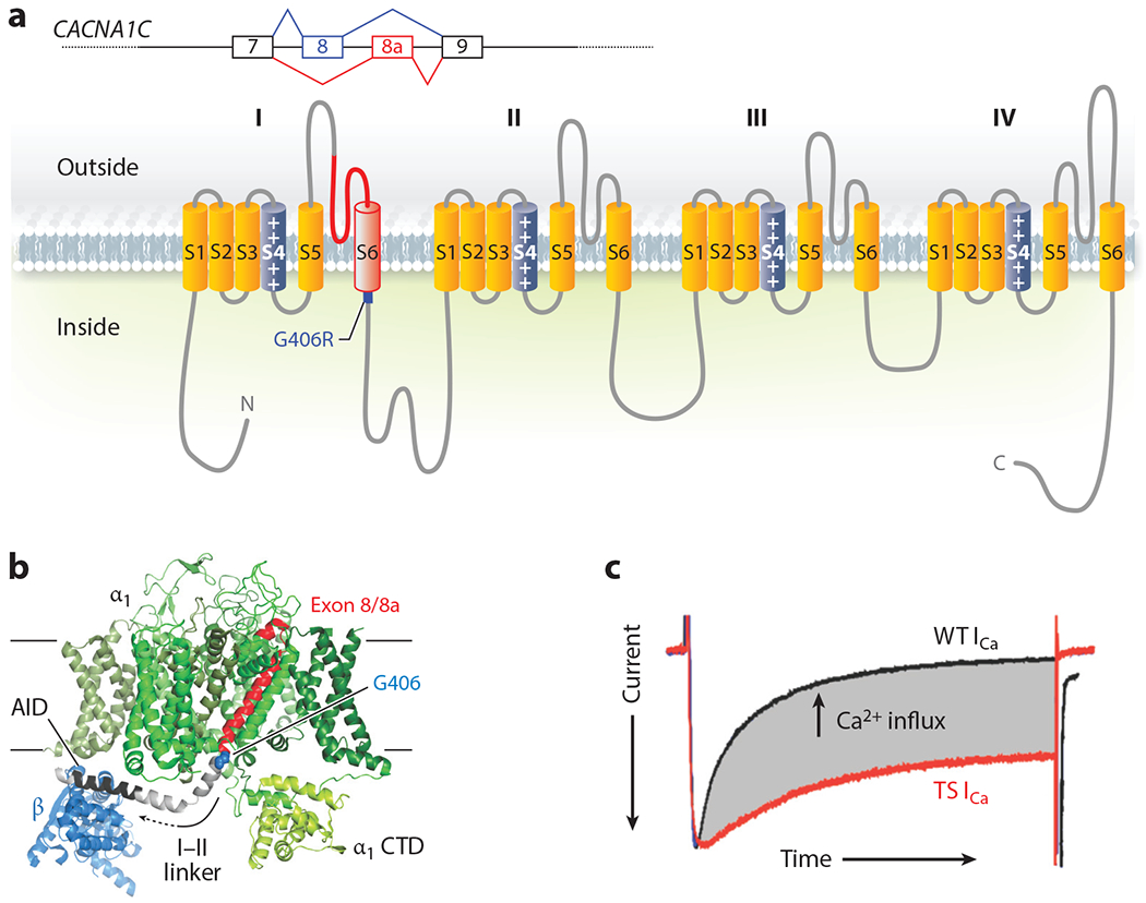
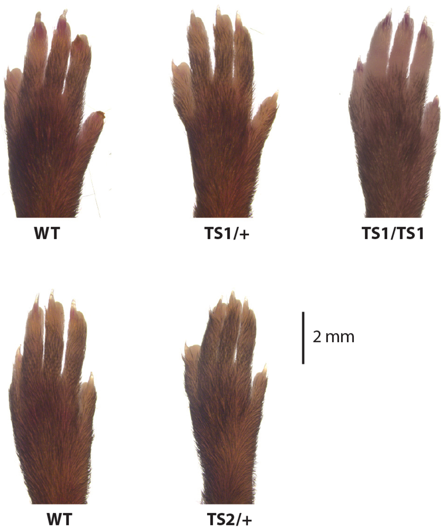
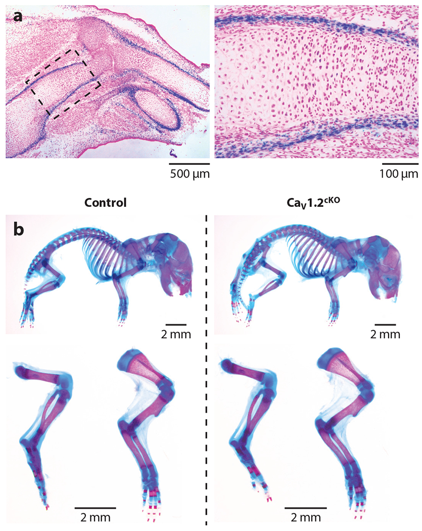
The identification of a gain-of-function mutation in CACNA1C as the cause of Timothy syndrome, a rare disorder characterized by cardiac arrhythmias and syndactyly, highlighted roles for the L-type voltage-gated Ca2+ channel CaV1.2 in nonexcitable cells. Previous studies in cells and animal models had suggested that several voltage-gated Ca2+ channels (VGCCs) regulated critical signaling events in various cell types that are not expected to support action potentials, but definitive data were lacking. VGCCs occupy a special position among ion channels, uniquely able to translate membrane excitability into the cytoplasmic Ca2+ changes that underlie the cellular responses to electrical activity. Yet how these channels function in cells not firing action potentials and what the consequences of their actions are in nonexcitable cells remain critical questions. The development of new animal and cellular models and the emergence of large data sets and unbiased genome screens have added to our understanding of the unanticipated roles for VGCCs in nonexcitable cells. Here, we review current knowledge of VGCC regulation and function in nonexcitable tissues and cells, with the goal of providing a platform for continued investigation.

Keywords: Timothy syndrome; nonexcitable cells; voltage-gated Ca2+ channel.


Figures

Figure 1
Figure 1
Voltage-gated Ca2+ channel structure. (a) Ribbon diagram of the pore-forming α1 subunit of a voltage-gated Ca2+ channel demonstrating the pseudo-fourfold symmetry and homology of each domain to a Kv channel monomer, as depicted in panel b. (c) Schematic of a domain-swapped architecture, shared by voltage-gated Ca2+ channels, and a nondomain-swapped architecture found in other cation channels for comparison. (d) Structure of rabbit CaV1.1 (Protein Data Bank: 5GVY) shown from the membrane. The pore-forming α1 subunit spans the membrane. The individual domains (DI–DIV) are colored in various shades of green, and the intracellular C-terminal domain (CTD) is brown. The extracellular α2δ subunit is turquoise, the γ subunit is dark purple, and the cytoplasmic β subunit is light purple. (e) View of the α1 and γ subunits from outside of the cell shows the domain-swapped architecture. For clarity, the α2δ subunit is not shown. (f) Zoomed image of the highlighted area in panel e, with the pore glutamate residues colored in purple and the Ca2+ ions in yellow. Abbreviation: VSD, voltage sensor domain.
Figure 2
Figure 2
Structure and function of Timothy syndrome (TS) mutation. (a) Schematic of the alternative splicing event for exon 8 or 8a in CACNA1C and the exon in the context of the CaV1.2 α1C subunit. (b) Structure of the homologous rabbit CaV1.1 (Protein Data Bank: 5GVY) shown from the membrane. Only the α1 subunit and the β subunit (blue) are shown. The individual α1 domains (DI–DIV) are colored in various shades of green except for the polypeptide encoded by exon 8 (or exon 8a) in red, with the G406 residue that is mutated to G406R highlighted in blue; the linker between domain I and domain II (I–II linker) is depicted in gray, in which the α interaction domain (AID) is highlighted in black. (c) Voltage-clamp Ca2+ current recording from a cardiomyocyte isolated from a mouse with the TS mutation (red) overlaid with a recording from a cardiomyocyte isolated from a wild-type (WT) littermate. The currents are scaled for comparison, and the enhanced Ca2+ influx through the TS mutant channel is highlighted in gray (E.Q. Wei and G.S. Pitt, unpublished data).
Figure 3
Figure 3
Tissue expression pattern of CaV1.2. (a) Figure and data are adapted from the ARCHS4 web resource (https://amp.pharm.mssm.edu/archs4/gene/CACNA1C) showing CACNA1C expression in various tissues. In red are nonexcitable cell types with expression of CaV1.2. Note that expression levels for many of these cell types are like the expression level in the heart, the canonical CaV1.2-expressing tissue. In contrast, skeletal muscle myoblasts, which express an alternative L-type voltage-gated Ca2+ channel (CaV1.1), show no expression (highlighted by gray box). (b) Figure and data for protein detection (adapted from https://www.proteinatlas.org/ENSG00000151067-CACNA1C/tissue) (55) also demonstrate broad tissue expression, with no CaV1.2 protein detected in skeletal muscle (highlighted by gray box).
Figure 4
Figure 4
Timothy syndrome (TS) mouse models do not display syndactyly. Images of forelimbs from TS1 and TS2 mice and their respective littermate wild-type (WT) controls.
Figure 5
Figure 5
CaV1.2 is expressed during mouse limb development. (a) A CaV1.2lacZ/+ reporter mouse detects CaV1.2 expression during mouse limb development. A frozen tissue section (10 μM) from CaV1.2lacZ/+ forelimb at E14.5 was stained with X-gal and counterstained with nuclear fast red. (b) Normal skeletal development of Col2a1-Cre;Cacna1cflox/flox conditional knockout (cKO) mouse embryos. Double staining with alizarin red and Alcian blue of the whole skeleton (top), hindlimbs and forelimbs (bottom) of Cacna1cflox/flox (control), and cKO littermate mouse embryos at P0.

References

    1. Yue DT. 2004. The dawn of high-resolution structure for the queen of ion channels. Neuron 42:357–59 - PubMed
    1. Hille B 2001. Ion Channels of Excitable Membranes. Oxford, UK: Oxford Univ. Press
    1. Splawski I, Timothy KW, Sharpe LM, Decher N, Kumar P, et al. 2004. CaV1.2 calcium channel dysfunction causes a multisystem disorder including arrhythmia and autism. Cell 119:19–31 - PubMed
    1. Catterall WA. 2011. Voltage-gated calcium channels. Cold Spring Harb. Perspect. Biol 3:a003947. - PMC - PubMed
    1. Catterall WA, Lenaeus MJ, Gamal El-Din TM. 2020. Structure and pharmacology of voltage-gated sodium and calcium channels. Annu. Rev. Pharmacol. Toxicol 60:133–54 - PubMed

Publication types

Supplementary concepts

LinkOut - more resources