. 2020 Dec 25;295(52):18213-18225.
doi: 10.1074/jbc.RA120.015882. Epub 2020 Oct 26.

A combinatorial native MS and LC-MS/MS approach reveals high intrinsic phosphorylation of human Tau but minimal levels of other key modifications

Affiliations

A combinatorial native MS and LC-MS/MS approach reveals high intrinsic phosphorylation of human Tau but minimal levels of other key modifications

Friedel Drepper et al. J Biol Chem. .

Abstract

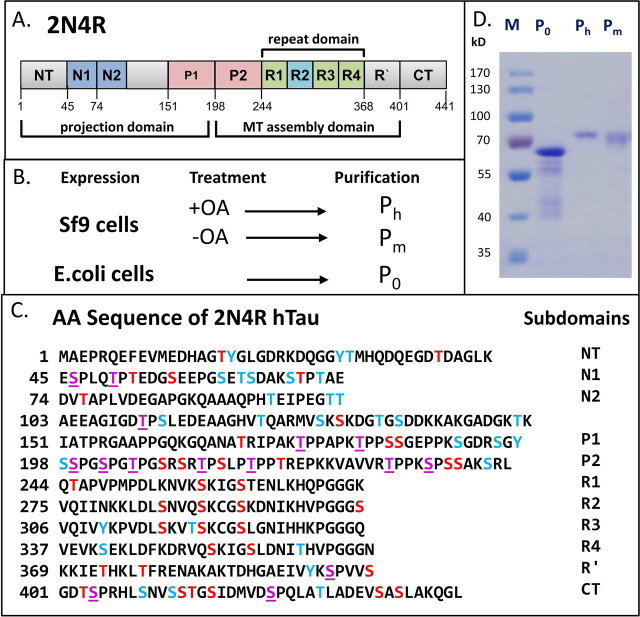
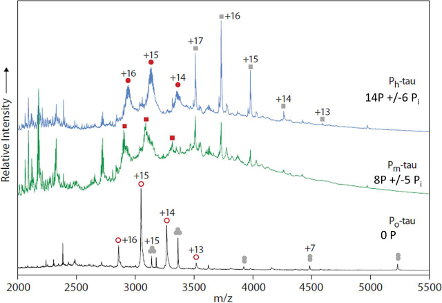
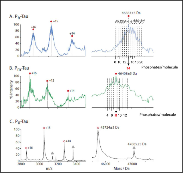
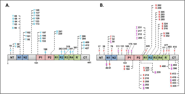
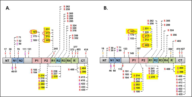
Abnormal changes of neuronal Tau protein, such as phosphorylation and aggregation, are considered hallmarks of cognitive deficits in Alzheimer's disease. Abnormal phosphorylation is thought to precede aggregation and therefore to promote aggregation, but the nature and extent of phosphorylation remain ill-defined. Tau contains ∼85 potential phosphorylation sites, which can be phosphorylated by various kinases because the unfolded structure of Tau makes them accessible. However, methodological limitations (e.g. in MS of phosphopeptides, or antibodies against phosphoepitopes) led to conflicting results regarding the extent of Tau phosphorylation in cells. Here we present results from a new approach based on native MS of intact Tau expressed in eukaryotic cells (Sf9). The extent of phosphorylation is heterogeneous, up to ∼20 phosphates per molecule distributed over 51 sites. The medium phosphorylated fraction Pm showed overall occupancies of ∼8 Pi (± 5) with a bell-shaped distribution; the highly phosphorylated fraction Ph had 14 Pi (± 6). The distribution of sites was highly asymmetric (with 71% of all P-sites in the C-terminal half of Tau). All sites were on Ser or Thr residues, but none were on Tyr. Other known posttranslational modifications were near or below our detection limit (e.g. acetylation, ubiquitination). These findings suggest that normal cellular Tau shows a remarkably high extent of phosphorylation, whereas other modifications are nearly absent. This implies that abnormal phosphorylations at certain sites may not affect the extent of phosphorylation significantly and do not represent hyperphosphorylation. By implication, the pathological aggregation of Tau is not likely a consequence of high phosphorylation.

Keywords: Alzheimer's disease; LC-MS; Tau protein (Tau); mass spectrometry (MS); native mass spectrometry; phosphorylation; protein aggregation.


Conflict of interest statement

Conflict of interest—The authors declare that they have no conflicts of interest with the contents of this article.

Figures

Figure 1
Figure 1
Overview: Phosphorylation of hTau-2N4R expressed in Sf9 cells and E. coli.A, diagram of domains of hTau40 (2N4R or Tau-F, Uniprot ID P10636-8), the largest human isoform of CNS Tau consisting of 441 residues with the three alternatively spliced inserts N1, N2, and R2. The N-terminal half represents the projection domain, and the C-terminal half contains the four pseudo-repeats (R1–R4) which, in combination with their flanking domains P2 and R′, represent the microtubule assembly domain. B, schematic representation of production of unphosphorylated Tau (P0) in E. coli (prokaryote) and hyperphosphorylated Tau (Pm and Ph) in Sf9 cells (eukaryote). Okadaic acid (OA) treatment increases the phosphorylation of Tau which yields Ph-Tau. C, amino acid sequence of Tau (2N4R, 441 residues) showing phosphorylation sites identified in this study (red and purple letters; purple = Pro directed)) and further potential phosphorylation sites (not detected so far, blue letters) (total of 85 sites, 45 Ser, 35 Thr, and 5 Tyr). Note that only a minority of phosphorylation sites of 37% was detected in the N-terminal part of the sequence (up to residue ∼150, compared with 71% in the C-terminal part of the protein). Note also that none of the five Tyr residues was phosphorylated. D, SDS-PAGE analysis stained with Coomassie Blue showing P0 Tau purified from E. coli and Pm- and Ph-Tau purified from Sf9 cells. Note the upward shift in Mr value with increasing phosphorylation, from 55 kDa (P0-Tau) to 68 kDa for Ph-Tau (compared with the theoretical molecular mass values of ∼45,850 Da and ∼46,863 Da). This shift is characteristic for AD-Tau.
Figure 2
Figure 2
Analysis of hTau-2N4R expressed in Sf9 cells by native MS. 20 µm of purified proteins in 200 mm ammonium acetate, pH 7.6, were analyzed by nanoflow electrospray ionization quadrupole TOF MS. Representative mass spectra for the highly phosphorylated states Ph-Tau (top) and Pm-Tau (middle) and unphosphorylated. P0-Tau (bottom) are shown. Top, main signals were assigned to charge state series of Ph-Tau (molecular mass of 46,883 Da; filled red circles) and a copurified component with a molecular mass of 59,642 Da (gray squares). Middle, spectrum of Pm-Tau displaying peaks assigned to a series of equivalent charge states but shifted toward lower m/z compared with those for Ph-Tau (red squares). Further signals match closely with those in the spectrum of Ph-Tau. Bottom, spectrum of control Tau P0 consisting of a series of charge states indicating a molecular mass of 45,724 Da (open red circles). A further series was assigned to a molecular mass of 47,085 Da. It was attributed to a trimeric species (gray circles) because it decomposes upon increasing collisional activation into highly charged (centered at +9, below m/z 2,000) monomeric and charge-stripped dimeric species (charges +6 to +8). Its monomeric mass of 15,691 Da matches the theoretical mass of the E. coli chaperone protein skp (see Table S4), taking into account removal of its N-terminal 20–amino acid signal peptide.
Figure 3
Figure 3
Phosphorylation of hTau-2N4R expressed in Sf9 cells analyzed by native MS.Left, signals in the range between 2800 and 3500 m/z attributed to Tau proteins. Right, corresponding charge state–deconvoluted spectra. A, the mass spectrum of Ph-Tau (top) displays a center mass of 46,883 Da (filled red circles) and additionally six peaks on both sides equally spaced by ∼80 Da. B, Pm-Tau (middle) displays peaks of similar width shifted toward lower m/z compared with those for Ph-Tau, which can be assigned to the equivalent charge states and indicate a molecular mass of ∼46,408 Da (red squares). C, the spectrum of control Tau P0 consists of a series of charge states resulting in a comparatively sharp peak at a mass of 45,724 Da (45,850 −131Da ± 5 because of cleavage of first residue Met-1; open red circles). A further series was assigned to a molecular mass of 47,085 Da and was attributed to a trimeric species (gray circles) because it decomposes upon increasing collisional activation into highly charged (centered at +9, below m/z 2000) monomeric and charge-stripped dimeric species (charges +6 to +8). Its monomeric mass of 15,691 Da matches the theoretical mass of the E. coli chaperone protein skp (see Table S4), taking into account removal of its N-terminal 20–amino acid signal peptide.
Figure 4
Figure 4
Bar diagram of full-length human Tau-2N4R protein with distribution of nonobserved versus observed phosphorylation sites in Sf9 cells. Thr residues shown above bar, Ser residues below. A, nonphosphorylated sites are located mainly in the N-terminal half (blue). B, observed phosphorylation sites are located mainly in the C-terminal half (red), especially in SP or TP motifs (purple underlined).
Figure 5
Figure 5
Bar diagram of full-length human Tau-2N4R protein with distribution of observed phosphorylation sites in Sf9 cells versus AD brain (>50 sites, mainly in C-terminal half, overlap 68%). A, 51 P-sites acquired by Tau in Sf9 cells (this study). B, 52 P-sites identified in AD-Tau; see collection of P-sites from AD brains (13). Red, Ser and Thr P-sites; purple underlined, SP or TP motifs; yellow highlight, high occupancy P-sites; red circles, P-sites important for AD diagnostics.

Similar articles

Cited by

References

    1. Wang Y., Mandelkow E. Tau in physiology and pathology. Nat. Rev. Neurosci. 2016;17:5–21. doi: 10.1038/nrn.2015.1. 26631930. - DOI - PubMed
    1. Grundke-Iqbal I., Iqbal K., Tung Y.C., Quinlan M., Wisniewski H.M., Binder L.I. Abnormal phosphorylation of the microtubule-associated protein tau (tau) in Alzheimer cytoskeletal pathology. Proc. Natl. Acad. Sci. U. S. A. 1986;83:4913–4917. doi: 10.1073/pnas.83.13.4913. 3088567. - DOI - PMC - PubMed
    1. Braak E., Braak H., Mandelkow E.M. A sequence of cytoskeleton changes related to the formation of neurofibrillary tangles and neuropil threads. Acta Neuropathol. 1994;87:554–567. doi: 10.1007/s004010050124. 7522386. - DOI - PubMed
    1. DeVos S.L., Miller R.L., Schoch K.M., Holmes B.B., Kebodeaux C.S., Wegener A.J., Chen G., Shen T., Tran H., Nichols B., Zanardi T.A., Kordasiewicz H.B., Swayze E.E., Bennett C.F., Diamond M.I. Tau reduction prevents neuronal loss and reverses pathological tau deposition and seeding in mice with tauopathy. Sci. Transl. Med. 2017;9 doi: 10.1126/scitranslmed.aag0481. 28123067. - DOI - PMC - PubMed
    1. Yanamandra K., Kfoury N., Jiang H., Mahan T.E., Ma S., Maloney S.E., Wozniak D.F., Diamond M.I., Holtzman D.M. Anti-tau antibodies that block tau aggregate seeding in vitro markedly decrease pathology and improve cognition in vivo. Neuron. 2013;80:402–414. doi: 10.1016/j.neuron.2013.07.046. 24075978. - DOI - PMC - PubMed

Publication types