. 2020 Oct 26;3(12):e202000879.
doi: 10.26508/lsa.202000879. Print 2020 Dec.

Plasmodium DEH is ER-localized and crucial for oocyst mitotic division during malaria transmission

Affiliations

Plasmodium DEH is ER-localized and crucial for oocyst mitotic division during malaria transmission

David S Guttery et al. Life Sci Alliance. .

Abstract

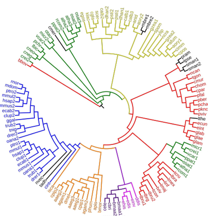
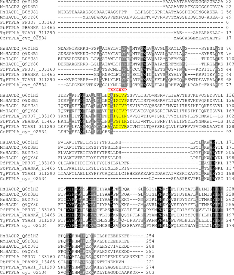
Cells use fatty acids (FAs) for membrane biosynthesis, energy storage, and the generation of signaling molecules. 3-hydroxyacyl-CoA dehydratase-DEH-is a key component of very long chain fatty acid synthesis. Here, we further characterized in-depth the location and function of DEH, applying in silico analysis, live cell imaging, reverse genetics, and ultrastructure analysis using the mouse malaria model Plasmodium berghei DEH is evolutionarily conserved across eukaryotes, with a single DEH in Plasmodium spp. and up to three orthologs in the other eukaryotes studied. DEH-GFP live-cell imaging showed strong GFP fluorescence throughout the life-cycle, with areas of localized expression in the cytoplasm and a circular ring pattern around the nucleus that colocalized with ER markers. Δdeh mutants showed a small but significant reduction in oocyst size compared with WT controls from day 10 postinfection onwards, and endomitotic cell division and sporogony were completely ablated, blocking parasite transmission from mosquito to vertebrate host. Ultrastructure analysis confirmed degeneration of Δdeh oocysts, and a complete lack of sporozoite budding. Overall, DEH is evolutionarily conserved, localizes to the ER, and plays a crucial role in sporogony.


Conflict of interest statement

The authors declare that they have no conflict of interest.

Figures

Figure S1.
Figure S1.. Schematic of the fatty acid synthase II (FASII) and fatty acid elongation (ELO) pathways.
The FASII pathway is localized to the apicoplast (green), whereby ketoacyl-acyl carrier protein is synthesized by condensation from CoA-activated precursors. The molecule is sequentially reduced, dehydrated and reduced to produce a fatty acyl chain. This cyclical process generates free fatty acids (FAs) as the final products. The FA elongation (ELO) pathway proceeds on the cytoplasmic face of the ER and catalyzes condensation a malonyl-CoA molecule with a fatty acyl-CoA derived from the FASII pathway. The products of ELO-A, ELO-B, and ELO-C are then cyclically reduced, dehydrated, and reduced by ketoacyl-CoA reductase (KCR), hydroxyacyl-CoA dehydratase (DEH), and enoyl-CoA reductase (ECR) to finally produce free FAs with two additional carbons with each cycle.
Figure S2.
Figure S2.. Phylogenetic analysis of DEH homologues across different species.
Phylogenetic analysis for PbDEH/PTPLA orthologs was performed using the neighbor joining method with MEGA6 software. Genome wide DEH/PTPLA analysis shows its presence in all organisms studied, including species from all major eukaryotic phyla. The analysis shows the clustering of organisms based on their evolutionary relatedness. Blue: Chordata; orange: non-Chordata; purple: Nematoda; pink: Ameobozoa; red: Apicomplexa, Euglenozoa, and Metamonada; green: Plants and Algae; yellow: Fungi; and black: others.
Figure S3.
Figure S3.. ClustalW alignment of Plasmodium DEH with human, mouse, and other apicomplexan homologues.
ClustalW alignment of Homo sapiens (Hs), Mus musculus (Ms), Plasmodium (Pf, Pb), Toxoplasma (Tg), and Cyclospora (Cc) homologues of DEH. For apicomplexans, the proteins are currently annotated as PTPLA. Highlighted in red is the CXXGXXP motif that defines PTP-like proteins.
Figure S4.
Figure S4.. Predicted 2D and 3D structures of Plasmodium DEH.
(A) Secondary structure of Plasmodium DEH. (B) Left: tertiary cartoon view of PbDEH showing the presence of six major hydrophobic membrane-spanning helices followed by coils and an absence of beta sheets, confirming the secondary structure. Right: tertiary cartoon with surface view of PbDEH.
Figure S5.
Figure S5.. Predicted interacting partners of PfDEH.
STRING analysis of PfDEH for potential interacting partners. The table on the right gives the annotation and combined score.
Figure 1.
Figure 1.. DEH-GFP protein expression in stages of the parasite life cycle.
Expression of DEH-GFP in rings, trophozoites, schizonts, gametocytes, zygotes, ookinetes, oocysts, and sporozoites. P28, a cy3-conjugated antibody which recognizes P28 on the surface of zygotes, and ookinetes was used as a marker of the sexual stages. Note that the female gametocyte in this figure has not been activated and is not expressing P28. Scale bar = 5 μm.
Figure 2.
Figure 2.. Co-localization of DEH-GFP and ER tracker.
(A) Analysis of DEH-GFP localization using ER tracker Red in merozoites, zygotes, ookinetes, and sporozoites. Scale bar = 5 μm. (B) Anti-GFP Western blot for subcellular localization of DEH-GFP. For WT-GFP, cytosolic GFP is shown. IMF, integral membrane protein fraction; PMF, peripheral membrane protein fraction.
Figure 3.
Figure 3.. Phenotypic analysis of Δdeh lines.
(A) Ookinete conversion as a percentage in Δdeh and WT lines. Ookinetes were identified using the marker P28 and defined as those cells that successfully differentiated into elongated “banana shaped” ookinetes. Bar is the mean ± SEM. n = 3 independent experiments. (B) Total number of GFP-positive oocysts per infected mosquito, including normal and smaller oocysts, at 7, 10, 14, and 21 d postinfection for Δdeh and WT parasite lines. Bar is the mean ± SEM. n = 3 independent experiments (20 mosquitoes for each). P < 0.001 for all time points. (C) Individual Δdeh and WT oocyst diameters (μm) at 7, 10, 14, and 21 d postinfection. Horizontal line indicates the mean from three independent experiments (20 mosquitoes for each) of Δdeh and WT. *P < 0.05, **P < 0.01, ***P < 0.001. (D) Total number of sporozoites per mosquito from 21 d postinfection salivary glands for Δdeh and WT lines. Three independent experiments, n = 20 mosquitoes for each replicate. ***P < 0.001. (E) Representative examples of Δdeh and WT oocysts (63× magnification) at 21 dpi showing fragmented GFP and Hoechst staining. Scale bar = 20 μm.
Figure S6.
Figure S6.. Representative examples of oocyst degeneration in the mosquito.
Oocysts (63× magnification) in Δdeh and WT lines. DIC and GFP images at 7, 10, 14, and 21 dpi. Scale bar = 20 μm.
Figure 4.
Figure 4.. Ultrastructure analysis of oocyst development in Δdeh lines.
(A, B, C, D, E, F, G, H, I, J, K, L) Electron micrographs of Δdeh (A, B, C, D, E, F) and WT (G, H, I, J, K, L) parasites at 10 d (A, B, G, H), 14 d (C, D, I, J), and 21 d (E, F, K, L) postinfection. (A, B, C, D, E, F, G, H, I, J, K, L) Bars represent 10 μm (A, C, E, G, I, K) and 1 μm (B, D, F, H, J, L). (A) Low-power image of an early oocyst showing lucent area made up numerous vacuoles (V). (A, B) Detail from a similar stage to that in (A) showing part of the cytoplasm containing a nucleus with a nuclear pole (NP). Note the lucent area due to the separation of the inner (inm) and outer (onm) nuclear membranes. (C) Low power image of a mid-stage oocyst showing nuclear swelling (N) and increased numbers of lucent cytoplasmic vacuoles (V). (D) Detail part of the cytoplasm showing a swollen nucleus (N), membrane bound lucent vacuoles (V), and mitochondria (Mi) with vesicles embedded in electron dense material. (E) Low power image of a late stage oocyst with abnormal nuclei (N) and the cytoplasm filled with electron lucent vacuoles. (E, F) Detail from the central region of (E) showing the peripheral location of electron dense chromatin (Ch) typical of apoptotic changes, whereas the cytoplasm consists of numerous vacuoles (V). (G) Low power image of an early oocyst (end of growth phase) in which the cytoplasm completely fills the oocyst and contains many nuclear profiles (N). (H) Detail of the peripheral cytoplasm limited by the plasmalemma (P) containing mitochondria (M) and nuclei (N). (I) Mid-stage oocyst showing the surface formation of numerous sporozoites (Sp). N, nucleus. (J) Detail showing partially formed sporozoites (Sp) budding from the surface of the cytoplasmic mass. N, nucleus; R, rhoptry. (K) Mature oocysts containing large number of fully form sporozoites (Sp). (L) Detail of cross sections through mature sporozoites (Sp) containing rhoptries (R) and micronemes (MN). OW, oocyst wall.

Similar articles

Cited by

References

    1. Aly ASI, Vaughan AM, Kappe SHI (2009) Malaria parasite development in the mosquito and infection of the mammalian host. Annu Rev Microbiol 63: 195–221. 10.1146/annurev.micro.091208.073403 - DOI - PMC - PubMed
    1. Andreeva AV, Kutuzov MA (2008) Protozoan protein tyrosine phosphatases. Int J Parasitol 38: 1279–1295. 10.1016/j.ijpara.2008.04.003 - DOI - PubMed
    1. Aurrecoechea C, Brestelli J, Brunk BP, Dommer J, Fischer S, Gajria B, Gao X, Gingle A, Grant G, Harb OS, et al. (2009) PlasmoDB: A functional genomic database for malaria parasites. Nucleic Acids Res 37: D539–D543. 10.1093/nar/gkn631 - DOI - PMC - PubMed
    1. Bach L, Michaelson LV, Haslam R, Bellec Y, Gissot L, Marion J, Da Costa M, Boutin JP, Miquel M, Tellier F, et al. (2008) The very-long-chain hydroxy fatty acyl-CoA dehydratase PASTICCINO2 is essential and limiting for plant development. Proc Natl Acad Sci U S A 105: 14727–14731. 10.1073/pnas.0805089105 - DOI - PMC - PubMed
    1. Beaudoin F, Wu X, Li F, Haslam RP, Markham JE, Zheng H, Napier JA, Kunst L (2009) Functional characterization of the Arabidopsis beta-ketoacyl-coenzyme A reductase candidates of the fatty acid elongase. Plant Physiol 150: 1174–1191. 10.1104/pp.109.137497 - DOI - PMC - PubMed

Publication types

MeSH terms