

Association of Dilated Perivascular Spaces and Disease Severity in Patients With Huntington Disease
- PMID: 33106388
- PMCID: PMC8105900
- DOI: 10.1212/WNL.0000000000011121
Association of Dilated Perivascular Spaces and Disease Severity in Patients With Huntington Disease
Abstract
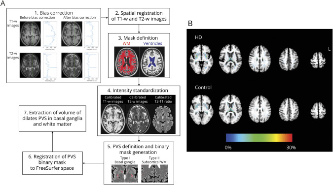
Objective: To quantify the percent volume of dilated perivascular space (PVS) in the subcortical forebrain in patients with early Huntington disease (HD) and to explore the relationship between PVS and disease severity.
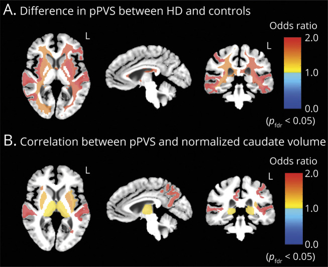
Methods: MRI scans were performed on 25 patients with HD and 23 healthy age-matched controls at Massachusetts General Hospital. The imaging data were analyzed with a novel algorithm to determine regional PVS volume. A fractional logistic regression analysis was used to quantify the association between regional percent PVS volume and (1) disease designation (HD or control) and (2) disease severity as assessed by normalized caudate volume.
Results: Patients with HD had the greatest percent volume of dilated PVS in the putamen (left putamen: odds ratio 2.06 [95% confidence interval (CI) 1.62-2.62], HD 3.27% [95% CI 2.83-3.78] vs controls 1.62% [95% CI 1.32-1.97], p fdr < 0.001; right putamen: odds ratio 1.66 [95% CI 1.33-2.08], HD 3.43% [95% CI 2.94-4.01] vs controls 2.09% [95% CI 1.79-2.45], p fdr < 0.001) and several subcortical white matter regions compared to controls. Dilated PVS increased with disease severity.
Conclusions: The objective quantification of dilated PVS suggests that PVS burden is high, is associated with disease severity, and may affect the distribution and success of treatments administered either intrathecally such as antisense oligonucleotides or by intraparenchymal administration such as cell and gene therapies.
Classification of evidence: This study provides Class II evidence that increased dilated PVS is associated with worse HD severity. The study is rated Class II because of the cross-sectional design.
Copyright © 2020 The Author(s). Published by Wolters Kluwer Health, Inc. on behalf of the American Academy of Neurology.
Figures
References
-
- Robin C. Recherches sur quelques particularites de la structure des capillaires de l'encephale. J Physiol Homme Anim 1859;2:537–548.
-
- Wuerfel J, Haertle M, Waiczies H, et al. . Perivascular spaces: MRI marker of inflammatory activity in the brain? Brain 2008;131:2332–2340. - PubMed
Publication types
MeSH terms
Grants and funding
LinkOut - more resources
Full Text Sources
Medical
Research Materials