. 2020 Nov 10;117(45):28092-28101.
doi: 10.1073/pnas.2008479117. Epub 2020 Oct 26.

RNA-binding protein DDX3 mediates posttranscriptional regulation of androgen receptor: A mechanism of castration resistance

Affiliations

RNA-binding protein DDX3 mediates posttranscriptional regulation of androgen receptor: A mechanism of castration resistance

Jordan E Vellky et al. Proc Natl Acad Sci U S A. .

Abstract

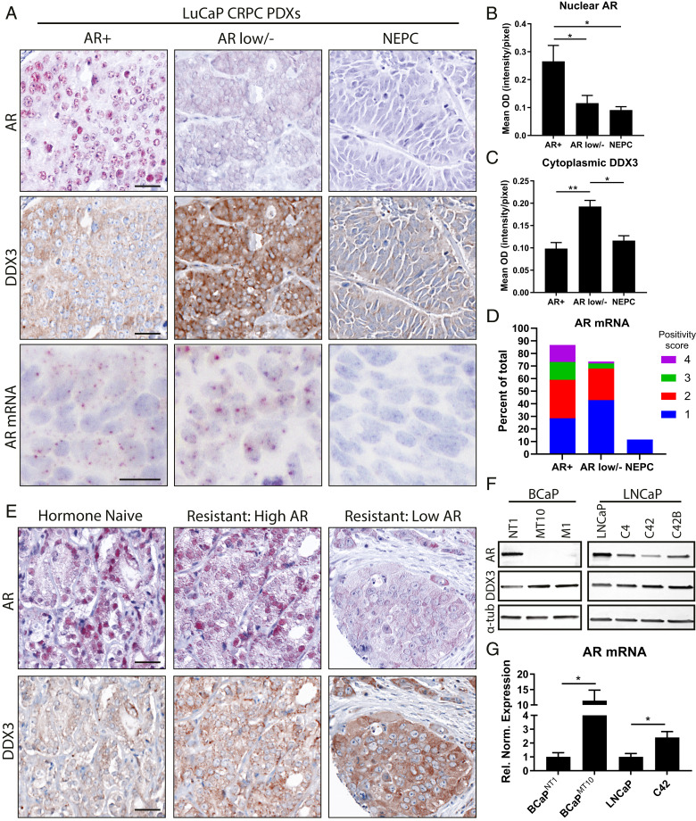
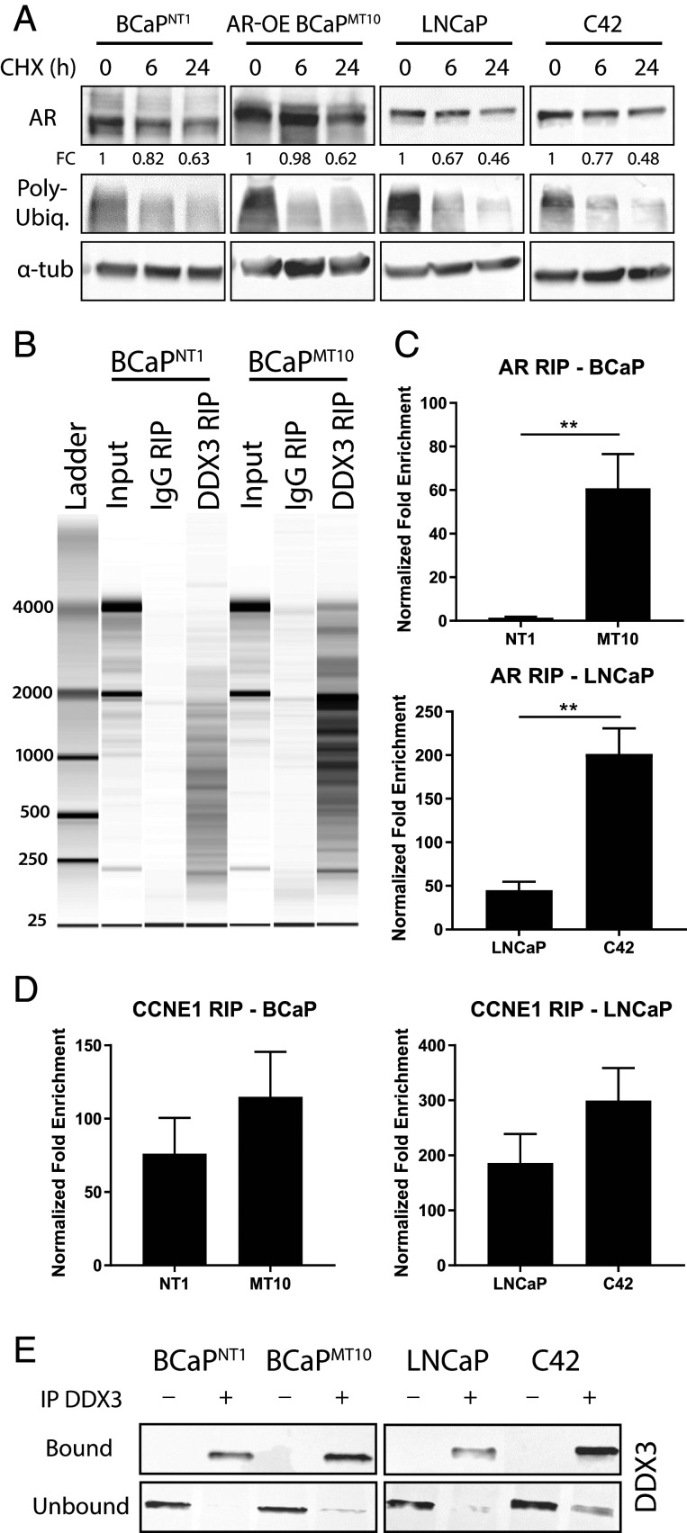
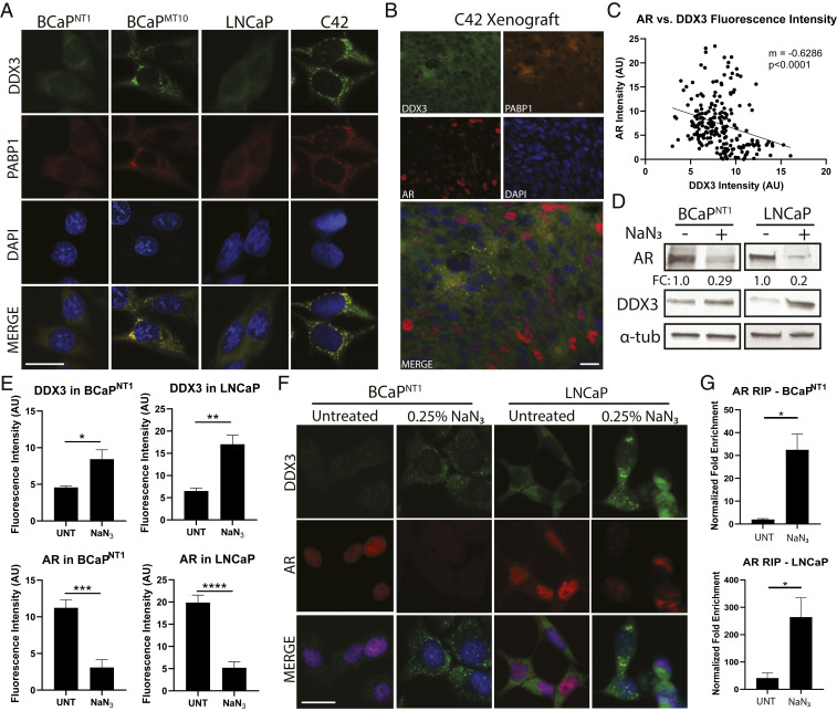
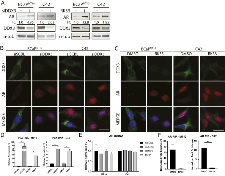
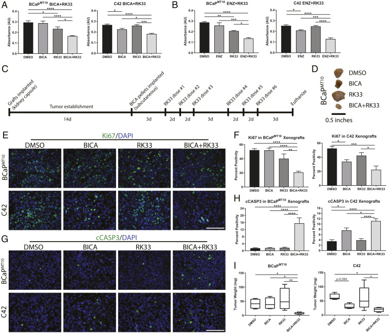
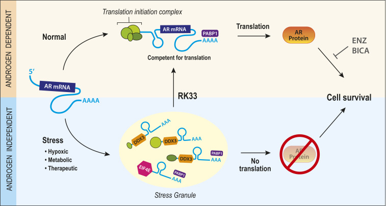
Prostate cancer (CaP) driven by androgen receptor (AR) is treated with androgen deprivation; however, therapy failure results in lethal castration-resistant prostate cancer (CRPC). AR-low/negative (ARL/-) CRPC subtypes have recently been characterized and cannot be targeted by hormonal therapies, resulting in poor prognosis. RNA-binding protein (RBP)/helicase DDX3 (DEAD-box helicase 3 X-linked) is a key component of stress granules (SG) and is postulated to affect protein translation. Here, we investigated DDX3-mediated posttranscriptional regulation of AR mRNA (messenger RNA) in CRPC. Using patient samples and preclinical models, we objectively quantified DDX3 and AR expression in ARL/- CRPC. We utilized CRPC models to identify DDX3:AR mRNA complexes by RNA immunoprecipitation, assess the effects of DDX3 gain/loss-of-function on AR expression and signaling, and address clinical implications of targeting DDX3 by assessing sensitivity to AR-signaling inhibitors (ARSI) in CRPC xenografts in vivo. ARL/- CRPC expressed abundant AR mRNA despite diminished levels of AR protein. DDX3 protein was highly expressed in ARL/- CRPC, where it bound to AR mRNA. Consistent with a repressive regulatory role, DDX3 localized to cytoplasmic puncta with SG marker PABP1 in CRPC. While induction of DDX3-nucleated SGs resulted in decreased AR protein expression, inhibiting DDX3 was sufficient to restore 1) AR protein expression, 2) AR signaling, and 3) sensitivity to ARSI in vitro and in vivo. Our findings implicate the RBP protein DDX3 as a mechanism of posttranscriptional regulation for AR in CRPC. Clinically, DDX3 may be targetable for sensitizing ARL/- CRPC to AR-directed therapies.

Keywords: androgen independence; castration-resistant prostate cancer; double-negative prostate cancer; posttranscriptional regulation; prostate cancer.


Conflict of interest statement

The authors declare no competing interest.

Figures

Fig. 1.
Fig. 1.
ARL/− CRPC retains AR mRNA and expresses high levels of DDX3. AR and DDX3 expression were assessed in human CRPC samples. (A) Representative images of AR protein (red, first row), DDX3 protein (brown, second row), and AR mRNA (brown, third row) in PDXs from three subtypes of CRPC: AR+, ARL/−, and NEPC. Nuclei were counterstained with hematoxylin (blue). (Scale bar, 25 µm.) (B) Mean optical density of nuclear AR expression from PDXs showed nuclear AR was significantly higher in the AR+ compared to ARL/− (P = 0.046) and NEPC (P = 0.017). AR+, n = 5; ARL/− (DNPC, ARLPC), n = 3; NEPC, n = 4. (C) Mean optical density of cytoplasmic DDX3 expression from prostate cancer PDX model (LuCaP) showed DDX3 was significantly higher in DNPC compared to AR+ and NEPC (P = 0.002 and 0.011, respectively). AR+, n = 5; ARL/− (DNPC, ARLPC), n = 3; NEPC, n = 4. (D) AR mRNA positivity scoring in CRPC where “1” indicates 1–3 dots per cell, “2” indicates 4–9 dots per cell, “3” indicates 10–15 dots per cell, and “4” indicates >15 dots/cell. (E) Representative images of IHC colocalization for AR protein (red) and DDX3 protein (brown) in hormone-naive and hormone-resistant (CRPC) patient specimens. Nuclei were counterstained with hematoxylin (blue). (Scale bar, 10 µm.) (F) Representative Western blot analysis in two models of CaP progression (BCaP, LNCaP-C4) showed decreased amounts of AR protein coincident with high amounts of DDX3 protein in CRPC; α-tubulin (α-tub) was used as a loading control. (G) Analysis of AR mRNA expression via qPCR in cell line models of CRPC (BCaPMT10 and C42) showed significantly higher amounts compared to parental cell lines (BCaPNT1, P = 0.011; LNCaP, P = 0.045, n = 3) after normalization to reference genes TBP and YWHAZ. Bar graphs represent mean ± SEM. Significance is represented by *P ≤ 0.05, **P ≤ 0.01.
Fig. 2.
Fig. 2.
DDX3 binds AR mRNA in ARL/− CRPC. Posttranscriptional regulation of AR was assessed via degradation and DDX3-dependent mechanisms, where AR was identified as a target mRNA of RBP DDX3. (A) Degradation rates of AR protein were determined using CHX pulse–chase assays. Expression of AR was determined at 0, 6, and 24 h post-CHX treatment. Poly-ubiquitin expression decreased when translation was inhibited, as expected, and α-tub as a loading control. AR protein expression fold change (FC) was determined relative to the 0-h time point using densitometry (n = 3). (B) Analysis of RNA binding using DDX3 RIP followed by bioanalyzer visualization of RNA in BCaPNT1 and BCaPMT10 showed robust RNA yields for the RNA input control and DDX3 RIP, but lower RNA yields for the IgG RIP controls, suggesting significant RNA binding to DDX3 and little nonspecific binding to IgG control. (C) RIP followed by qPCR for AR mRNA showed significantly increased binding of DDX3 to AR mRNA in CRPC cell lines BCaPMT10 and C42 compared to parental cell lines BCaPNT1 and LNCaP (BCaP, P = 0.009; LNCaP, P = 0.002). These results were normalized to input RNA, and represented as the FC for specific antibody pulldown over the IgG control pulldown. (D) Validation of RIP-DDX3 RNA binding using qPCR for CCNE1 showed mRNA was bound to DDX3 in all cell lines. (E) Western analysis showed “bound” samples, taken directly after the IP before RNA isolation, exhibited robust DDX3 pulldown in samples given DDX3 antibody (IP DDX3 +), while IgG controls (IP DDX3 -) did not pull down DDX3. Conversely, the “unbound”, i.e., the remainder of the lysate after the magnetic beads were removed, showed a decrease of DDX3 protein in the DDX3 IP pulldowns, while the IgG controls retained high DDX3 protein content, as expected. Bar graphs represent mean ± SEM. Significance is represented by **P ≤ 0.01.
Fig. 3.
Fig. 3.
DDX3 localizes to SGs in CRPC and represses AR protein expression. Consistent with translational repression, DDX3-mediated regulation of AR occurred when DDX3 was localized to SGs. Induction of SGs resulted in increased AR mRNA bound to DDX3 and decreased AR protein expression. (A) Immunofluorescence for DDX3 (green) showed localization to cytoplasmic puncta (SGs) in CRPC lines BCaPMT10 and C42 grown in vitro, as compared to diffuse cytoplasmic staining in the parental cell lines BCaPNT1 and LNCaP. Merged images show colocalization (yellow) of DDX3 with SG marker PABP1 (red). Nuclei were counterstained with DAPI (blue). (B) IHC of a C42 xenograft grown in vivo showed colocalization of DDX3 (green) and PABP1 (orange) puncta and AR protein expression in red. In these xenografts, DDX3/PABP1 puncta are only present in AR protein negative cells, despite robust AR protein expression in the surrounding area (red nuclei). Nuclei were counterstained with DAPI (blue). (C) Linear regression for DDX3 and AR protein fluorescence intensity in C42 xenografts showed a significant negative correlation between DDX3 and AR expression (n = 3, P < 0.0001). (D) Western blot analysis of AR protein expression in BCaPNT1 and LNCaP bulk populations after induced hypoxic stress by 3 h treatment with 0.25% sodium azide (NaN3) showed a decrease of overall AR protein (FC = 0.29 for BCaPNT1 and 0.2 for LNCaP), with a concurrent increase of DDX3 protein expression. α-tub was used as a loading control. (E) Quantification of DDX3 and AR protein fluorescence intensity after treatment with NaN3 showed a significant increase of DDX3 intensity (P = 0.037 in BCaPNT1 and P = 0.002 in LNCaP) and a significant decrease of AR intensity (P = 0.0001 in BCaPNT1 and P < 0.0001 in LNCaP) compared to UNTs. Fluorescence intensity was averaged between at least three separate experiments. (F) Representative images for hypoxia-induced stress from E showed NaN3 treatment increased DDX3 expression (green) and induced localization to cytoplasmic puncta concurrent with decreased AR protein expression (red) in parental cell lines BCaPNT1 and LNCaP. Nuclei were counterstained with DAPI (blue). (G) RIP analysis following treatment with NaN3 significantly increased AR mRNA bound to DDX3 compared to UNT (BCaPNT1, P = 0.043; LNCaP, P = 0.039). (Scale bars, 10 µm.) Bar graphs represent mean ± SEM. Significance is represented by *P ≤ 0.05, **P ≤ 0.01, ***P ≤ 0.001, ****P ≤ 0.0001.
Fig. 4.
Fig. 4.
Genetic and pharmacological inhibition of DDX3 increases AR protein expression and signaling. Inhibition of DDX3 resulted in increased AR protein expression and AR signaling in CRPC. (A) Western blot analysis of AR protein expression in BCaPMT10 and C42 bulk populations after inhibition of DDX3 by siRNA and small-molecule inhibitor RK33 showed an increase of overall AR protein and decrease of DDX3 protein. With siRNA, AR protein increased 4.86-fold in BCaPMT10 and 2.03-fold in C42 compared to scramble control. With RK33 treatment, AR protein increased 12.8-fold in BCaPMT10 and 1.85-fold in C42 compared to DMSO control. α-tub was used as a loading control. (B) Immunofluorescence analysis of DDX3 expression and localization after inhibition with siRNAs showed decreased cytoplasmic DDX3 expression (green) and increased AR expression (red), compared to scramble control (siSCBL) in two CRPC models (BCaPMT10 and C42). Nuclei were counterstained with DAPI (blue). (C) Immunofluorescence analysis of DDX3 expression and localization after pharmacologic inhibition using 2 µM RK33 showed decreased cytoplasmic DDX3 expression (green) and increased AR expression (red) compared to DMSO control in BCaPMT10 and C42. Nuclei were counterstained with DAPI (blue). (D) Assessment of AR signaling, using PSA, a transcriptional target of AR, was significantly increased with siDDX3 compared to siSCBL (BCaPMT10, P = 0.036; C42, P = 0.021) and with RK33 compared to DMSO (BCaPMT10, P = 0.019; C42, P = 0.029) in both CRPC models. (E) qPCR for AR mRNA showed no significant difference in expression between siSCBL vs. siDDX3 and RK33 vs. DMSO. (F) RIP analysis of AR mRNA bound to DDX3 after RK33 treatment (RK33) showed a significant decrease in DDX3 binding to AR mRNA compared to control (DMSO) in CRPC cell lines BCaPMT10 (P = 0.02) and C42 (P = 0.004). (Scale bars, 10 µm.) Bar graphs represent mean ± SEM. Significance is represented by *P ≤ 0.05, **P ≤ 0.01.
Fig. 5.
Fig. 5.
DDX3 inhibition resensitizes ARL/− cells to anti-androgen therapy. By increasing AR protein expression with DDX3 inhibition, ARL/− CRPC was sensitized to AR-targeted therapy. Cotreatment with a DDX3 inhibitor and BICA resulted in decreased cell viability, decreased proliferation, and increased apoptosis. (A) MTT assay in BCaPMT10 and C42 showed cotreatment with BICA, and RK33 significantly decreased cell viability compared to BICA alone (BCaPMT10, P < 0.0001; C42, P = 0.014) and RK33 alone (BCaPMT10, P = 0.011; C42, P = 0.0001). RK33 treatment alone was sufficient to decrease cell viability in BCaPMT10 compared to DMSO control (P = 0.041). (B) Cotreatment with ENZ and RK33 significantly decreased cell viability in BCaPMT10 and C42 compared to ENZ alone (BCaPMT10, P = 0.0003; C42, P = 0.0002) and RK33 alone (BCaPMT10, P = 0.021; C42, P < 0.0001). RK33 treatment alone was sufficient to decrease cell viability in BCaPMT10 (P = 0.007s). (C) Schematic for experimental design using RK33 and BICA cotreatment in vivo. d = days. (D) Representative images of tumors from each treatment group harvested from mice at necropsy. (E) Representative images of proliferation (Ki67, green) in each treatment group in BCaPMT10 and C42 xenografts. Nuclei were counterstained with DAPI (blue). (F) Labeling index of Ki67-positivity showed a significant decrease in cotreatment compared to DMSO control (BCaPMT10, P < 0.0001; C42, P = 0.0006). In BCaPMT10, cotreatment significantly decreased Ki67 compared to BICA alone and RK33 alone (P < 0.0001 and P = 0.007, respectively). In C42, cotreatment significantly decreased Ki67 compared to RK33 alone (P = 0.015) but was not significantly different from BICA alone (P = 0.512). (G) Representative images of apoptosis (cCASP3, green) from each treatment group in BCaPMT10 and C42 xenografts. Nuclei were counterstained with DAPI (blue). (H) Calculation of percent positivity of cCASP3 showed a significant increase in cotreatment compared to DMSO in both BCaPMT10 and C42 (P < 0.0001 for both). In BCaPMT10, cotreatment significantly increased cCASP3 compared to BICA alone (P < 0.0001) and RK33 alone (P < 0.0001). In C42, cotreatment significantly increased cCASP3 compared to BICA alone (P = 0.034) and RK33 alone (P < 0.0001). (I) Quantification of tumor weight at necropsy showed mice receiving cotreatment (BICA+RK33) had significantly smaller tumors than the control mice for both BCaPMT10 (P = 0.049) and C42 (P = 0.0255). In BCaPMT10, tumor weight after cotreatment was significantly smaller than BICA alone (P = 0.046) and RK33 alone (P = 0.008). In C42, tumor weight after cotreatment was significantly smaller than RK33 alone (P = 0.0263), but not BICA alone (P = 0.534). (Scale bars in E and G, 100 µm.) Bar graphs represent mean ± SEM. Significance is represented by *P ≤ 0.05, **P ≤ 0.01, ***P ≤ 0.001, ****P ≤ 0.0001.
Fig. 6.
Fig. 6.
Model for DDX3 regulation of AR in CRPC. Schematic for DDX3-mediated regulation of AR in CRPC. Under normal conditions, AR mRNA is translated into AR protein, resulting in AR protein and androgen-mediated cell survival, which can be inhibited using androgen/AR targeting therapies BICA or ENZ. Under stress conditions (hypoxic, metabolic, therapeutic), DDX3 is up-regulated and localizes to SGs. Here, within SGs, AR mRNA is bound to DDX3 causing repression of AR protein translation. This process is associated with low sensitivity to anti-androgens due to the absence of targetable AR protein, resulting in AR-independent cell survival. Therefore, by pharmacologically inhibiting the formation of SGs with chemical DDX3 inhibitors, one can resensitize castration-resistant (stressed) cells to hormonal therapy through the restoration of AR protein translation.

Comment in

  • Uro-Science.
    Atala A. Atala A. J Urol. 2021 Aug;206(2):480-482. doi: 10.1097/JU.0000000000001849. Epub 2021 May 12. J Urol. 2021. PMID: 33975458 No abstract available.

References

    1. Heinlein C. A., Chang C., Androgen receptor in prostate cancer. Endocr. Rev. 25, 276–308 (2004). - PubMed
    1. Kirby M., Hirst C., Crawford E. D., Characterising the castration-resistant prostate cancer population: A systematic review. Int. J. Clin. Pract. 65, 1180–1192 (2011). - PubMed
    1. Deng Q., Tang D. G., Androgen receptor and prostate cancer stem cells: Biological mechanisms and clinical implications. Endocr. Relat. Cancer 22, T209–T220 (2015). - PMC - PubMed
    1. Bluemn E. G., Coleman I. M., Lucas J. M. et al. , Androgen receptor pathway-independent prostate cancer is sustained through FGF signaling. Cancer Cell. 32, 474–489.e6 (2017). - PMC - PubMed
    1. Labrecque M. P., et al. , Molecular profiling stratifies diverse phenotypes of treatment-refractory metastatic castration-resistant prostate cancer. J. Clin. Invest. 129, 4492–4505 (2019). - PMC - PubMed

Publication types

MeSH terms