. 2020 Oct 26;11(1):5393.
doi: 10.1038/s41467-020-19241-7.

Presence of low virulence chytrid fungi could protect European amphibians from more deadly strains

Affiliations

Presence of low virulence chytrid fungi could protect European amphibians from more deadly strains

Mark S Greener et al. Nat Commun. .

Abstract

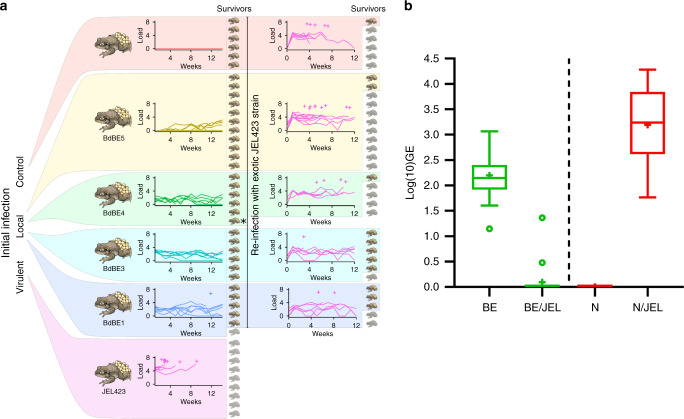
Wildlife diseases are contributing to the current Earth's sixth mass extinction; one disease, chytridiomycosis, has caused mass amphibian die-offs. While global spread of a hypervirulent lineage of the fungus Batrachochytrium dendrobatidis (BdGPL) causes unprecedented loss of vertebrate diversity by decimating amphibian populations, its impact on amphibian communities is highly variable across regions. Here, we combine field data with in vitro and in vivo trials that demonstrate the presence of a markedly diverse variety of low virulence isolates of BdGPL in northern European amphibian communities. Pre-exposure to some of these low virulence isolates protects against disease following subsequent exposure to highly virulent BdGPL in midwife toads (Alytes obstetricans) and alters infection dynamics of its sister species B. salamandrivorans in newts (Triturus marmoratus), but not in salamanders (Salamandra salamandra). The key role of pathogen virulence in the complex host-pathogen-environment interaction supports efforts to limit pathogen pollution in a globalized world.


Conflict of interest statement

The authors declare no competing interests.

Figures

Fig. 1
Fig. 1. Experimental results for phenotypic traits of B. dendrobatidis isolates.
ad Boxes indicate 25th and 75th percentiles, central lines the mean, bars the 95th percentiles, and points indicate individual samples. N of sporangia = number of sporangia in the central 1000 × 1000 pixels, n = 4 independent experiments; area of sporangia = area of the largest 10 sporangia, n = 10 technical replicates per independent experiment; N of zoospores = number of spores in the central 1000 × 1000 pixels of the well/image, n = 4 independent experiments; fecundity was calculated following the formula (average NZOOSPORE/average NSPOR)/average ASPOR, n = 4 independent experiments. e Non-metric multidimensional scaling across the four variables in (ad); labels indicate one point per group as a colour legend. BdBE1-10 are local isolates isolated in this study. JEL423 is a BdGPL isolate isolated from an episode of amphibian mortality in the neotropics. Source data are provided as a Source data file.
Fig. 2
Fig. 2. BdGPL virulence coincides with growth ability in mucosomes, Crinkler (CRN) gene expression and adhesion and invasion capacities.
a Growth (expressed in corrected GE values) of BdBE1 and BdJEL423 in mucosomes collected from different amphibian species. PC: positive control; Ao: Alytes obstetricans; Ss: Salamandra salamandra; NC: negative control. Significant differences are shown with an asterisk and were assessed by a Kruskal–Wallis analysis, followed by pairwise Mann–Whitney U-test (two-tailed) with a Bonferroni-corrected P value of 0.017 (Ao vs Ss: P value = 0.001 (BdJEL423) and 0.009 (BdBE1); Ao vs PC: P value < 0.001 (BdJEL423) and 0.003 (BdBE1)). The experiment was carried out in tenfold and individual data points are shown (blue = BdJEL423; red = BdBE1), with the mean ± s.e.m. depicted by the horizontal bars. b Box plots of mean fold changes in mRNA expression of CRN genes (CRN_22492, CRN_25085 and CRN_23176). The data in the left panel show the normalized target gene amount in spores of each isolate (n = 4) relative to spores of BdJEL423 (n = 4) which is considered 1. The data in the right panel show the normalized target gene amount in spores that were incubated with skin tissue of A. obstetricans for 2 h (n = 4) relative to spores of the respective isolate (n = 4) which is considered 1. Boxes indicate 25th and 75th percentiles, central lines the median, bars the minima and maxima, and points indicate individual samples. Target genes were based on Farrer et al.. Left panel: an asterisk indicates a significant difference compared to BdJEL423 spores (Kruskal–Wallis analysis, followed by pairwise Mann–Whitney U-tests (two-tailed) with a Benjamini–Hochberg-adjusted P value < 0.05). Right panel: an asterisk indicates a significant difference compared to spores of the respective isolate (Kruskal–Wallis analysis, followed by pairwise Mann–Whitney U-tests (two-tailed). Individual P values are shown in Supplementary Tables 6–7. c Representative image showing germ tube formation of BdJEL423. Fluorescent signals of Bd (Calcofluor White (blue)) and A6 cells (green cell tracker) were merged to assess the ability of germ tube formation, 4 h after contact with the cells. d, e Representative images demonstrating endobiotic growth (d: BdJEL423) and epibiotic growth (e: BdBE4) of Bd. The cell wall of extracellular Bd was coloured using Calcofluor White (blue). Bd was visualized using Alexa Fluor 568 targeting a polyclonal antibody against Bd (Thomas et al.), resulting in red fluorescence of both intracellular and extracellular Bd. The Bd-exposed A6 cells were stained with a cell tracker (green). d New intracellular chytrid thalli are formed and the cell content of the mother thallus is transferred into the new daughter thallus (DT). e Epibiotic growth is limited to Bd development outside the host cells. GT germ tube, EX GT extracellular germ tube, IN GT intracellular germ tube, MT mother thallus, DT daughter thallus. Scale bar = 20 µm. Three independent in vitro experiments were conducted with every condition being tested in triplicate, with similar results. Source data are provided as a Source data file.
Fig. 3
Fig. 3. Local BdGPL isolates protect midwife toads against infection with virulent BdGPL.
a (Left) Toads exposed to local (BdBE) isolates had survival comparable to the control (non-exposed) group; toads exposed to hypervirulent BdJEL423 had 100% mortality. (Right) When all survivors were re-infected with BdJEL423, toads previously exposed to BdBE1 and BdBE3 had higher survival than naive toads (control) or toads previously exposed to BdBE4-5. For each group, curves indicate infection load (log10[GE+1]), crosses indicate times of individual deaths (crosses offset vertically for readability, dead individuals shaded). The asterisk * indicates one animal that died of non-treatment-related causes between the two experiments. b Tukey-style box plots of Bd peak loads in midwife toads either pre-exposed (BE: green) or not pre-exposed (N: red) to the low-virulence BdBE3 isolate and 4 weeks later challenged with the highly virulent BdJEL423 (BE/JEL and N/JEL). n = 20 biologically independent animals per condition. Source data are provided as a Source data file.
Fig. 4
Fig. 4. Low-virulence BdGPL isolates protect some urodelan species, but not others, against infection with B. salamandrivorans.
a For Triturus marmoratus, low virulent BdGPL exposure reduced Bsal-induced mortality but increased the length of the infectious period. b For Pleurodeles waltl, low-virulence BdGPL exposure reduced Bsal-induced mortality but had no effect on the length of infectious period or on infection loads. c For Salamandra salamandra, low virulent BdGPL exposure had no effect on Bsal-induced mortality or infection course. For each species, Bsal exposure occurred 21 days after Bd exposure. For all groups, curves indicate infection load (log10[GE+1]), crosses the time of individual deaths (offset vertically for readability), boxes the number of survivors in each treatment group by species (dead individuals shaded). Source data are provided as a Source data file.

References

    1. O’Hanlon SJ, et al. Recent Asian origin of chytrid fungi causing global amphibian declines. Science. 2018;360:621–627. doi: 10.1126/science.aar1965. - DOI - PMC - PubMed
    1. Scheele BC, et al. Amphibian fungal panzootic causes catastrophic and ongoing loss of biodiversity. Science. 2019;363:1459–1463. doi: 10.1126/science.aav0379. - DOI - PubMed
    1. Berger L, et al. History and recent progress on chytridiomycosis in amphibians. Fungal Ecol. 2016;19:89–99. doi: 10.1016/j.funeco.2015.09.007. - DOI
    1. Martel A, et al. Batrachochytrium salamandrivorans sp. nov. causes lethal chytridiomycosis in amphibians. Proc. Natl Acad. Sci. USA. 2013;110:15325–15329. doi: 10.1073/pnas.1307356110. - DOI - PMC - PubMed
    1. Farrer RA, et al. Genomic innovations linked to infection strategies across emerging pathogenic chytrid fungi. Nat. Commun. 2017;8:14742. doi: 10.1038/ncomms14742. - DOI - PMC - PubMed

Publication types