. 2020 Oct 26;10(1):18284.
doi: 10.1038/s41598-020-75385-y.

Osteogenic potential of human adipose derived stem cells (hASCs) seeded on titanium trabecular spinal cages

Affiliations

Osteogenic potential of human adipose derived stem cells (hASCs) seeded on titanium trabecular spinal cages

Laura Caliogna et al. Sci Rep. .

Abstract

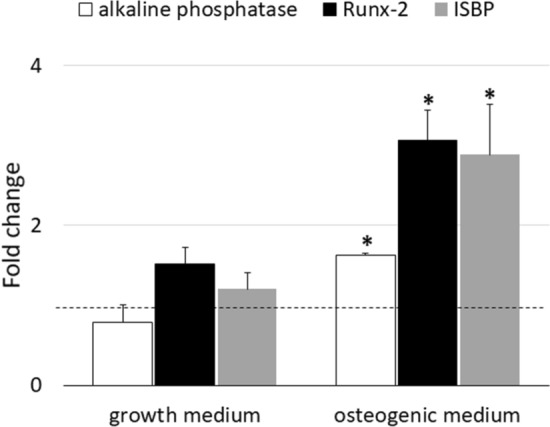
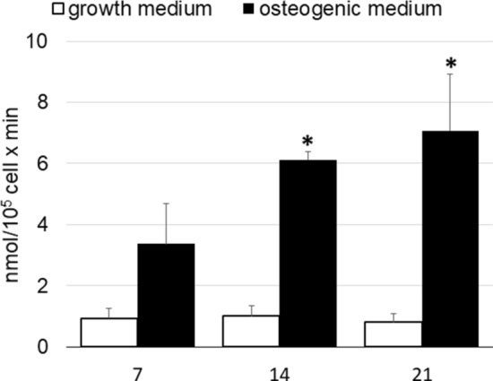
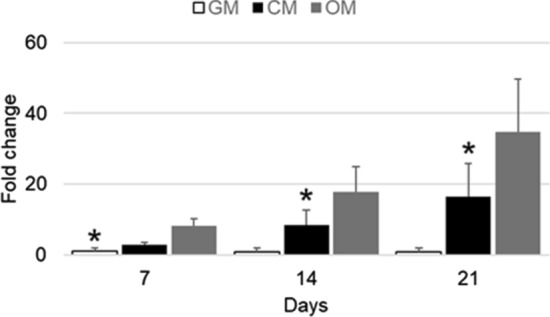
Spine degenerative conditions are becoming increasingly prevalent, affecting about 5.7% of the population in Europe, resulting in a significant reduction of life's quality. Up to now, many materials have been used in manufacturing cage implants, used as graft substitutes, to achieve immediate and long-term spinal fixation. Particularly, titanium and its alloys are emerging as valuable candidates to develop new types of cages. The aim of this in vitro study was to evaluate the adhesion, proliferation and osteogenic differentiation of adipose derived mesenchymal stem cells (ASCs) seeded on trabecular titanium cages. ASCs adhered, proliferated and produced an abundant extracellular matrix during the 3 weeks of culture. In the presence of osteogenic medium, ASCs differentiated into osteoblast-like cells: the expression of typical bone genes, as well as the alkaline phosphatase activity, was statistically higher than in controls. Furthermore, the dispersive spectrometry microanalysis showed a marked increase of calcium level in cells grown in osteogenic medium. Plus, our preliminary data about osteoinduction suggest that this titanium implant has the potential to induce the ASCs to produce a secretome able to trigger a shift in the ASCs phenotype, possibly towards the osteogenic differentiation, as illustrated by the qRT-PCR and ALP biochemical assay results. The trabecular porous organization of these cages is rather similar to the cancellous bone structure, thus allowing the bone matrix to colonize it efficiently; for these reasons we can conclude that the architecture of this cage may play a role in modulating the osteoinductive capabilities of the implant, thus encouraging its engagement in in vivo studies for the treatment of spinal deformities and diseases.


Conflict of interest statement

The authors declare no competing interests.

Figures

Figure 1
Figure 1
hASC proliferation after seeding on cage. Each point is the mean ± SEM of 4 different experiments. When not present the standard error was within the symbol. *, p ≤ 0.05 versus 1 day (one-way ANOVA method followed by Newman-Keuls’Q test).
Figure 2
Figure 2
SEM analysis of a cage cultured in osteogenic medium. The arrow highlights the presence of a little capillary. Magnification: 1, 75 kx.
Figure 3
Figure 3
SEM analysis of hASC/cage constructs cultured with growth (GM) and osteogenic (OM) medium. The BSE detection showed the organic parts (cells and their matrix) that appeared dark, while the metallic scaffold appeared clear. The matrix of constructs in OM appeared thicker.
Figure 4
Figure 4
alp, runx-2 and ibsp transcript expression in hASCs seeded on cage in the presence (OM) and absence (GM) of osteogenic factors after 14 days from differentiation. The values obtained were reported as ΔΔCT (fold change) versus 7 days expression (horizontal line). Each bar is the mean ± SEM of four different preparations. *, p ≤ 0.05 versus growth medium (Student t test).
Figure 5
Figure 5
Alkaline phosphatase activity in hASCs grown on cage cultured with growth medium and osteogenic medium. Each bar represents the mean ± S.E.M. of four different preparations; each assay was repeated three times. *, p ≤ 0.05 vs. growth medium (one-way ANOVA method followed by Newman-Keuls’Q test).
Figure 6
Figure 6
Calcium content in hASC/cage constructs cultured with growth (GM) and osteogenic (OM) medium. Each bar is the mean ± SEM of ten measurements. *, p ≤ 0.05 vs GM (Student t test).
Figure 7
Figure 7
alp gene expression of hASCs grown in monolayer cultured with GM, CM and OM after 7, 14 and 21 days of in vitro culture. The values obtained were reported as ΔΔCT (fold change) versus 7 days expression. Each bar is the mean ± SEM of four different preparations. *, p ≤ 0.05 versus CM and OM (one-way ANOVA method followed by Newman-Keuls’Q test).
Figure 8
Figure 8
Alkaline phosphatase activity of hASCs grown in monolayer and cultured with GM, CM and OM at 7, 14 and 21 days of in vitro culture. Each bar represents the mean ± S.E.M. of four different preparations; each assay was repeated three times. *, p ≤ 0.05 vs. GM and OM (one-way ANOVA method followed by Newman-Keuls’Q test).

References

    1. Chau AMT, Mobbs RJ. Bone graft substitutes in anterior cervical discectomy and fusion. Eur. Spine J. 2009;18:449–464. doi: 10.1007/s00586-008-0878-4. - DOI - PMC - PubMed
    1. Jain S, Eltorai AEM, Ruttiman R, Daniels AH. Advances in spinal interbody cages. Orthop. Surg. 2016;8:278–284. doi: 10.1111/os.12264. - DOI - PMC - PubMed
    1. Cloward RB. The anterior approach for removal of ruptured cervical disks. J. Neurosurg. Spine. 2007;6:496–508. doi: 10.3171/spi.2007.6.5.496. - DOI - PubMed
    1. McConnell, J.R., Freeman, B.J.C., Debnath, U.K., Grevitt, M.P., Prince, H.G., Webb, J.K. A prospective randomized comparison of coralline hydroxyapatite with autograft in cervical interbody fusion. Spine (Phila. Pa. 1976)28 (2003). - PubMed
    1. Bagby GW. Arthrodesis by the distraction-compression method using a stainless steel implant. Orthopedics. 1988;11:931–934. doi: 10.3928/0147-7447-19880601-13. - DOI - PubMed

Publication types