[Preprint]. 2020 Oct 20:2020.10.15.20213512.
doi: 10.1101/2020.10.15.20213512.

COVID-19 neutralizing antibodies predict disease severity and survival

Affiliations

COVID-19 neutralizing antibodies predict disease severity and survival

Wilfredo F Garcia-Beltran et al. medRxiv. .

Update in

  • COVID-19-neutralizing antibodies predict disease severity and survival.
    Garcia-Beltran WF, Lam EC, Astudillo MG, Yang D, Miller TE, Feldman J, Hauser BM, Caradonna TM, Clayton KL, Nitido AD, Murali MR, Alter G, Charles RC, Dighe A, Branda JA, Lennerz JK, Lingwood D, Schmidt AG, Iafrate AJ, Balazs AB. Garcia-Beltran WF, et al. Cell. 2021 Jan 21;184(2):476-488.e11. doi: 10.1016/j.cell.2020.12.015. Epub 2020 Dec 15. Cell. 2021. PMID: 33412089 Free PMC article.

Abstract

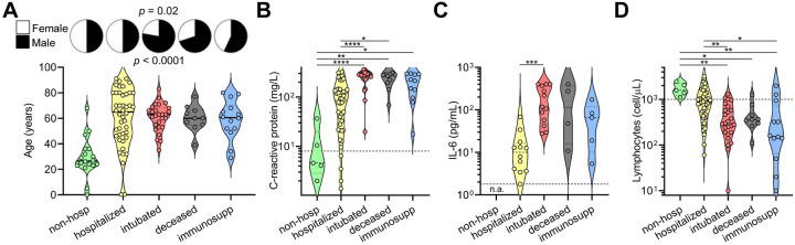
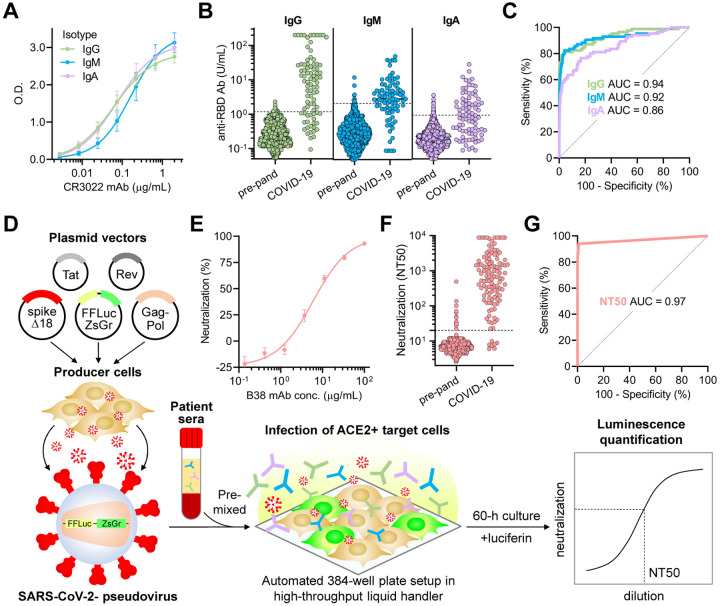
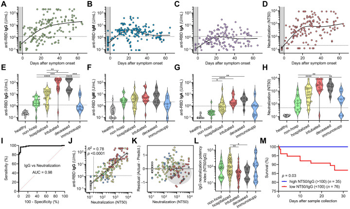
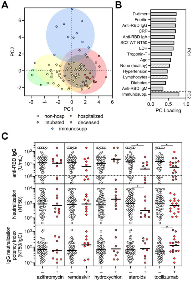
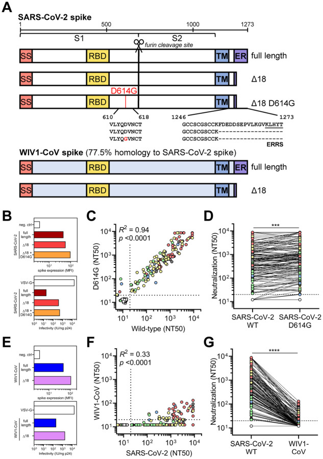
COVID-19 exhibits variable symptom severity ranging from asymptomatic to life-threatening, yet the relationship between severity and the humoral immune response is poorly understood. We examined antibody responses in 113 COVID-19 patients and found that severe cases resulting in intubation or death exhibited increased inflammatory markers, lymphopenia, and high anti-RBD antibody levels. While anti-RBD IgG levels generally correlated with neutralization titer, quantitation of neutralization potency revealed that high potency was a predictor of survival. In addition to neutralization of wild-type SARS-CoV-2, patient sera were also able to neutralize the recently emerged SARS-CoV-2 mutant D614G, suggesting protection from reinfection by this strain. However, SARS-CoV-2 sera was unable to cross-neutralize a highly-homologous pre-emergent bat coronavirus, WIV1-CoV, that has not yet crossed the species barrier. These results highlight the importance of neutralizing humoral immunity on disease progression and the need to develop broadly protective interventions to prevent future coronavirus pandemics.


Figures

Figure 1:
Figure 1:. Clinical severity of SARS-CoV-2 infection is influenced by patient characteristics and coupled to clinical laboratory data.
(A) A cross-sectional cohort of COVID-19 patients (n = 113) was divided into groups of varying clinical severity, i.e., non-hospitalized (n = 18), hospitalized (n = 45), intubated (n = 27), deceased (n = 10), and immunosuppressed (n = 13) and analyzed for their age and sex. Median age was 28 years in patients who were never hospitalized (n = 20; includes 2 from immunosuppressed group) and 63 years in all patients who were admitted to the hospital (n = 93), with statistical significance of p < 0.0001 with t test. Fisher’s exact test on the number of males who were intubated or deceased (n = 31 males out of 42 total; includes 5 from immunosuppressed group who were intubated) versus not (n = 36 males out of 71 total) demonstrated a significant enrichment with p = 0.02. (B-D) Peak levels of C-reactive protein and IL-6 as well as lymphocyte count nadir are presented in violin plots when data was available. In C, none of the non-hospitalized patients had serum IL-6 levels measured (n.a., not assessed). For B and C, clinical laboratory-defined cut-offs of the upper limit of normal are indicated with a dotted line; for D, the dotted line represents the lower limit of normal. For each parameter, a non-parametric ANOVA was performed; statistical significance is indicated with the following notations: **** p < 0.0001, *** p < 0.001, ** p < 0.01, and * p < 0.05.
Figure 2:
Figure 2:. Quantitative SARS-CoV-2 receptor binding domain ELISA and high-throughput SARS-CoV-2 pseudovirus neutralization assay reveal highly variable IgG, IgM, and IgA responses and neutralization potency after SARS-CoV-2 infection.
(A) For quantitation of anti-RBD IgG, IgM, and IgA antibodies, a 7-point standard curve consisting of a SARS-CoV and -CoV-2 RBD-binding monoclonal antibody, CR3022, in all three isotypes (CR3022-IgG, CR3022-IgM, and CR3022-IgA) was used as a reference to interpolate O.D. values from samples and calculate units/mL (U/mL), with 1 U/mL defined as the equivalent reactivity caused by 1 μg/mL of the corresponding CR3022 monoclonal antibody (B) Anti-RBD IgG, IgM, and IgA antibodies were measured in both pre-pandemic samples (n = 1,257) and COVID-19 patient samples (n = 85). Dotted lines indicate the threshold of seropositivity that achieves >99.0% specificity on ROC analyses. (C) ROC analyses for each assay were done to assess how seropositivity predicted COVID-19 status. Area under the curve (AUC) was 0.94 for IgG, 0.92 for IgM, and 0.86 for IgA. Cut-offs of 1.18 U/mL for IgG achieved a sensitivity of 73%, 2.14 U/mL for IgM achieved 66%, and 0.95 U/mL for IgA achieved 48%, with >99.0% specificity for all three. (D) A schematic of the high-throughput SARS-CoV-2 pseudovirus neutralization assay is shown. (E) Validation of the neutralization assay using a recently discovered neutralizing monoclonal antibody, B38, was performed and showed an IC50 of 6 μg/mL. (F) Neutralization titers that achieved 50% neutralization (NT50) were calculated for pre-pandemic samples (n = 1,220, individuals on antiretroviral therapy excluded) and COVID-19 patient samples (n = 118). (G) An ROC analysis demonstrated an AUC of 0.97, with an NT50 cut-off of 20 achieving sensitivity of 94% and specificity of >99.0%.
Figure 3:
Figure 3:. SARS-CoV-2 antibody levels and neutralization potency predict clinical severity and survival.
(A-C) Anti-RBD IgG, IgM, and IgA levels were plotted over days after symptom onset for confirmed COVID-19 cases for which data of symptom onset was known (n = 98 patients, n = 147 samples total). Healthy blood donors (n = 37) are included as a negative control within the gray region. The dotted lines indicate the cut-offs for anti-RBD IgG, IgM, and IgA seropositivity. (D) Titers that achieve 50% neutralization (NT50) were plotted over days after symptom onset for each patient sample. (E-H) Patient samples were selected for collection between 14 and 42 days after symptom onset (earliest time point for each patient), and for each cohort of healthy blood donors, non-hospitalized, hospitalized, intubated, deceased, and immunosuppressed patients, anti-RBD IgG, IgM, IgA, and neutralization (NT50) was plotted. Non-parametric multivariate ANOVA was performed for each (excluding healthy blood donors); statistical significance is indicated as follows: **** p < 0.0001, *** p < 0.001, ** p < 0.01, and * p < 0.05. (I-J) An ROC and log-log regression analyses were performed on IgG versus neutralization. For J, the severity cohort is indicated as follows: healthy (white), non-hospitalized (green), hospitalized (yellow), intubated (red), deceased (gray), and immunosuppressed (blue). For J, Pearson correlations were performed and R2 and p values are indicated. (K) A residual plot for neutralization titer was generated from the log-log correlation. The gray ellipse indicates a cluster of samples from intubated (red) and deceased (gray) patients. (L) Neutralization potency index (NT50/IgG) was calculated for all 113 patients (at earliest time point) and plotted by cohort. A non-parametric multivariate ANOVA was performed without correction for multiple comparisons; unadjusted p values are indicated as follows: ** p < 0.01, * p < 0.05. (M) Survival analysis of COVID-19 patients classified as having a high (≥100) (n = 30) or low (<100) (n = 68) neutralization potency index (NT50/IgG) was performed using Kaplan-Meier method and revealed significantly decreased risk of death in low neutralization potency individuals (p = 0.03).
Figure 4:
Figure 4:. Corticosteroid and tocilizumab therapy decrease humoral immune responses to SARS-CoV-2.
(A) Principle components analysis was performed using the following variables: age, sex language, pre-existing medical conditions, treatments received, clinical laboratory data (ferritin, CRP, D-dimer, LDH, troponin-T, and lymphocyte nadir), anti-RBD antibody levels, and neutralization titers. The severity cohort of each patient is indicated by color. Patients with missing data were excluded. (B) Loading of principle components (PC) is shown. Hatched bars indicate negative loading. (C) Sub-analyses on COVID-19 patients that were in the hospital for at least 3 days to (n = 69) were performed on the last collected specimen to show the effect of azithromycin (n = 10 treated), remdesivir (n = 9 treated), hydroxychloroquine (n = 8 treated), corticosteroids (n = 9 treated), and tocilizumab (n = treated as part of a trial with 2:1 randomization to placebo) on anti-RBD IgG levels (upper panel), neutralization titer (middle panel), and neutralization potency index (NT50/IgG) (lower panel). A t test was performed for each comparison; * indicates unadjusted p < 0.05.
Figure 5:
Figure 5:. SARS-CoV-2-infected patient sera cross-neutralizes both wild-type and D614G mutant SARS-CoV-2 spike but not the highly homologous pre-emergent bat coronavirus WIV1-CoV.
(A) A schematic of the SARS-CoV-2 and WIV1-CoV spike proteins, including full-length, truncated (Δ18), and mutant (D614G) forms is shown. Full-length WIV1-CoV spike has 77.5% sequence homology to SARS-CoV-2 spike and has the same putative ER retention signal (ERRS) as SARS-CoV-2. (B) Expression of full-length, Δ18, and Δ18 D614G SARS-CoV-2 spike constructs in 293T cells in comparison to empty vector (neg. ctrl) was measured by flow cytometry (upper panel). Infectivity of lentivirus, which was defined as the infectious units divided by the quantity of p24 in lentiviral supernatant, was also measured and compared to VSV-G-pseudotyped lentivirus (lower panel). (C-D) Cross-neutralization of serum samples from COVID-19 patients that were non-hospitalized (green, n = 16), hospitalized (yellow, n = 67), intubated (red, n = 43), deceased (gray, n = 15), or immunosuppressed (blue, n = 21) and healthy blood donors (n = 35) was measured for wild-type versus D614G mutant SARS-CoV-2 Δ18 spike pseudovirus. For C, Pearson correlations were performed and R2 and p values are indicated; for D, paired, non-parametric t test was performed; *** indicates p < 0.001. (E) Similar to B, expression and infectivity of full length and Δ18 WIV1-CoV spike was measured. (F-G) Similar to C-D, cross-neutralization of serum samples from COVID-19 patients was measured for wild-type SARS-CoV-2 versus WIV1-CoV pseudovirus. For F, Pearson correlations were performed and R2 and p values are indicated; for G, paired, non-parametric t test was performed; **** indicates p < 0.0001.

References

    1. Arvin A. M. et al. (2020) ‘A perspective on potential antibody-dependent enhancement of SARS-CoV-2’, Nature, 584(7821), pp. 353–363. - PubMed
    1. Beigel J. H. et al. (2020) ‘Remdesivir for the Treatment of Covid-19 - Preliminary Report’, The New England journal of medicine. doi: 10.1056/NEJMoa2007764. - DOI - PubMed
    1. Boulware D. R. et al. (2020) ‘A Randomized Trial of Hydroxychloroquine as Postexposure Prophylaxis for Covid-19’, The New England journal of medicine, 383(6), pp. 517–525. - PMC - PubMed
    1. Case J. B. et al. (2020) ‘Replication-Competent Vesicular Stomatitis Virus Vaccine Vector Protects against SARS-CoV-2-Mediated Pathogenesis in Mice’, Cell host & microbe, 28(3), pp. 465–474.e4. - PMC - PubMed
    1. Chandrashekar A. et al. (2020) ‘SARS-CoV-2 infection protects against rechallenge in rhesus macaques’, Science, 369(6505), pp. 812–817. - PMC - PubMed

Publication types