. 2020 Dec 31;383(27):2628-2638.
doi: 10.1056/NEJMoa2026834. Epub 2020 Oct 27.

Somatic Mutations in UBA1 and Severe Adult-Onset Autoinflammatory Disease

Affiliations

Somatic Mutations in UBA1 and Severe Adult-Onset Autoinflammatory Disease

David B Beck et al. N Engl J Med. .

Abstract

Background: Adult-onset inflammatory syndromes often manifest with overlapping clinical features. Variants in ubiquitin-related genes, previously implicated in autoinflammatory disease, may define new disorders.

Methods: We analyzed peripheral-blood exome sequence data independent of clinical phenotype and inheritance pattern to identify deleterious mutations in ubiquitin-related genes. Sanger sequencing, immunoblotting, immunohistochemical testing, flow cytometry, and transcriptome and cytokine profiling were performed. CRISPR-Cas9-edited zebrafish were used as an in vivo model to assess gene function.

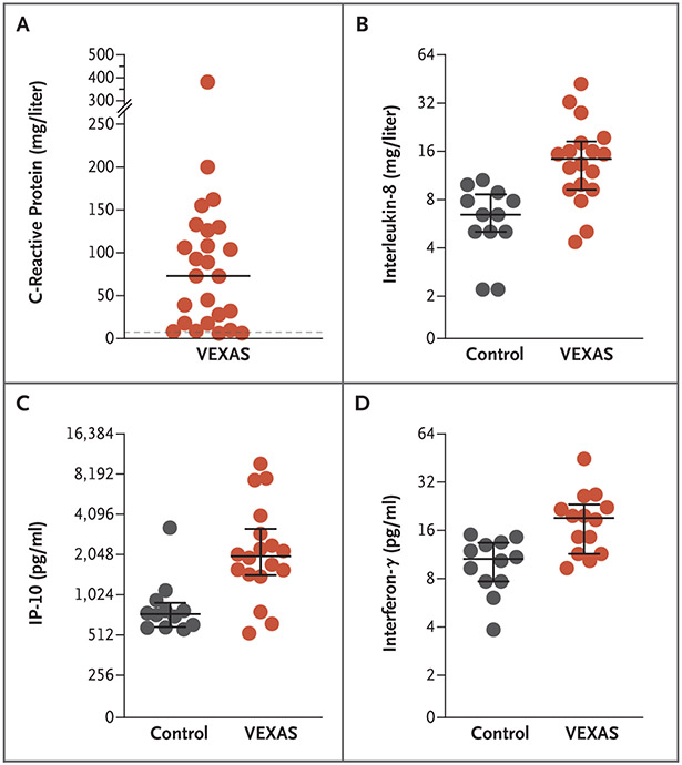
Results: We identified 25 men with somatic mutations affecting methionine-41 (p.Met41) in UBA1, the major E1 enzyme that initiates ubiquitylation. (The gene UBA1 lies on the X chromosome.) In such patients, an often fatal, treatment-refractory inflammatory syndrome develops in late adulthood, with fevers, cytopenias, characteristic vacuoles in myeloid and erythroid precursor cells, dysplastic bone marrow, neutrophilic cutaneous and pulmonary inflammation, chondritis, and vasculitis. Most of these 25 patients met clinical criteria for an inflammatory syndrome (relapsing polychondritis, Sweet's syndrome, polyarteritis nodosa, or giant-cell arteritis) or a hematologic condition (myelodysplastic syndrome or multiple myeloma) or both. Mutations were found in more than half the hematopoietic stem cells, including peripheral-blood myeloid cells but not lymphocytes or fibroblasts. Mutations affecting p.Met41 resulted in loss of the canonical cytoplasmic isoform of UBA1 and in expression of a novel, catalytically impaired isoform initiated at p.Met67. Mutant peripheral-blood cells showed decreased ubiquitylation and activated innate immune pathways. Knockout of the cytoplasmic UBA1 isoform homologue in zebrafish caused systemic inflammation.

Conclusions: Using a genotype-driven approach, we identified a disorder that connects seemingly unrelated adult-onset inflammatory syndromes. We named this disorder the VEXAS (vacuoles, E1 enzyme, X-linked, autoinflammatory, somatic) syndrome. (Funded by the NIH Intramural Research Programs and the EU Horizon 2020 Research and Innovation Program.).


Figures

Figure 1 (facing page).
Figure 1 (facing page).. Identification of Lineage-Restricted UBA1 Somatic Variants in the VEXAS Syndrome.
Panel A shows a schematic representation of a genotype-first screening approach to identify novel disease-causing variants and Sanger-sequencing chromatograms for mosaic variants in Patients 1 through 3 at UBA1 methionine-41 (p.Met41); the black triangles indicate the bases with distinct nucleotides. The term gnomAD denotes Genome Aggregation Database, pLI probability of being loss-of-function intolerant (scores on the pLI range from 0.0 to 1.0, with a higher score indicating a greater degree of intolerance for loss-of-function variants in healthy persons), and VEXAS vacuoles, E1 enzyme, X-linked, autoinflammatory, somatic. Panel B shows a dendrogram of hematopoietic differentiation with overlying Sanger sequencing of sorted bone marrow progenitors and peripheral-blood lineages from a representative case (in Patient 1). UBA1 mosaic variants were enriched in progenitor cells and myeloid lineages and were absent in lymphocytes. GMPs denotes granulocyte–monocyte progenitors, HSCs hematopoietic stem cells, LPs lymphoid progenitors, MEPs megakaryocyte–erythroid progenitors, and MPPs multipotent progenitors. Panel C shows quantification of variant allele fraction with the use of digital droplet polymerase-chain-reaction assay in isolated cell lines including peripheral blood, fibroblasts, HSCs and MPPs, LPs, GMPs, and MEPs sorted from bone marrow–biopsy samples and neutrophils, monocytes, and T cells and B cells sorted from peripheral-blood samples.
Figure 2.
Figure 2.. Clinical Manifestations of the VEXAS Syndrome.
Lung involvement included pulmonary infiltrates and pleural effusions (Panel A), vasculitis of medium-sized bronchial arteries (Panel B), and neutrophilic alveolitis (Panel C). Characteristic vacuoles were present in myeloid precursor cells from bone marrow aspirates (Panel D). Cutaneous manifestations included neutrophilic dermatosis with small- to medium-vessel vasculitis (Panel E) and tender plaques (Panel F). Cartilaginous involvement included auricular chondritis (Panel G) and nasal chondritis (Panel H), which were sometimes associated with periorbital inflammation. Hematoxylin and eosin staining was used in Panels B, C, and E, and Wright–Giemsa staining was used in Panel D.
Figure 3.
Figure 3.. Serum Cytokines in Men with the VEXAS Syndrome.
Cytokine profiling was performed with the use of serum samples obtained from patients with the VEXAS syndrome, and the results were compared with those from controls. Enzyme-linked immunosorbent assay revealed higher inflammatory markers in the patients with the VEXAS syndrome than in the controls. Panel A shows the results for C-reactive protein values in the 25 participants with the VEXAS syndrome (values for the controls are not shown). The dashed line indicates the upper limit of the normal range (<5 mg per deciliter). Panel B shows the results for interleukin-8, with median values of 14.4 mg per liter (interquartile range, 9.2 to 18.5) among 18 samples from 13 participants with the VEXAS syndrome and 6.5 mg per liter (interquartile range, 5.1 to 8.6) among 12 controls. Panel C shows the results for interferon-inducible protein 10 (IP-10), with median values of 1995 pg per milliliter (interquartile range, 1443 to 3201) among 18 samples from 13 participants with the VEXAS syndrome and 734 pg per milliliter (interquartile range, 564 to 893) among 12 controls. Panel D shows the results for interferon-γ, with median values of 19.2 pg per milliliter (interquartile range, 11.4 to 23.3) among 14 samples from 13 participants with the VEXAS syndrome and 10.6 pg per milliliter (7.7 to 13.4) among 12 controls. In Panels B, C, and D, the long horizontal line represents the median value, and shorter bars represent the interquartile range.
Figure 4 (facing page).
Figure 4 (facing page).. Loss of Cytoplasmic UBA1 Function Leading to Inflammation.
Panel A shows a schematic diagram with protein isoforms and domains for UBA1. UBA1a is a long isoform with a nuclear localization sequence (NLS) and a translation start site at p.Met1, and UBA1b is a short isoform without an NLS with a translation start site at p.Met41. AAD denotes active adenylation domain, FCCH first catalytic cysteine half-domain, IAD inactive adenylation domain, SCCH second catalytic cysteine half-domain, Ub ubiquitin, and UFD Ub fold domain. Panel B shows immunoblots of transfected human embryonic kidney (HEK293T) cells. Hemagglutinin (HA)-tagged UBA1 Met41 constructs lead to expression of a novel short isoform, abrogated by the introduction of a Met67Ala mutation. The term αHA denotes anti-HA, and WT wild type. Panel C shows a representative Ub thioester formation assay with the use of purified recombinant UBA1 isoforms. UBA1c shows impaired activity, whereas UBA1a p.Met41Val activity is similar to that in a UBA1a WT control. Ub thioester–charged forms of UBA1 variants are labeled with “~Ub.” The quantification of five replicates is shown below in a bar chart. Thioester formation activity differed significantly between UBA1c and UBA1a WT (adjusted P<0.05). Panel D shows immunoblots of isolated T cells and monocytes from three participants with the VEXAS syndrome and age-matched controls. In the participants with the VEXAS syndrome, T cells are primarily WT and monocytes are primarily mutant. Mutant cells show decreased UBA1b and polyubiquitylation (Poly-Ub) levels, an increased free Ub level, and increased eIF2α phosphorylation (p-EIF2α) and LC3 levels. Glyceraldehyde-3-phosphate dehydrogenase (GAPDH) was used as a loading control. Panel E shows higher levels of inflammatory gene expression in Δuba1 and Δuba1b embryos than in Δuba1a and sibling control heterozygous or WT control embryos (calculated as log10 factor change in the messenger RNA [mRNA] level). A Δ indicates loss of protein, and T bars standard errors. EF1-α denotes elongation factor 1α, ISG15 interferon-stimulated gene 15, and TNF tumor necrosis factor.

Comment in

References

    1. Briggs TA, Rice GI, Daly S, et al. Tartrate-resistant acid phosphatase deficiency causes a bone dysplasia with autoimmunity and a type I interferon expression signature. Nat Genet 2011;43:127–31. - PMC - PubMed
    1. Manthiram K, Zhou Q, Aksentijevich I, Kastner DL. The monogenic autoinflammatory diseases define new pathways in human innate immunity and inflammation. Nat Immunol 2017;18:832–42. - PubMed
    1. Beck DB, Aksentijevich I. Biochemistry of autoinflammatory diseases: catalyzing monogenic disease. Front Immunol 2019;10:101. - PMC - PubMed
    1. Okada Y, Wu D, Trynka G, et al. Genetics of rheumatoid arthritis contributes to biology and drug discovery. Nature 2014; 506:376–81. - PMC - PubMed
    1. Cooper JN, Young NS. Clonality in context: hematopoietic clones in their marrow environment. Blood 2017;130: 2363–72. - PMC - PubMed

Publication types

MeSH terms