. 2021 Jan 4;131(1):e139481.
doi: 10.1172/JCI139481.

Neutrophilic inflammation during lung development disrupts elastin assembly and predisposes adult mice to COPD

Affiliations

Neutrophilic inflammation during lung development disrupts elastin assembly and predisposes adult mice to COPD

John T Benjamin et al. J Clin Invest. .

Abstract

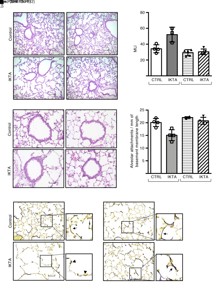
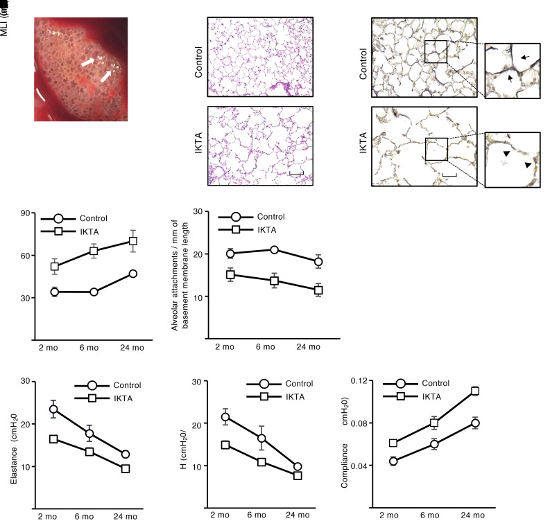
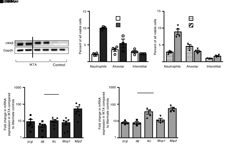
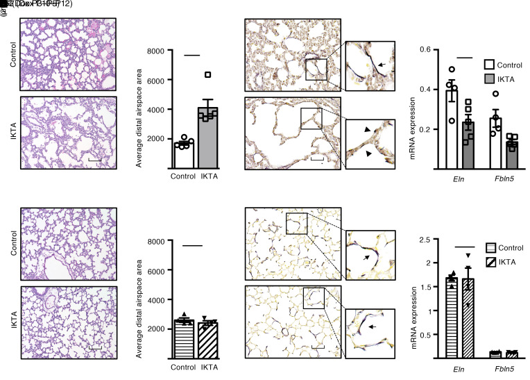
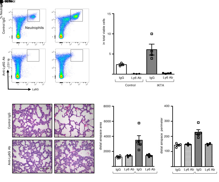
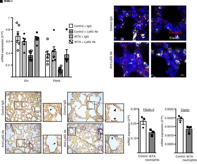
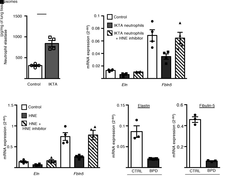
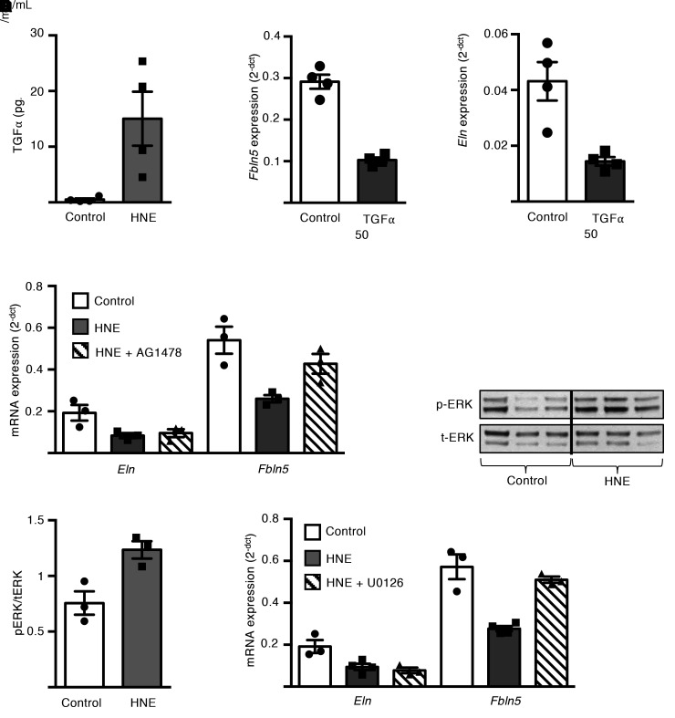
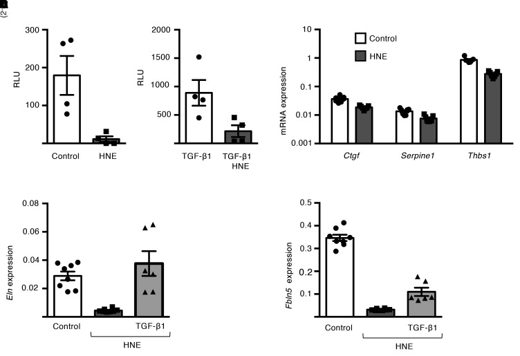
Emerging evidence indicates that early life events can increase the risk for developing chronic obstructive pulmonary disease (COPD). Using an inducible transgenic mouse model for NF-κB activation in the airway epithelium, we found that a brief period of inflammation during the saccular stage (P3-P5) but not alveolar stage (P10-P12) of lung development disrupted elastic fiber assembly, resulting in permanent reduction in lung function and development of a COPD-like lung phenotype that progressed through 24 months of age. Neutrophil depletion prevented disruption of elastic fiber assembly and restored normal lung development. Mechanistic studies uncovered a role for neutrophil elastase (NE) in downregulating expression of critical elastic fiber assembly components, particularly fibulin-5 and elastin. Further, purified human NE and NE-containing exosomes from tracheal aspirates of premature infants with lung inflammation downregulated elastin and fibulin-5 expression by saccular-stage mouse lung fibroblasts. Together, our studies define a critical developmental window for assembling the elastin scaffold in the distal lung, which is required to support lung structure and function throughout the lifespan. Although neutrophils play a well-recognized role in COPD development in adults, neutrophilic inflammation may also contribute to early-life predisposition to COPD.

Keywords: COPD; Extracellular matrix; Inflammation; Neutrophils; Pulmonology.


Conflict of interest statement

Conflict of interest: The authors have declared that no conflict of interest exists.

Figures

Figure 1
Figure 1. Epithelial NF-κB activation in saccular-stage lungs results in emphysema, loss of alveolar attachments, and disruption of elastic fiber organization.
Dams were treated with Dox from P3 to P5 (saccular stage) or P10 to P12 (alveolar stage), after which lungs from IKTA and littermate control mice were harvested at 2 months of age. (A) Representative H&E staining and (B) morphometric evaluation of emphysema as measured by mean linear intercept (MLI). Data are expressed as mean SEM, n = 3–4 lungs per group, 5–6 imaged lung fields per lung. *P < 0.05 by 1-way ANOVA and post hoc Tukey test. (C) Representative H&E-stained sections showing small airways (arrows denote alveolar attachments) and (D) alveolar attachment counts in control and IKTA lungs. Data are expressed as mean ± SEM, n = 3 lungs per group, P < 0.05 by 1-way ANOVA and post hoc Tukey test. (E) Representative photomicrographs of Hart’s elastin-stained lung sections. Arrows denote elastic fibers around airspaces. Arrowhead denotes fragmented elastic fibers. Scale bar: 100 μm in A, 50 μm in C and E.
Figure 2
Figure 2. Epithelial NF-κB activation in saccular-stage lungs results in lifelong abnormalities in lung structure and function.
Dams were treated with Dox from P3 to P5 (saccular stage), after which lungs from IKTA and littermate control mice were harvested at 2, 6, or 24 months. (A) Representative image showing gross appearance of a 24-month IKTA lung. Arrows denote areas of emphysema. (B and C) Representative images showing H&E-stained (B) and Hart’s elastin–stained (C) lung sections from 24-month control and IKTA lungs. Arrows denote intact elastic fibers. Arrowhead denotes fragments of elastic fibers. (D and E) Quantification of mean linear intercept (MLI) (D) and alveolar attachments (E) in control and IKTA lungs at 2, 6, and 24 months. Data are expressed as mean ± SEM, n = 3–4 lungs per group, 6 imaged lung fields per lung *P < 0.05 by 2-tailed Student’s t test comparing IKTA to littermate controls at each age. (FH) Lung function, including total respiratory system elastance (F), tissue elastance (G), and dynamic compliance (H), was measured at 2, 6, and 24 months in control and IKTA mice. Data are expressed as mean ± SEM, n = 4–6 mice per group, *P < 0.05 by 2-tailed Student’s t test comparing IKTA to littermate controls at each age, #P < 0.05 by 1-way ANOVA and post hoc linear trend test for each group (control and IKTA). Scale bar: 100 μm in B, 50 μm in C.
Figure 3
Figure 3. Transgene activation results in neutrophilic inflammation during both the saccular and alveolar stages of lung development.
Dams were treated with Dox from P3 to P5 (saccular stage) or P10 to P12 (alveolar stage), after which lungs from IKTA and littermate control mice were harvested at P5 or P12. (A) Expression of constitutively active IKK-β (cIkk-β) transgene and Gapdh was evaluated by PCR followed by agarose gel electrophoresis. (B and C) Quantification of immune cells in P5 (B) and P12 (C) control and IKTA lungs by flow cytometry. Data are expressed as mean ± SEM, n = 3–4 per group. *P < 0.05 by 2-tailed Student’s t test. (D and E) mRNA expression of select inflammatory cytokines in P5 (D) and P12 (E) control and IKTA lungs. Data are expressed as mean ± SEM, n = 4–6 per group.
Figure 4
Figure 4. Neutrophilic inflammation disrupts saccular-stage (but not alveolar-stage) elastic fiber assembly and results in a BPD-like phenotype.
Dams were treated with Dox from P3 to P5 (saccular stage) or P10 to P12 (alveolar stage), after which lungs from IKTA and littermate control mice were harvested at P5 or P12. (A, B, E, and F) Representative H&E-stained lung sections (A and E) and morphometric measurements (B and F) demonstrated dilated terminal saccules in P5 IKTA lungs, whereas P12 IKTA lungs appeared similar to controls. Data are expressed as mean ± SEM, n = 4–5 lungs per group, 5–6 imaged lung fields per lung, *P < 0.05 by 2-tailed Student’s t test. (C and G) Representative photomicrographs of Hart’s elastin–stained sections of P5 (C) and P12 (G) control and IKTA lungs demonstrating disorganized and fragmented elastic fibers in P5 IKTA lungs. Arrows denote normal elastic fibers in controls. Arrowhead denotes fragmented elastic fibers in IKTA lungs. (D and H) mRNA expression of elastic fiber assembly components Eln and Fbln5 expression in P5 (D) and P12 (H) IKTA lungs. Data are expressed as mean ± SEM, n = 4–5 per group, *P < 0.05 by 2-tailed Student’s t test. Scale bar: 100 μm in A and E, 50 μm in C and G.
Figure 5
Figure 5. Neutrophil depletion rescues lung development in saccular-stage lungs with NF-κB activation.
Control and IKTA pups were administered anti-ly6G antibodies or control IgG by i.p. injection on P2 prior to placing dams on Dox from P3 to P5 for transgene activation. (A and B) Representative flow plots (A) and quantification of CD11b+ Ly6G+ neutrophils (B) showing reduction in neutrophils with anti-ly6G antibody treatment. Data are expressed as mean ± SEM, n = 3–4 lungs per group, *P < 0.05 by 1-way ANOVA and post hoc Tukey test. (CE) Representative H&E-stained lung sections (C), morphometric evaluation of average distal airspace area (D), and average distal airspace perimeter (E) in P5 lungs. Data are expressed as mean ± SEM, n = 4 lungs per group, 4–6 imaged fields per lung (D and E). *P < 0.05 by 1-way ANOVA and post hoc Tukey test. Scale bar: 100 μm in C.
Figure 6
Figure 6. Neutrophil depletion prevents disruption of elastic fibers in saccular-stage IKTA lungs.
Control and IKTA pups were administered anti-ly6G antibodies or control IgG IP on P2 prior to placing dams on Dox from P3 to P5 for transgene activation. (A) Expression of Eln and Fbln5 in P5 lungs. Data are expressed as mean ± SEM, n = 6 per group, *P < 0.05 by 1-way ANOVA and post hoc Tukey test. (B) Representative photomicrographs of RNAscope-labeled sections from P5 lungs. Fbln5- and Eln-expressing cells were reduced in IKTA lungs with control IgG treatment, but Fbln5 and Eln expression appeared similar to controls in IKTA mice with anti-Ly6G antibody treatment. (C) Representative Hart’s elastin–stained sections from P5 lungs showing normal cord-like structures around airspaces in IKTA mice treated with anti-Ly6G antibodies. Arrows denote elastic fibers around airspaces. Arrowhead denotes fragmented elastic fibers. (D and E) CD11b+ Ly6G+ neutrophils were isolated from IKTA lungs and cocultured with wild-type saccular-stage lung fibroblast for 4 hours. Expression of Fbln5 (D) and Eln (E) was reduced in saccular-stage lung fibroblasts cocultured with IKTA neutrophils. Data are expressed as mean ± SEM, n = 3 per group. Data are representative of 3 independent experiments. (D and E), *P < 0.05 by 2-tailed Student’s t test. Scale bar: 10 μm in B, 50 μm in C.
Figure 7
Figure 7. Neutrophil elastase inhibits expression of elastic fiber assembly components by saccular-stage mouse lung fibroblasts.
(A) Quantification of neutrophil elastase (NE) by ELISA in P5 control and IKTA lung homogenates. Data are expressed as mean ± SEM, n = 4–6 per group, *P < 0.05 by 2-tailed Student’s t test. (B and C) P5 mouse lung fibroblasts were cultured with or without IKTA neutrophils for 4 hours (B) or purified human neutrophil elastase (HNE) 1 μg/mL for 24 hours (C) and Eln and Fbln5 expression was quantified. Where indicated, cells were also pretreated with 10 μM N-methoxysuccinyl-Ala-Ala-Pro-Val-chloromethyl ketone (HNE inhibitor). Data expressed as mean ± SEM, n = 4 per group. Data are representative of 4 independent experiments. *P < 0.05 compared with control by 1-way ANOVA and post hoc Tukey test. (D and E) Saccular-stage mouse lung fibroblasts were cultured with tracheal aspirate exosomes (109 exosomes/mL) isolated from preterm infants with severe BPD or from term-born control infants (CTRL) and Eln (D) and Fbln5 (E) expression was quantified. Data are expressed as mean ± SEM, n = 3 per group, *P < 0.05 by 2-tailed Student’s t test.
Figure 8
Figure 8. Neutrophil elastase downregulates mRNA expression of fibulin-5 in saccular-stage mouse lung fibroblasts through EGFR/MEK/ERK signaling.
P5 mouse lung fibroblasts were grown to confluence and cultured with or without purified human neutrophil elastase (HNE) at 1 or 4 μg/mL or TGF-α (50 ng/mL) for up to 24 hours. In some experiments, fibroblasts were treated with AG1478 20 μM (EGFR inhibitor) or U0126 25 μM (MEK inhibitor) prior to HNE treatment. (A) TGF-α ELISA from conditioned media samples after 15 minutes of HNE treatment of saccular stage lung fibroblasts. Data are expressed as mean ± SEM, n = 4 per group. (B and C) Quantification of Fbln5 (B) and Eln (C) expression by qPCR in saccular-stage fibroblasts 24 hours after treatment with TGF-α. Data are expressed as mean ± SEM, n = 4 per group. (D) Expression of Eln and Fbln5 in fibroblasts treated with HNE ± AG1478 20 μM for 24 hours. Data are expressed as mean ± SEM, n = 3–4 per group. (E and F) Western blot analysis of phospho-Erk1/2 and total-Erk1/2 in cell lysates from fibroblasts treated with HNE for 15 minutes. Data are expressed as mean ± SEM, n = 3 per group. (G) Expression of Eln and Fbln5 in fibroblasts treated with HNE ± U0126 25 μM for 24 hours. Data are expressed as mean ± SEM, n = 3–4 per group. Data are representative of 3 independent experiments. *P < 0.05 compared with control by 2-tailed student’s t test (A, B, C, and F); *P < 0.05 compared with control by 1-way ANOVA and post hoc Tukey test (D and G).
Figure 9
Figure 9. Neutrophil elastase downregulates TGF-β signaling and reduces elastin expression in saccular-stage lung fibroblasts.
(A and B) P5 mouse lung fibroblasts were transfected with a Smad LUC reporter (SBE4-LUC) and treated with or without HNE (1 μg/mL) and/or recombinant TGF-β1 (0.1 ng/mL) for 12 hours. Luminescence measurements are reported as relative light units (RLU) above background (untransfected control) in (A) control cells and (B) TGF-β1–treated cells. Data are expressed as mean ± SEM, n = 4 per group. *P < 0.05 by 2-tailed Student’s t test. (CE) Mouse lung fibroblasts were cultured with or without HNE (1 μg/mL) and TGF-β1(0.1 ng/mL) for 24 hours. (C) mRNA expression of Ctgf, Serpine1, and Thbs1 in fibroblasts treated with or without HNE. Data are from 3 independent experiments and expressed as mean ± SEM, n = 7 per group. *P < 0.05 by 2-tailed Student’s t test. (D and E) mRNA expression of Eln (D) and Fbln5 (E) in saccular-stage fibroblasts treated with HNE ± recombinant TGF-β1. Data are from 3 independent experiments and expressed as mean ± SEM, n = 6–8 per group, *P < 0.05 compared with control by 1-way ANOVA and post hoc Tukey test.

References

    1. Lange P, et al. Lung-function trajectories leading to chronic obstructive pulmonary disease. N Engl J Med. 2015;373(2):111–122. doi: 10.1056/NEJMoa1411532. - DOI - PubMed
    1. Martinez FD. Early-life origins of chronic obstructive pulmonary disease. N Engl J Med. 2016;375(9):871–878. doi: 10.1056/NEJMra1603287. - DOI - PubMed
    1. Postma DS, Bush A, van den Berge M. Risk factors and early origins of chronic obstructive pulmonary disease. Lancet. 2015;385(9971):899–909. doi: 10.1016/S0140-6736(14)60446-3. - DOI - PubMed
    1. Islam JY, Keller RL, Aschner JL, Hartert TV, Moore PE. Understanding the short- and long-term respiratory outcomes of prematurity and bronchopulmonary dysplasia. Am J Respir Crit Care Med. 2015;192(2):134–156. doi: 10.1164/rccm.201412-2142PP. - DOI - PMC - PubMed
    1. Doyle LW, et al. Increasing airway obstruction from 8 to 18 years in extremely preterm/low-birthweight survivors born in the surfactant era. Thorax. 2017;72(8):712–719. doi: 10.1136/thoraxjnl-2016-208524. - DOI - PubMed

Publication types

MeSH terms