. 2020 Dec;7(12):2342-2355.
doi: 10.1002/acn3.51183. Epub 2020 Oct 27.

Tau pathology associates with in vivo cortical thinning in Lewy body disorders

Affiliations

Tau pathology associates with in vivo cortical thinning in Lewy body disorders

Nicola Spotorno et al. Ann Clin Transl Neurol. 2020 Dec.

Abstract

Objectives: To investigate the impact of Alzheimer's disease (AD) co-pathology on an in vivo structural measure of neurodegeneration in Lewy body disorders (LBD).

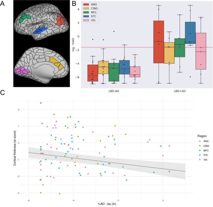
Methods: We studied 72 LBD patients (Parkinson disease (PD) = 2, PD-MCI = 25, PD with dementia = 10, dementia with Lewy bodies = 35) with either CSF analysis or neuropathological examination and structural MRI during life. The cohort was divided into those harboring significant AD co-pathology, either at autopsy (intermediate/high AD neuropathologic change) or with CSF signature indicating AD co-pathology (t-tau/Aβ1-42 > 0.3) (LBD+AD, N = 19), and those without AD co-pathology (LBD-AD, N = 53). We also included a reference group of 25 patients with CSF biomarker-confirmed amnestic AD. We investigated differences in MRI cortical thickness estimates between groups, and in the 21 autopsied LBD patients (LBD-AD = 14, LBD+AD = 7), directly tested the association between antemortem MRI and post-mortem burdens of tau, Aβ, and alpha-synuclein using digital histopathology in five representative neocortical regions.

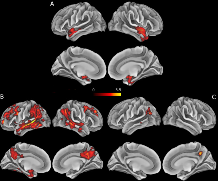
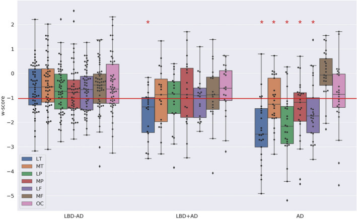
Results: The LBD+AD group was characterized by cortical thinning in anterior/medial and lateral temporal regions (P < 0.05 FWE-corrected) relative to LBD-AD. In LBD+AD, cortical thinning was most pronounced in temporal neocortex, whereas the AD reference group showed atrophy that equally encompassed temporal, parietal and frontal neocortex. In autopsied LBD, we found an inverse correlation with cortical thickness and post-mortem tau pathology, while cortical thickness was not significantly associated with Aβ or alpha-synuclein pathology.

Interpretation: LBD+AD is characterized by temporal neocortical thinning on MRI, and cortical thinning directly correlated with post-mortem histopathologic burden of tau, suggesting that tau pathology influences the pattern of neurodegeneration in LBD.


Conflict of interest statement

The authors report no competing interests.

Figures

Figure 1
Figure 1
Results of the voxel‐wise analysis directly comparing patient groups. 1. Clusters display the t‐values for the significant results (P < 0.05 FWE) of the between‐group voxel‐wise analyses. Age, sex, and imaging protocol were included in every model as nuisance covariates. (A) contrast LBD+AD < LBD−AD. (B) contrast AD < LBD−AD. (C) contrast AD < LBD+AD. For visualization purposes, the results have been overlapped to an inflated mesh of the MNI space (Montreal neurological institute template) ICBM‐152 using the Connectome Workbench (v.1.3; https://github.com/Washington‐University/workbench).
Figure 2
Figure 2
W‐score of macro‐ROIs across patients groups. Abbreviations: LT, lateral temporal ROI; MT, ventro‐medial temporal ROI; LP, lateral parietal ROI; MT, medial parietal ROI; LF, lateral frontal ROI; MF, medial frontal ROI; OC, occipital ROI. The plot represent the w‐score for each macro‐ROI in each group of patients. Each box extends from lower to upper quartile values, with line at median. Whiskers extend from box to show the range of the data. * = region in which more of the 50% of the cases lie below the threshold of w‐score = −1. Following the cut‐off of −1 (red line) for pathological thinning, it appeared that in the AD group more than 50% of the participants lay below this cut‐off in all regions except the medial frontal and the occipital ROIs while in the LBD+AD group 50% of the participants lay below the −1 cut‐off only in the lateral temporal ROI [lateral temporal vs. ventro‐medial temporal: x 2 = 4.07, P < 0.05; lateral temporal vs. lateral parietal: x 2 = 4.07, P < 0.05; lateral temporal vs. medial parietal: x 2 = 5.40, P < 0.05; lateral temporal vs. lateral frontal: x 2 = 5.40, P < 0.05; lateral temporal vs. medial frontal: x2 = 5.40, P < 0.05; lateral temporal vs. medial parietal: x 2 = 8.61, P < 0.01]. In no region did more of the 50% of LBD−AD participant lay below the cut‐off.
Figure 3
Figure 3
Association between the in vivo estimates of cortical thinning and tau accumulation. Abbreviations: %AO, percentage of area occupied; ANG, angular gyrus; CING, anterior cingulate cortex; MFC, medial frontal cortex; STC, superior temporal cortex; VIS, visual cortex. (A) representation of the MRI‐ROIs on an inflated brain (Green = MFC, Blue = STC, Red = ANG, Yellow = CING and Pink = VIS). (B) Box‐plots depict the ln %AO distribution of tau pathology across the sampled regions. (C) The scatter plot represents the w‐score of median cortical thickness values plotted as a function of the percentage of area occupied of reactivity for tau in the tissue sample (log transformed). The regression line represents the fixed effect of the %AO and the gray area around the regression line covers the standard error around the regression estimate.

References

    1. Irwin DJ, Lee VM‐Y, Trojanowski JQ. Parkinson’s disease dementia: convergence of α‐synuclein, tau and amyloid‐β pathologies. Nat Rev Neurosci 2013;14:626–636. - PMC - PubMed
    1. Toledo JB, Gopal P, Raible K, et al. Pathological α‐synuclein distribution in subjects with coincident Alzheimer’s and Lewy body pathology. Acta Neuropathol 2016;131:393–409. - PMC - PubMed
    1. Graff‐Radford J, Lesnick TG, Boeve BF, et al. Predicting survival in dementia with Lewy bodies with hippocampal volumetry. Mov Disord 2016;31:989–994. - PMC - PubMed
    1. Kantarci K, Boeve BF, Weigand SD, et al. Focal atrophy on MRI and neuropathologic classification of dementia with Lewy bodies. Neurology 2012;79:553–560. - PMC - PubMed
    1. Irwin DJ, Grossman M, Weintraub D, et al. Neuropathological and genetic correlates of survival and dementia onset in synucleinopathies: a retrospective analysis. Lancet Neurol 2017;16:55. - PMC - PubMed

Publication types

MeSH terms