. 2020 Dec 9;108(5):876-886.e4.
doi: 10.1016/j.neuron.2020.09.016. Epub 2020 Oct 26.

Robust Myelination of Regenerated Axons Induced by Combined Manipulations of GPR17 and Microglia

Affiliations

Robust Myelination of Regenerated Axons Induced by Combined Manipulations of GPR17 and Microglia

Jing Wang et al. Neuron. .

Abstract

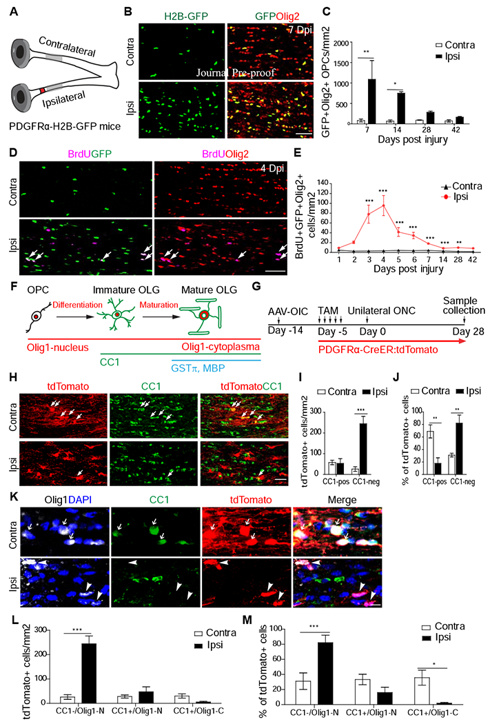
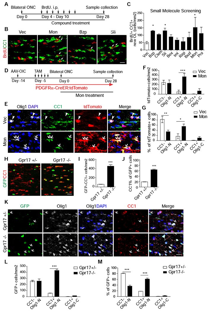
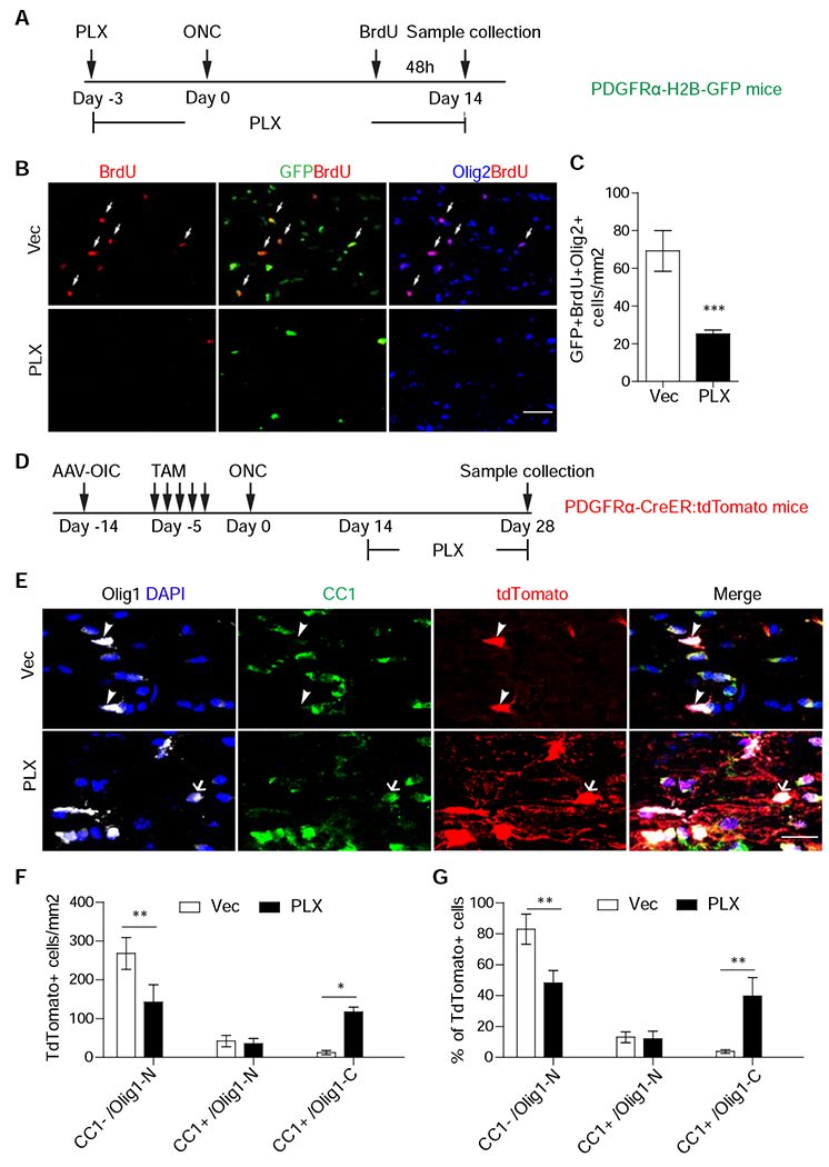
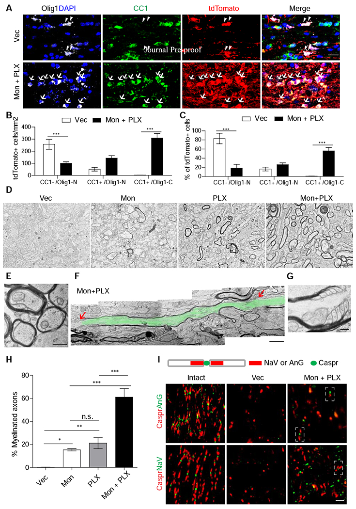
Myelination facilitates rapid axonal conduction, enabling efficient communication across different parts of the nervous system. Here we examined mechanisms controlling myelination after injury and during axon regeneration in the central nervous system (CNS). Previously, we discovered multiple molecular pathways and strategies that could promote robust axon regrowth after optic nerve injury. However, regenerated axons remain unmyelinated, and the underlying mechanisms are elusive. In this study, we found that, in injured optic nerves, oligodendrocyte precursor cells (OPCs) undergo transient proliferation but fail to differentiate into mature myelination-competent oligodendrocytes, reminiscent of what is observed in human progressive multiple sclerosis. Mechanistically, we showed that OPC-intrinsic GPR17 signaling and sustained activation of microglia inhibit different stages of OPC differentiation. Importantly, co-manipulation of GPR17 and microglia led to extensive myelination of regenerated axons. The regulatory mechanisms of stage-dependent OPC differentiation uncovered here suggest a translatable strategy for efficient de novo myelination after CNS injury.

Keywords: GPR17; axon regeneration; microglia; myelination; oligodendrocyte precursor cells.


Conflict of interest statement

Declaration of Interests A patent based on the results in this manuscript was filed by Boston Children’s Hospital (Z.H., J.W., and X.H. are co-inventors).

Figures

Figure 1.
Figure 1.. Increased proliferation and failed differentiation of OPCs in injured optic nerves.
(A) Scheme for experiments in Figure 1A-C. The red spot indicates crush injury site and the gray region indicates the regions that were analyzed for this study. (B-C) Images and quantification of OPC numbers in injured optic nerves at different time points after injury. N = 3-8 mice per group. (D-E) Images and quantification of BrdU+/Olig2+ cells in injured optic nerves. (F) Illustration of the differentiation stages of OPCs and their respective markers. (G) Scheme for experiments in Figure 1H-M. AAV-OIC: AAVs expressing osteopontin/IGF1/CNTF1. TAM: tamoxifen. ONC: optic nerve crush. (H-J) Images (H) and quantitation (cell number in I and proportion in J) of CC1+ and tdTomato+ cells. N = 6 mice per group. (K-M) Images (K) and quantitation of three different populations. In K, arrows in the contralateral side indicate tdTomato+/CC1+/Olig1-C, arrowheads in ipsilateral side indicate tdTomato+/CC1−/Olig1-N (un-differentiated cells). N = 6 mice per group. Scale bar: 100 μm (B, D), 50 μm (H), 10 μm (K). *, **, *** P < 0.05, 0.01, 0.001, respectively. Olig1-N: nuclear Olig1; Olig1-C: cytoplasmic Olig1.
Figure 2.
Figure 2.. GPR17 is an intrinsic blocker of early oligodendrocyte differentiation of OPCs in injured optic nerves.
(A) Scheme of compound screening. (B, C) Images of injured optic nerves stained with anti-CC1 and BrdU (B) and quantification (C). N = 4-13 mice per group. Vec (Vehicle). Mon (Montelukast), Bzp (Benztropine Mesylate), and Sli (Solifenacin succinate). (D) Design for experiments in Figure 2E-G. N = 6 mice per group. AAV-OIC: AAVs expressing osteopontin/IGF1/CNTF1. TAM: tamoxifen. ONC: optic nerve crush. (E-G) Images of injured or intact optic nerves stained with antibodies against Olig1, CC1, tdTomato, and DAPI (E) and quantification results of the densities (F) or proportions (G) of different populations. In E, arrowheads indicate CC1−/ Olig1-N cells in Vec, while arrows indicate CC1+/Olig1-N cells after treatment. (H-J) Images of injured optic nerves at 28 days after injury (H) and quantification results of the densities of GFP+/CC1+ cells (I) or proportion of CC1+ among GFP+ cells (J). (K-M) Images (K) of injured optic nerves stained with indicated antibodies and BrdU and quantification results of the densities (L) or proportions (M) of different populations. N = 6 mice per group. Scale bar: 20 μm (B, H), 10 μm (E), 20 μm (K). *, **, *** P < 0.05, 0.01, 0.001, respectively. Olig1-N: nuclear Olig1; Olig1-C: cytoplasmic Olig1.
Figure 3.
Figure 3.. Microglia are required for OPC proliferation but detrimental for their maturation.
(A) Scheme for experiments in Figure 3B, C. (B, C). Images of injured optic nerves stained with GFP, Olig2, or BrdU (B) and quantification of the densities of GFP+/Olig2+/BrdU+ cells (C). N = 6 mice per group. (D) Scheme for experiments in Figure 3E-G. (E-G) Images of injured optic nerves stained with indicated antibodies or DAPI (E) and quantification results of the densities (F) or proportions (G) of different populations. E, arrowheads indicate CC1−/Olig1-N cells in Vec, while arrows indicate CC1+/Olig1-C cells after PLX treatment. N = 6 mice per group. Scale bar: 100 μm (B), 25 μm (E). *, **, *** P < 0.05, 0.01, 0.001, respectively.
Figure 4.
Figure 4.. Combinatorial treatment of Montelukast and PLX3397 lead to robust myelination of regenerated axons in injured optic nerves.
(A-C) Images of injured optic nerves stained with antibodies against Olig1, CC1, tdTomato, and DAPI (A) and quantification results of the densities (B) or proportions (C) of different populations. Arrowheads: CC1−/Olig1-N cells; arrows: CC1+/Olig1-N cells. N = 6 mice per group. (D-H) Transmission electron microscopic images (D-G) and quantification (H) of myelination of regenerated axons of injured optic nerves from the mice with the treatment of Montelukast and/or PLX3397. (D, H): Low magnification of coronal sections (D) and quantification (H) of different groups. n=4 for each group. An enlarged image (E) showing ongoing myelination, an image montage showing a complete internode highlighted in green color (F), and an enlarged image of half of nodes of Ranvier (G) from the mice with combined treatments. (I) Images of injured optic nerves with the combined treatments stained with nodes of Ranvier markers. Scale bar: 20 μm (A), 2 μm (D), 500 μm (E), 1400 nm (F), 3.5 μm (I). *, **, *** P < 0.05, 0.01, 0.001, respectively.

Comment in

References

    1. Abiraman K, Pol SU, O’Bara MA, Chen G. Di Khaku ZM, Wang J, Thorn D, Vedia BH, Ekwegbalu EC, Li JX, et al. (2015). Anti-muscarinic adjunct therapy accelerates functional human oligodendrocyte repair. J. Neurosci 35, 3676–3688. - PMC - PubMed
    1. Arenkiel BR, Hasegawa H, Yi JJ, Larsen RS, Wallace ML, Philpot BD, Wang F, and Ehlers MD (2011). Activity-induced remodeling of olfactory bulb microcircuits revealed by monosynaptic tracing. PLoS One 6. - PMC - PubMed
    1. Arnett HA, Fancy SPJ, Alberta JA, Zhao C, Plant SR, Kaing S, Raine CS, Rowitch DH, Franklin RJM, and Stiles CD (2004). bHLH transcription factor Oligl is required to repair demyelinated lesions in the CNS. Science 306, 2111–2115. - PubMed
    1. Bei F, Lee HHC, Liu X, Gunner G, Jin H, Ma L, Wang C, Hou L, Hensch TK, Frank E, et al. (2016). Restoration of Visual Function by Enhancing Conduction in Regenerated Axons. Cell 164, 219–232. - PMC - PubMed
    1. Benowitz LI, He Z, Goldberg JL. (2017). Reaching the brain: Advances in optic nerve regeneration. 287, 365–373. - PubMed

Publication types

MeSH terms

Substances