. 2020 Oct 27;20(1):324.
doi: 10.1186/s12866-020-01964-8.

The role of the msaABCR operon in implant-associated chronic osteomyelitis in Staphylococcus aureus USA300 LAC

Affiliations

The role of the msaABCR operon in implant-associated chronic osteomyelitis in Staphylococcus aureus USA300 LAC

Gyan S Sahukhal et al. BMC Microbiol. .

Abstract

Background: The msaABCR operon regulates several staphylococcal phenotypes such as biofilm formation, capsule production, protease production, pigmentation, antibiotic resistance, and persister cells formation. The msaABCR operon is required for maintaining the cell wall integrity via affecting peptidoglycan cross-linking. The msaABCR operon also plays a role in oxidative stress defense mechanism, which is required to facilitate persistent and recurrent staphylococcal infections. Staphylococcus aureus is the most frequent cause of chronic implant-associated osteomyelitis (OM). The CA-MRSA USA300 strains are predominant in the United States and cause severe infections, including bone and joint infections.

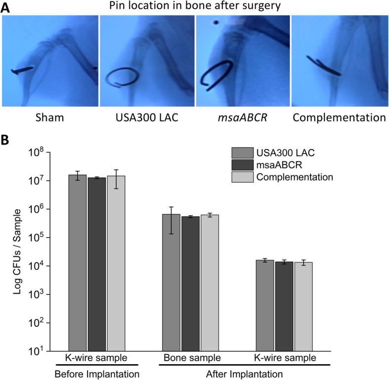
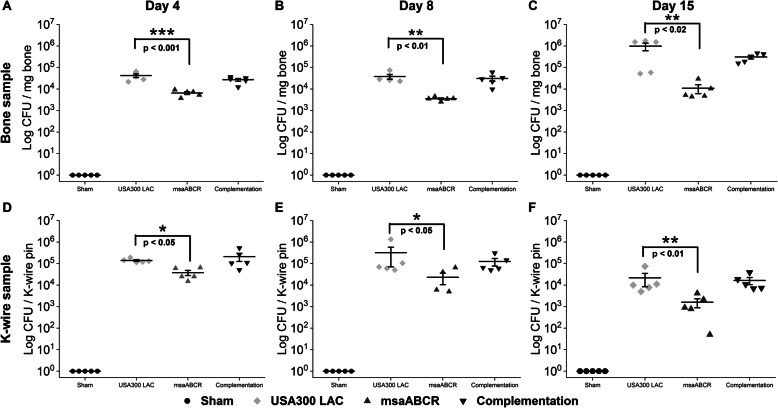
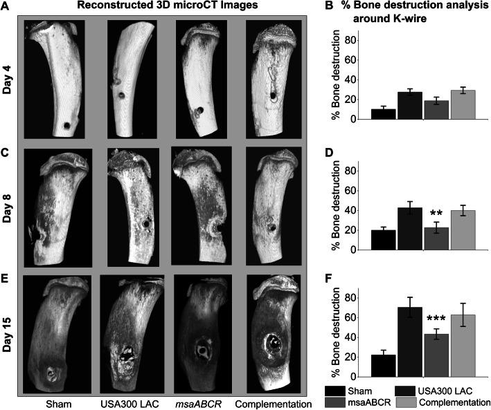
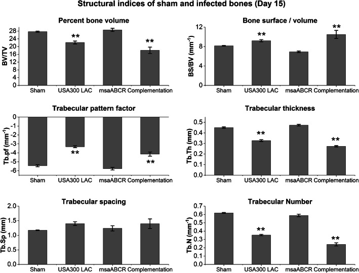
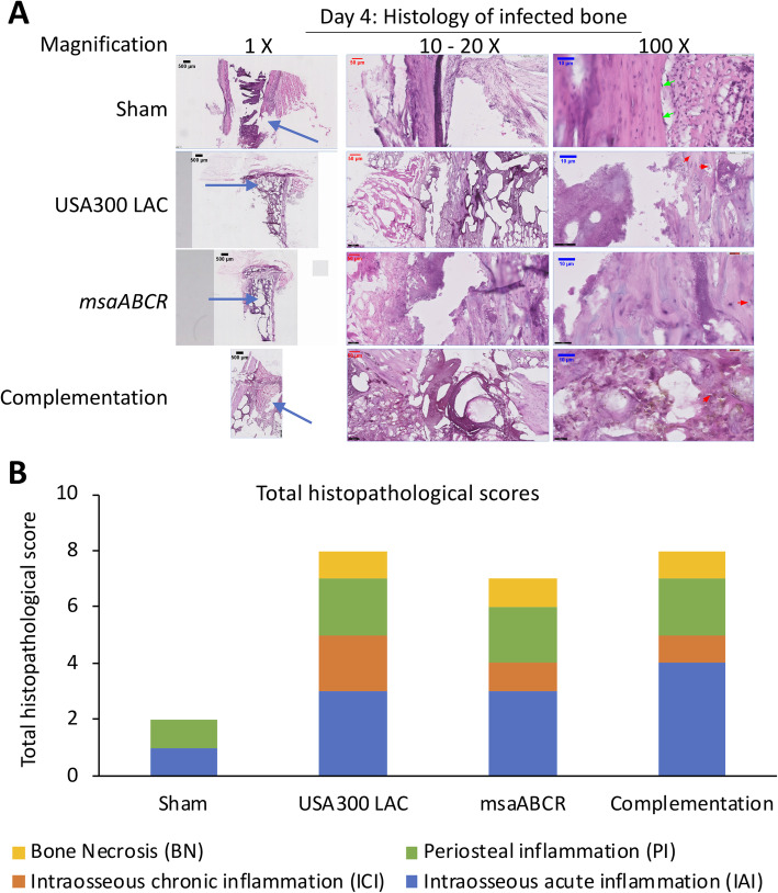
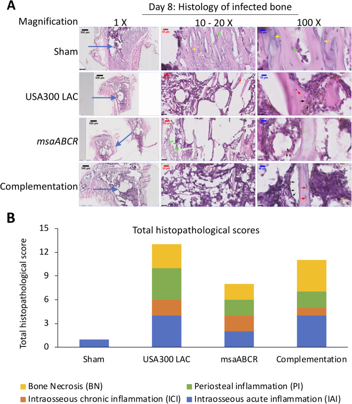
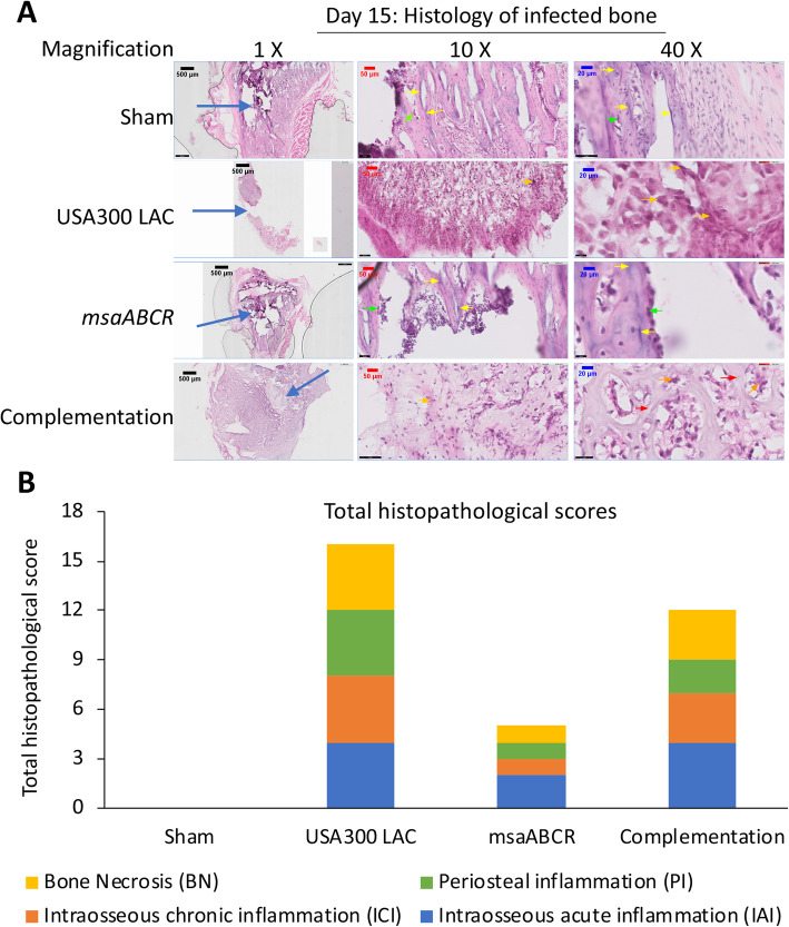
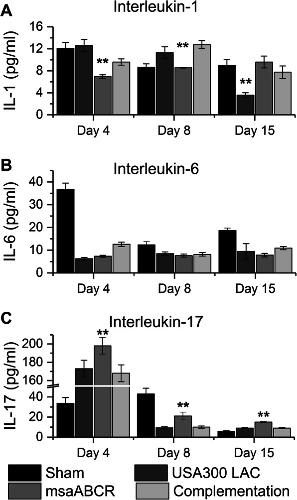
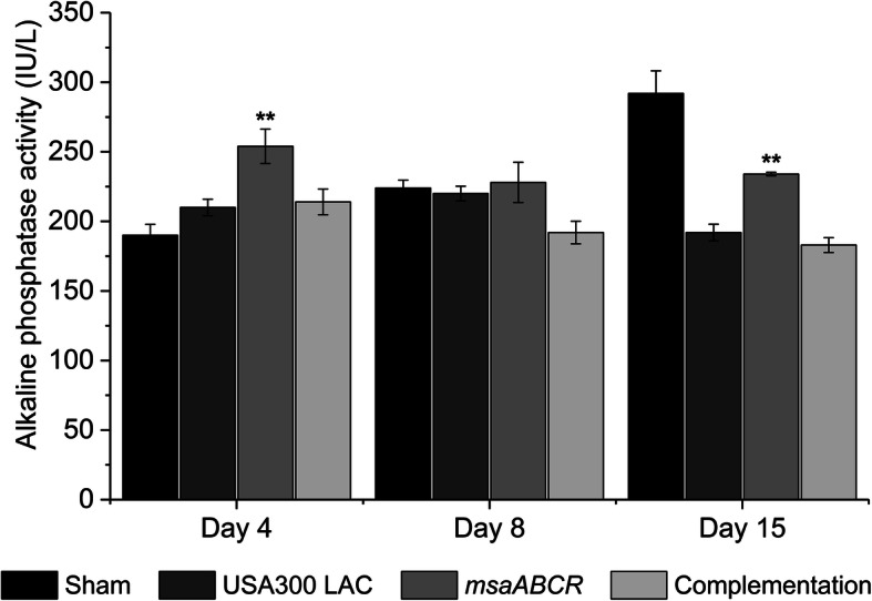
Results: The USA300 LAC strain caused significant bone damage, as evidenced by the presence of severe bone necrosis with multiple foci of sequestra and large numbers of multinucleated osteoclasts. Intraosseous survival and biofilm formation on the K-wires by USA300 LAC strains was pronounced. However, the msaABCR deletion mutant was attenuated. We observed minimal bone necrosis, with no evidence of intramedullary abscess and/or fibrosis, along reduced intraosseous bacterial population and significantly less biofilm formation on the K-wires by the msaABCR mutant. microCT analysis of infected bone showed significant bone loss and damage in the USA300 LAC and complemented strain, whereas the msaABCR mutant's effect was reduced. In addition, we observed increased osteoblasts response and new bone formation around the K-wires in the bone infected by the msaABCR mutant. Whole-cell proteomics analysis of msaABCR mutant cells showed significant downregulation of proteins, cell adhesion factors, and virulence factors that interact with osteoblasts and are associated with chronic OM caused by S. aureus.

Conclusion: This study showed that deletion of msaABCR operon in USA300 LAC strain lead to defective biofilm in K-wire implants, decreased intraosseous survival, and reduced cortical bone destruction. Thus, msaABCR plays a role in implant-associated chronic osteomyelitis by regulating extracellular proteases, cell adhesions factors and virulence factors. However additional studies are required to further define the contribution of msaABCR-regulated molecules in osteomyelitis pathogenesis.

Keywords: K-wire implants; Osteomyelitis; Staphylococcus aureus; Virulence factors; msaABCR operon.


Conflict of interest statement

The authors declare that they have no competing interests.

Figures

Fig. 1
Fig. 1
Examination of K-wire localization and microbial load in bone and K-wires after surgery. The K-wire was inserted in the tibia of Sprague Dawley rats surgically and examined by X-ray to assure its positioning (a). The microbial load before K-wire implantation and after surgery (b). The microbial load was estimated for both K-wires and bone (n = 3) to ensure an equal starting microbial load (USA300 LAC, msaABCR mutant, and complementation strains) prior to the infection process. Error bars indicate standard errors of the means. The differences in the numbers of microbial CFUs between the test strains were not statistically significant when analyzed by one-way ANOVA followed a post-hoc Tukey test
Fig. 2
Fig. 2
Microbial count from bone and K-wires samples. Microbial enumeration from post-mortem bone samples (a-c) and K-wire samples (n = 5) were obtained to determine intraosseous bacterial survivability and biofilm formation on the K-wires (d-f) on days 4, 8, and 15 post-infection (left to right). The infected bone sample and K-wires were harvested after each infection period (days 4, 8, and 15). Error bars indicate standard errors of the means. The bacterial CFUs difference between the USA300 LAC and msaABCR mutant strains was analyzed using one-way ANOVA followed by a post-hoc Tukey test. A P-value of < 0.05 was considered statistically significant (*p-value < 0.05, **p-value < 0.01, and ***p-value < 0.001)
Fig. 3
Fig. 3
Microbial count from blood, heart, and spleen samples (left to right). The infected blood (a), heart (b), and spleen (c) samples were processed to enumerate the bacterial CFUs, as described in methods, after each infection period (on days 4, 8, and 15). Error bars indicate standard errors of the means. The difference in microbial counts between the USA300 LAC and msaABCR mutant strains was analyzed using one-way ANOVA followed by a post-hoc Tukey test. A P-value of < 0.05 was considered statistically significant
Fig. 4
Fig. 4
Reconstructed 3D microCT images of bone samples (sham and infected) and cortical bone destruction analysis. The sham and infected bone samples were harvested after each infection period (on days 4, 8, and 15). The 3D microCT images of sham and infected bone samples (a, c, and e) and cortical bone destruction analysis of sham and infected bones samples (b, d, and f). Reconstructed 3D images showed significant bone damage and/or loss by USA300 LAC and complementation strains, which was also revealed by cortical bone destruction analysis. Bone destruction analysis was subjected to one-way ANOVA followed by a post-hoc Tukey test. Error bars indicate standard errors of the means. A P-value of < 0.05 was considered statistically significant (**p-value < 0.01, and ***p-value < 0.001)
Fig. 5
Fig. 5
Structural indices of sham and infected bones after day 15 post-infection. Analysis of trabecular bone microarchitecture (percentage bone volume, BV/TV; bone surface/volume ratio, BS/BV; trabecular pattern factor, Tb.Pf; trabecular thickness, Tb.Th; trabecular spacing, Tb.Sp; and trabecular number, Tb.N) were performed. All values were analyzed using one-way ANOVA followed by a post-hoc Tukey test. Error bars indicate standard errors of the means. A P-value of < 0.05 was considered statistically significant (**p-value < 0.01)
Fig. 6
Fig. 6
Hematoxylin and eosin (H&E) stained sections of representative bone samples after day 4 post-infection. Bone infected with sham (control) pins or with the USA300 LAC (wild type), msaABCR mutant, or complementation strains (a) at low (left) and high (right) magnification. Blue arrows show the points of insertion of K-wires, green arrows show osteoblast cells, and red arrows show osteoclasts cells. Total histopathological score of infected bone (b). Histopathological scores were assessed on the basis of intraosseous acute inflammation (IAI, blue), intraosseous chronic inflammation (ICI, orange), periosteal inflammation (PI, green), and bone necrosis (BN, yellow)
Fig. 7
Fig. 7
Hematoxylin and eosin (H&E) stained sections of representative bone samples after day 8 post-infection. Bone infected with sham (control) pins or with the USA300 LAC, msaABCR mutant, or complementation strains (a) at low (left) and high (right) magnification. Blue arrows show the points of insertion of K-wires, green arrows show osteoblast cells, red arrows show osteoclasts cells, black arrows show bacterial cells, and yellow arrows show newly formed bones. Total histopathological score of infected bone (b). Histopathological scores were assessed on the basis of intraosseous acute inflammation (IAI, blue), intraosseous chronic inflammation (ICI, orange), periosteal inflammation (PI, green), and bone necrosis (BN, yellow)
Fig. 8
Fig. 8
Hematoxylin and eosin (H&E) stained sections of representative bone samples after day 15 post-infection. Bone infected with sham pins (control) or with the USA300 LAC, msaABCR mutant, or complementation strains (a) at low (left) and high (right) magnification. Blue arrows show the points of insertion of the K-wires, green arrows show osteoblast cells, red arrows show osteoclasts cells, yellow arrows show newly formed bones, and orange arrows show bone necrosis. Total histopathological scores of infected bone (b). Histopathological scores were assessed on the basis of intraosseous acute inflammation (IAI, blue), intraosseous chronic inflammation (ICI, orange), periosteal inflammation (PI, green), and bone necrosis (BN, yellow)
Fig. 9
Fig. 9
Immune response against staphylococcal bone infection. Blood samples were collected after each infection period (on days 4, 8 and 15) and were processed to obtain serum. Quantitative measurements of IL-1 (a), IL-17 (b), and IL-6 (c) were performed. All values were analyzed using one-way ANOVA followed by a post-hoc Tukey test. Error bars indicate standard errors of the means. A P-value of < 0.05 was considered statistically significant (**p-value < 0.01)
Fig. 10
Fig. 10
Alkaline phosphatase (ALP) activity measurements in serum. Alkaline phosphatase activity in serum was measured after each infection period (on days 4, 8, and 15) as an indicator of bone metabolic activity. All values were analyzed using one-way ANOVA followed by a post-hoc Tukey test. Error bars indicate standard errors of the means. A P-value of < 0.05 was considered statistically significant (**p-value < 0.01)

Similar articles

Cited by

References

    1. Crum NF. The emergence of severe, community-acquired methicillin-resistant Staphylococcus aureus infections. Scand J Infect Dis. 2005;37(9):651–656. doi: 10.1080/00365540510033636. - DOI - PubMed
    1. Gerber JS, Coffin SE, Smathers SA, Zaoutis TE. Trends in the incidence of methicillin-resistant Staphylococcus aureus infection in children’s hospitals in the United States. Clin Infect Dis. 2009;49(1):65–71. doi: 10.1086/599348. - DOI - PMC - PubMed
    1. Iwamoto M, Mu Y, Lynfield R, Bulens SN, Nadle J, Aragon D, Petit S, Ray SM, Harrison LH, Dumyati G, et al. Trends in invasive methicillin-resistant Staphylococcus aureus infections. Pediatrics. 2013;132(4):e817–e824. doi: 10.1542/peds.2013-1112. - DOI - PMC - PubMed
    1. Arnold SR, Elias D, Buckingham SC, Thomas ED, Novais E, Arkader A, Howard C. Changing patterns of acute hematogenous osteomyelitis and septic arthritis: emergence of community-associated methicillin-resistant Staphylococcus aureus. J Pediatr Orthop. 2006;26(6):703–708. doi: 10.1097/01.bpo.0000242431.91489.b4. - DOI - PubMed
    1. Gwynne-Jones DP, Stott NS. Community-acquired methicillin-resistant Staphylococcus aureus: a cause of musculoskeletal sepsis in children. J Pediatr Orthop. 1999;19(3):413–416. - PubMed

Publication types

LinkOut - more resources