. 2020 Oct 27;20(1):1029.
doi: 10.1186/s12885-020-07478-w.

Upregulation of miR-205 induces CHN1 expression, which is associated with the aggressive behaviour of cervical cancer cells and correlated with lymph node metastasis

Affiliations

Upregulation of miR-205 induces CHN1 expression, which is associated with the aggressive behaviour of cervical cancer cells and correlated with lymph node metastasis

Jianbing Liu et al. BMC Cancer. .

Abstract

Background: Cervical cancer is the leading cause of cancer-related death in women worldwide. However, the mechanisms mediating the development and progression of cervical cancer are unclear. In this study, we aimed to elucidate the roles of microRNAs and a1-chimaerin (CHN1) protein in cervical cancer progression.

Methods: The expression of miR-205 and CHN1 protein was investigated by in situ hybridisation and immunohistochemistry. We predicted the target genes of miR-205 using software prediction and dual luciferase assays. The expression of mRNAs and proteins was tested by qRT-PCR and western blotting respectively. The ability of cell growth, migration and invasion was evaluated by CCK-8 and transwell. Cell apoptosis was analysed by flow cytometry analysis.

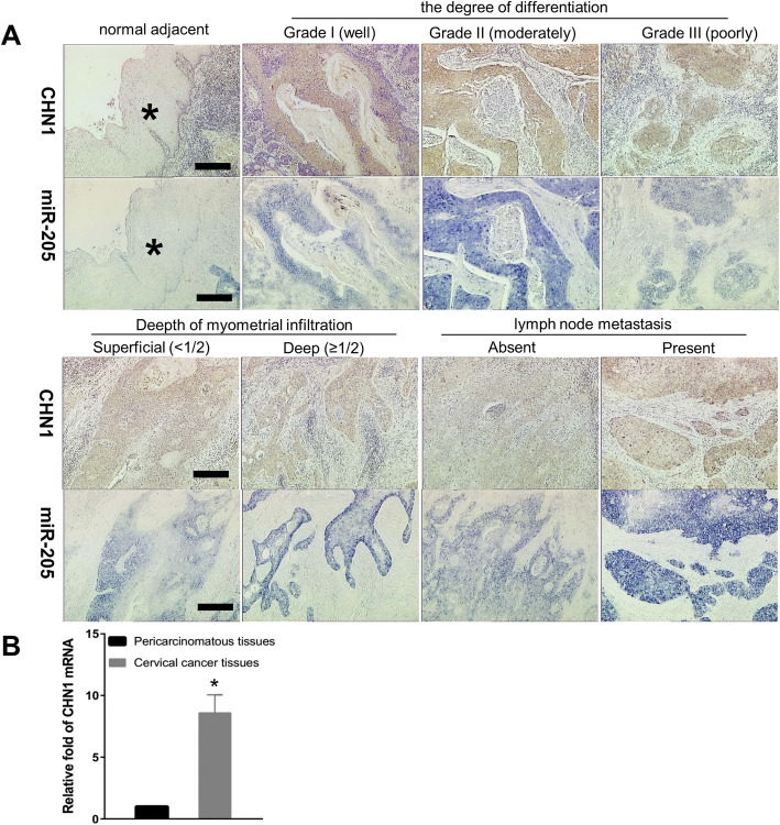
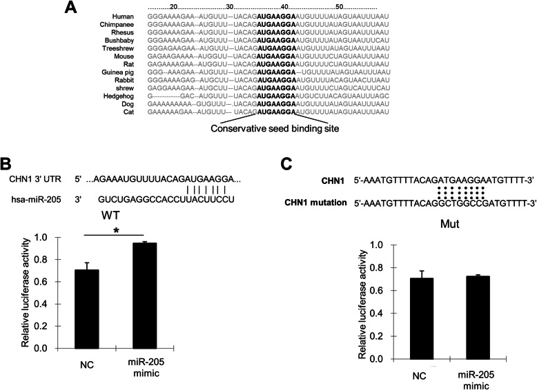
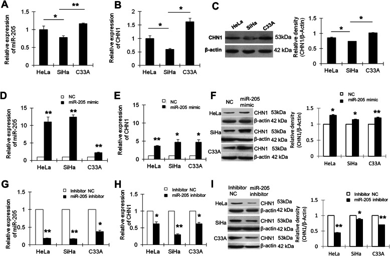
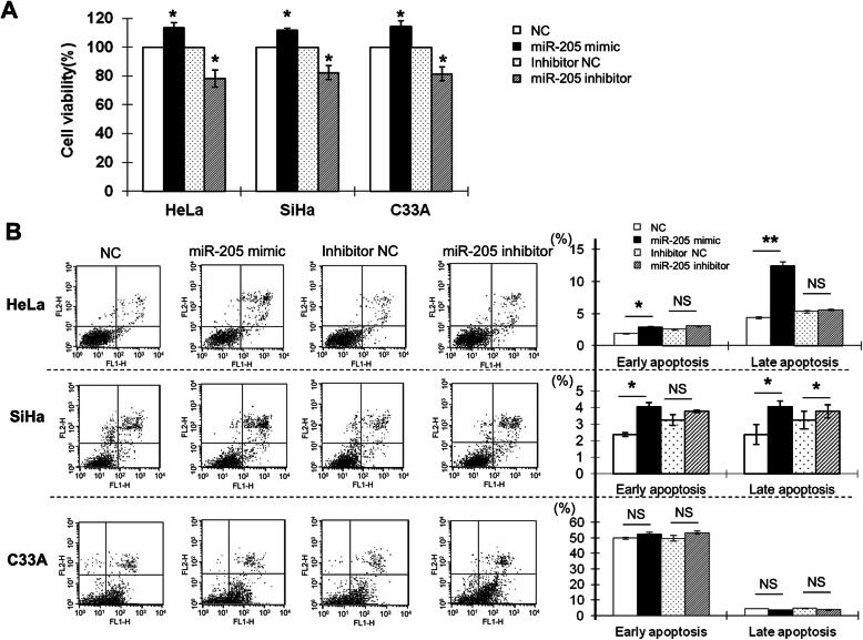
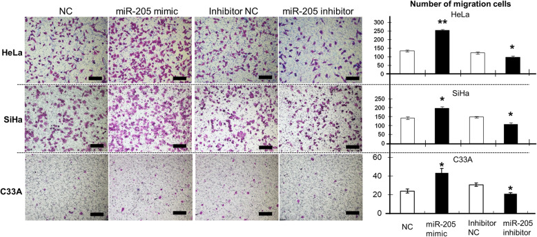
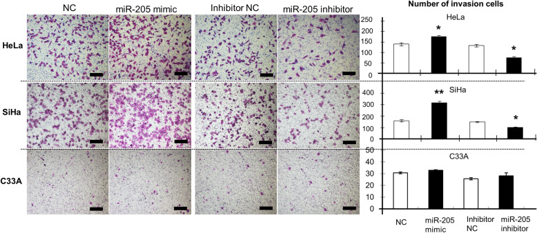
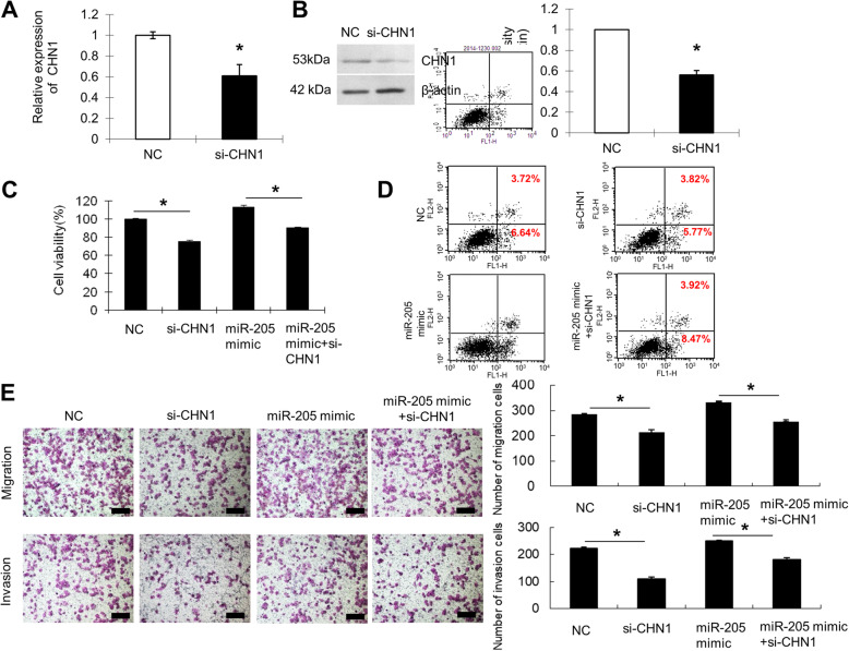
Results: We found that miR-205 and CHN1 were highly expressed in human cervical cancer tissue compared with paired normal cervical tissues. The CHN1 gene was shown to be targeted by miR-205 in HeLa cells. Interestingly, transfection with miR-205 mimic upregulated CHN1 mRNA and protein, while miR-205 inhibitor downregulated CHN1 in high-risk and human papilloma virus (HPV)-negative human cervical cancer cells in vitro,. These data suggested that miR-205 positively regulated the expression of CHN1. Furthermore, the miR-205 mimic promoted cell growth, apoptosis, migration, and invasion in high-risk and HPV-negative cervical cancer cells, while the miR-205 inhibitor blocked these biological processes. Knockdown of CHN1 obviously reduced the aggressive cellular behaviours induced by upregulation of miR-205, suggesting that miR-205 positively regulated CHN1 to mediate these cell behaviours during the development of cervical cancer. Furthermore, CHN1 was correlated with lymph node metastasis in clinical specimens.

Conclusions: Our findings showed that miR-205 positively regulated CHN1 to mediate cell growth, apoptosis, migration, and invasion during cervical cancer development, particularly for high-risk HPV-type cervical cancer. These findings suggested that dysregulation of miR-205 and subsequent abnormalities in CHN1 expression promoted the oncogenic potential of human cervical cancer.

Keywords: Cancer gene; Cervical cancer; Invasion; Migration; a1-chimaerin; microRNA-205.


Conflict of interest statement

The authors declare that they have no competing interests.

Figures

Fig. 1
Fig. 1
The expression of miR-205 and CHN1 in cervical cancer. a:Distribution and expression of miR-205 and CHN1 protein in human cervical tissues. The expression and localisation of miR-205 in human cervical cancer tissues and adjacent normal cervical tissues was determined using in situ hybridisation. The stain was developed with BCIP/NBT. The expression and localisation of CHN1 protein in human cervical cancer tissues and adjacent normal cervical tissues was analysed by immunohistochemistry. The stain was developed with DAB, and nuclei were stained with hematoxylin. *Normal cervical epithelial tissue adjacent to carcinoma. Bar = 200 μm. b: The expression of CHN1 mRNA in the cervical cancer tissues and the para-carcinoma tissues was detected by qRT-PCR. GAPDH served as an internal reference gene. *P < 0.05
Fig. 2
Fig. 2
Prediction and detection of miR-205 targets. a miR-205 binding sites in the 3′UTR of CHN1 in cross-species. b Confirmation of the miR-205 target gene. HeLa cells were cotransfected with negative control (NC), miR-205 mimic, and pmirGLO-CHN1-wt for dual luciferase assays. PRL-TK, containing Renilla luciferase, was cotransfected with the 3′UTR of CHN1 for data normalisation. c Mutation of the miR-205 binding site. HeLa cells were cotransfected with NC, miR-205 mimic, and pmirGLO-CHN1-mt for dual luciferase assays. *P < 0.05
Fig. 3
Fig. 3
Confirmation of the relationship between miR-205 and CHN1. a The expression of miR-205 in HeLa, SiHa, and C33A cells was detected by qRT-PCR. U6 served as an internal reference and was used to normalise miR-205 expression. The y-axis displays the relative expression of miR-205 normalised to the expression of U6. b The expression of CHN1 mRNA in HeLa, SiHa, and C33A cells was detected by qRT-PCR. GAPDH served as an internal reference gene. c CHN1 protein expression was detected by western blotting. β-Actin was used as a loading control. The black histogram shows the optical densities of the signals quantified by densitometric analysis and represented as the CHN1 intensity/β-Actin intensity for normalisation of gel loading and transfer. d HeLa, SiHa, and C33A cells were transfected with NC or miR-205 mimic. The expression of miR-205 was detected by qRT-PCR. e The level of CHN1 mRNA was detected by qRT-PCR. GAPDH served as an internal reference gene. f CHN1 protein expression was detected by western blotting. β-Actin was used as a loading control. g HeLa, SiHa, and C33A cells were transfected with inhibitor NC or miR-205 inhibitor. The expression of miR-205 was detected by qRT-PCR. h CHN1 mRNA expression was detected by qRT-PCR after transfection of cells with inhibitor NC or miR-205 inhibitor. GAPDH served as an internal reference gene. i CHN1 protein expression was detected by western blotting after transfection of cells with inhibitor NC or miR-205 inhibitor. β-Actin was used as a loading control. *P < 0.05, **P < 0.01. The cropping of the blot was done. Full-length uncropped blots are presented in Supplementary Figure 1, which all the samples derived from the same experiment and blots were processed in parallel
Fig. 4
Fig. 4
The effects of miR-205 on the proliferation and apoptosis of human cervical cancer cells. a HeLa, SiHa, and C33A cells were transfected with the NC, miR-205 mimic, inhibitor NC, or miR-205 inhibitor. At 48 h after transfection, cell proliferation was determined by CCK-8 assay. All experiments were performed at least three times, and cell proliferation was determined as the stimulation index (SI; i.e., the ratio of absorbance at 450 nm of cells transfected with miR-205 mimic or inhibitor to that of cells transfected with NC or inhibitor NC). b HeLa, SiHa, and C33A cells were transfected with the NC, miR-205 mimic, inhibitor NC, or miR-205 inhibitor for 48 h. Cells were then stained with annexin V/PI and subjected to flow cytometry analysis. Lower left quadrant, viable cells (annexin V-FITC and PI negative); lower right quadrant, early apoptotic cells (annexin V-FITC positive and PI negative); upper right quadrant, late apoptotic/necrotic cells (annexin V-FITC and PI positive). The average percentage of apoptotic cells was analysed in cells transfected with miR-205 mimic or inhibitor at early and late stages. The histograms represent the average percentages of apoptotic cells in cells transfected with miR-205 mimic at early and late stages or miR-205 inhibitor at early and late stages. The experiment was repeated at least three times. *P < 0.05, **P < 0.01, NS: not significant
Fig. 5
Fig. 5
The effects of miR-205 on the migration of human cervical cancer cells. HeLa, SiHa, and C33A cells were transfected with the NC, miR-205 mimic, inhibitor NC, or miR-205 inhibitor for 48 h. Cells were then subjected to migration assays as described in the Methods. Cell migration was quantified by counting the number of cells passing through the membrane from five different randomly selected fields of view per sample at 100× magnification. Representative images are shown. The histograms represent the number of migrated HeLa, SiHa, and C33A cells. Data are expressed as the means of independent triplicate experiments. Magnification, 100×; scale bar =200 μm. *P < 0.05, **P < 0.01
Fig. 6
Fig. 6
The effects of miR-205 on the invasion of human cervical cancer cells. Cell invasion was quantified by counting the number of cells passing through the membrane from five different randomly selected fields of view per sample at 100× magnification. Representative images are shown. The histograms represent the number of invaded HeLa, SiHa, and C33A cells. Data are expressed as the means of independent triplicate experiments. Scale bar =200 μm. *P < 0.05, **P < 0.01
Fig. 7
Fig. 7
Knockdown of CHN1 attenuated the miR-205-mediated enhancement of cell growth and metastasis. a SiHa cells were transfected with NC or si-CHN1. At 48 h after transfection, CHN1 mRNA expression was determined by qRT-PCR. GAPDH served as an internal reference gene. b CHN1 protein levels were determined by western blotting at 48 h after transfection. β-Actin was used as a loading control. The cropping of the blot was done. Full-length uncropped blots are presented in Supplementary Figure 2, which all the samples derived from the same experiment and blots were processed in parallel. c Cell proliferation was determined by CCK-8 assay at 48 h after transfection. SiHa cells were transfected with NC, si-CHN1, or miR-205 mimic or cotransfected with miR-205 mimic and si-CHN1. d Apoptosis was detected by flow cytometry. The percentages of early and late apoptotic cells (representative of three separate experiments) are shown in the lower right and upper right panels, respectively. e Cells (0.5 × 106 cells/mL) were subjected to migration or invasion assays as described in the methods. Magnification, 100×; scale bar =200 μm. *P < 0.05

References

    1. Siegel RL, Miller KD, Jemal A. Cancer statistics, 2019. CA Cancer J Clin. 2019;69(1):7–34. doi: 10.3322/caac.21551. - DOI - PubMed
    1. Pimple S, Mishra G, Shastri S. Global strategies for cervical cancer prevention. Curr Opin Obstet Gynecol. 2016;28(1):4–10. doi: 10.1097/GCO.0000000000000241. - DOI - PubMed
    1. Bartel D. MicroRNAs: genomics, biogenesis, mechanism, and function. Cell. 2004;116(2):281–297. doi: 10.1016/S0092-8674(04)00045-5. - DOI - PubMed
    1. Lewis B, Burge C, Bartel D. Conserved seed pairing, often flanked by adenosines, indicates that thousands of human genes are microRNA targets. Cell. 2005;120(1):15–20. doi: 10.1016/j.cell.2004.12.035. - DOI - PubMed
    1. Orang AV, Safaralizadeh R, Hosseinpour Feizi MA. Insights into the diverse roles of miR-205 in human cancers. Asian Pac J Cancer Prev. 2014;15(2):577–583. doi: 10.7314/APJCP.2014.15.2.577. - DOI - PubMed