. 2020 Oct 27;22(1):256.
doi: 10.1186/s13075-020-02325-6.

Mesenchymal stem cell-derived exosomal microRNA-136-5p inhibits chondrocyte degeneration in traumatic osteoarthritis by targeting ELF3

Affiliations

Mesenchymal stem cell-derived exosomal microRNA-136-5p inhibits chondrocyte degeneration in traumatic osteoarthritis by targeting ELF3

Xue Chen et al. Arthritis Res Ther. .

Abstract

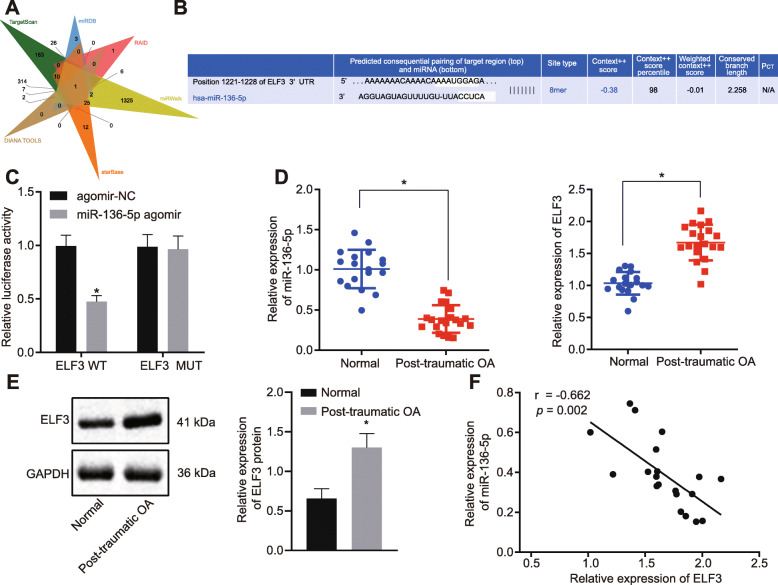
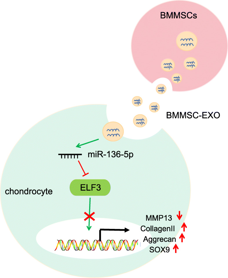
Background: Emerging evidence suggests that microRNAs (miRs) are associated with the progression of osteoarthritis (OA). In this study, the role of exosomal miR-136-5p derived from mesenchymal stem cells (MSCs) in OA progression is investigated and the potential therapeutic mechanism explored.

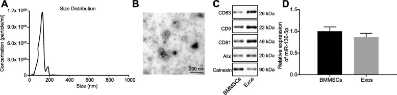
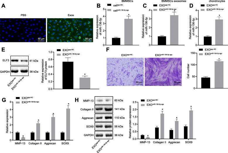
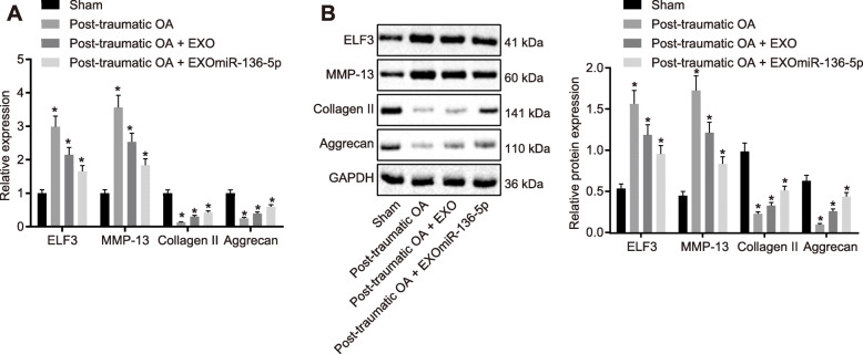
Methods: Bone marrow mesenchymal stem cells (BMMSCs) and their exosomes were isolated from patients and identified. The endocytosis of chondrocytes and the effects of exosome miR-136-5p on cartilage degradation were observed and examined by immunofluorescence and cartilage staining. Then, the targeting relationship between miR-136-5p and E74-like factor 3 (ELF3) was analyzed by dual-luciferase report assay. Based on gain- or loss-of-function experiments, the effects of exosomes and exosomal miR-136-5p on chondrocyte migration were examined by EdU and Transwell assay. Finally, a mouse model of post-traumatic OA was developed to evaluate effects of miR-136-5p on chondrocyte degeneration in vivo.

Results: In the clinical samples of traumatic OA cartilage tissues, we detected increased ELF3 expression, and reduced miR-136-5p expression was determined. The BMMSC-derived exosomes showed an enriched level of miR-136-5p, which could be internalized by chondrocytes. The migration of chondrocyte was promoted by miR-136-5p, while collagen II, aggrecan, and SOX9 expression was increased and MMP-13 expression was reduced. miR-136-5p was verified to target ELF3 and could downregulate its expression. Moreover, the expression of ELF3 was reduced in chondrocytes after internalization of exosomes. In the mouse model of post-traumatic OA, exosomal miR-136-5p was found to reduce the degeneration of cartilage extracellular matrix.

Conclusion: These data provide evidence that BMMSC-derived exosomal miR-136-5p could promote chondrocyte migration in vitro and inhibit cartilage degeneration in vivo, thereby inhibiting OA pathology, which highlighted the transfer of exosomal miR-136-5p as a promising therapeutic strategy for patients with OA.

Keywords: Bone marrow mesenchymal stem cells; Chondrocyte degeneration; ELF3; Exosome; Extracellular matrix secretion; Migration; Traumatic osteoarthritis; microRNA-136-5p.


Conflict of interest statement

None

Figures

Fig. 1
Fig. 1
Overexpression of ELF3 and underexpression of miR-136-5p were observed in cartilage tissues of traumatic OA. a The Venn map of the upstream miR of the ELF3 by database search via in TargetScan, miRDB, RAID, miRWalk, starBase, and DIANA TOOLS. The only intersected miR was miR-136-5p. b Binding sites of miR-136-5p and ELF3 in TargetScan. c Dual-luciferase reporter assay of the miR-136-5p binding to ELF3. d The expression of miR-136-5p and ELF3 in normal cartilage tissues and traumatic OA cartilage tissues examined by RT-qPCR. e The expression of ELF3 in normal cartilage tissues and traumatic OA cartilage tissues examined by Western blot. f Pearson’s correlation analysis of the relationship between miR-136-5p and ELF3. Asterisk symbol indicated comparison with the normal group or the cells transfected with agomir-NC. A p < 0.05 was considered to be statistically significant. The above data were measurement data and expressed as the mean ± standard deviation. The independent sample t test was used for comparison between the two groups
Fig. 2
Fig. 2
Identification of BMMSCs and exosomes. a DLS measuring the particle size and distribution of exosomes. b The morphology of exosomes (× 100,000) observed using a transmission electron microscope. c Western blot analysis of exosomes surface markers (CD63, CD9, CD81, and Alix) and endogenous proteins (TSG101). d The expression of miR-136-5p in BMMSCs and their secreted exosomes examined by RT-qPCR. The above data were measurement data and expressed as the mean ± standard deviation. The independent sample t test was used for comparison between the two groups
Fig. 3
Fig. 3
The migration and ECM secretion of chondrocytes were promoted by miR-136-5p through ELF3. a The expression of miR-136-5p in transfected chondrocytes examined by RT-qPCR. b Migration analysis of chondrocytes (× 200). c The mRNA expression of MMP-13, collagen II, aggrecan, and SOX9 in chondrocytes examined by RT-qPCR. d The protein expression of MMP-13, collagen II, aggrecan, and SOX9 in chondrocytes examined by Western blot. Asterisk symbol indicated p < 0.05 compared with the cells transfected with agomir-NC. Number sign indicated p < 0.05 compared with the cells transfected with antagomir-NC. Ampersand symbol indicated p < 0.05 compared with the cells transfected with miR-136-5p agomir. p < 0.05 indicated a significant difference. The above data were measurement data and expressed as the mean ± standard deviation. The data among groups were compared using one-way ANOVA: followed by Tukey’s post hoc test
Fig. 4
Fig. 4
The migration and ECM secretion of chondrocytes were promoted by miR-136-5p-exo. a A typical immunofluorescence pattern of DIO (green)-labeled exosomes absorbed by chondrocytes, whose nuclei were stained with DAPI (blue) (× 200). b The expression of miR-136-5p in transfected BMMSCs examined by RT-qPCR. c The expression of miR-136-5p in exosomes secreted from transfected BMMSCs examined by RT-qPCR. d The expression of miR-136-5p in chondrocytes after exosome treatment examined by RT-qPCR. e Western blot analysis of the expression of ELF3 in chondrocytes treated with exosomes. f Migration analysis of chondrocytes (× 200). g The mRNA expressions of MMP-13, collagen II, aggrecan, and SOX9 in chondrocytes examined by RT-qPCR. h Western blot analysis of the protein expressions of MMP-13, collagen II, aggrecan, and SOX9 in chondrocytes. Asterisk symbol indicated comparison with the EXOago-NC or cellago-NC group. A value of p < 0.05 was considered to be statistically significant. The above data were measurement data and expressed as mean ± standard deviation. The independent sample t test was used for comparison between the two groups. One-way ANOVA was used among multiple groups, followed by Tukey’s post hoc test
Fig. 5
Fig. 5
EXOmiR-136-5p inhibited cartilage degradation. a RT-qPCR examining the expressions of ELF3, collagen II, aggrecan, and MMP-13 in cartilage tissues of mice in each group. b Western blot examination of the expressions of ELF3, collagen II, aggrecan, and MMP-13 in cartilage tissues of each group. Asterisk symbol indicated comparison with the mice received sham operation. A value of p < 0.05 was considered to be statistically significant. The above data were measurement data and expressed as mean ± standard deviation. Data comparison among groups was performed using one-way ANOVA, followed by Tukey’s post hoc test
Fig. 6
Fig. 6
Schematic diagram illustrating that BMMSCs exosomal miR-136-5p could promote chondrocyte migration and ECM secretion by targeting ELF3, thereby inhibiting cartilage degradation in traumatic OA

References

    1. Martel-Pelletier J, Barr AJ, Cicuttini FM, Conaghan PG, Cooper C, Goldring MB, et al. Osteoarthritis. Nat Rev Dis Primers. 2016;2:16072. doi: 10.1038/nrdp.2016.72. - DOI - PubMed
    1. Hosseinzadeh A, Kamrava SK, Joghataei MT, Darabi R, Shakeri-Zadeh A, Shahriari M, et al. Apoptosis signaling pathways in osteoarthritis and possible protective role of melatonin. J Pineal Res. 2016;61:411–425. doi: 10.1111/jpi.12362. - DOI - PubMed
    1. Fang H, Beier F. Mouse models of osteoarthritis: modelling risk factors and assessing outcomes. Nat Rev Rheumatol. 2014;10:413–421. doi: 10.1038/nrrheum.2014.46. - DOI - PubMed
    1. Kolasinski SL, Neogi T, Hochberg MC, Oatis C, Guyatt G, Block J, et al. 2019 American College of Rheumatology/Arthritis Foundation Guideline for the Management of Osteoarthritis of the Hand, Hip, and Knee. Arthritis Rheumatol. 2020;72:220–233. doi: 10.1002/art.41142. - DOI - PMC - PubMed
    1. Sacitharan PK. Ageing and osteoarthritis. Subcell Biochem. 2019;91:123–159. doi: 10.1007/978-981-13-3681-2_6. - DOI - PubMed