. 2020 Oct 27;10(1):18400.
doi: 10.1038/s41598-020-75376-z.

Modulation of complement activation by pentraxin-3 in prostate cancer

Affiliations

Modulation of complement activation by pentraxin-3 in prostate cancer

Giovanni Stallone et al. Sci Rep. .

Abstract

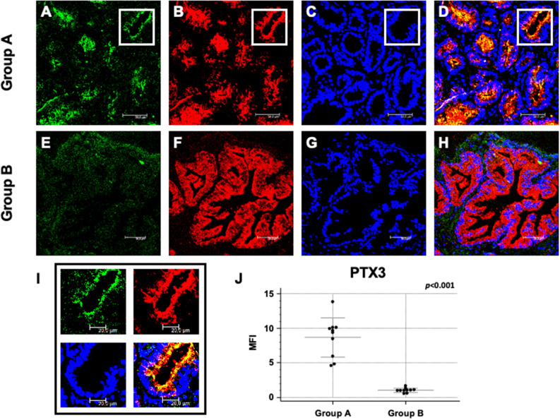
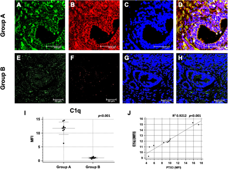
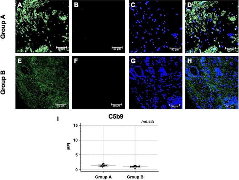
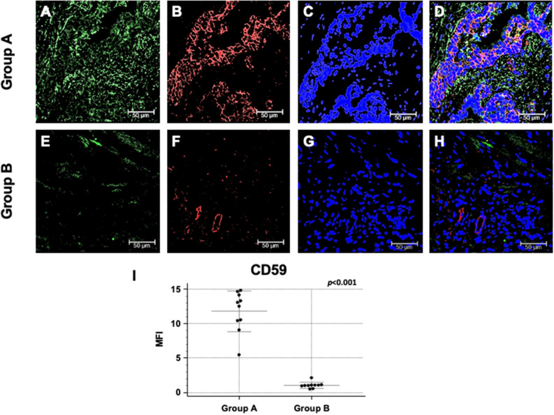
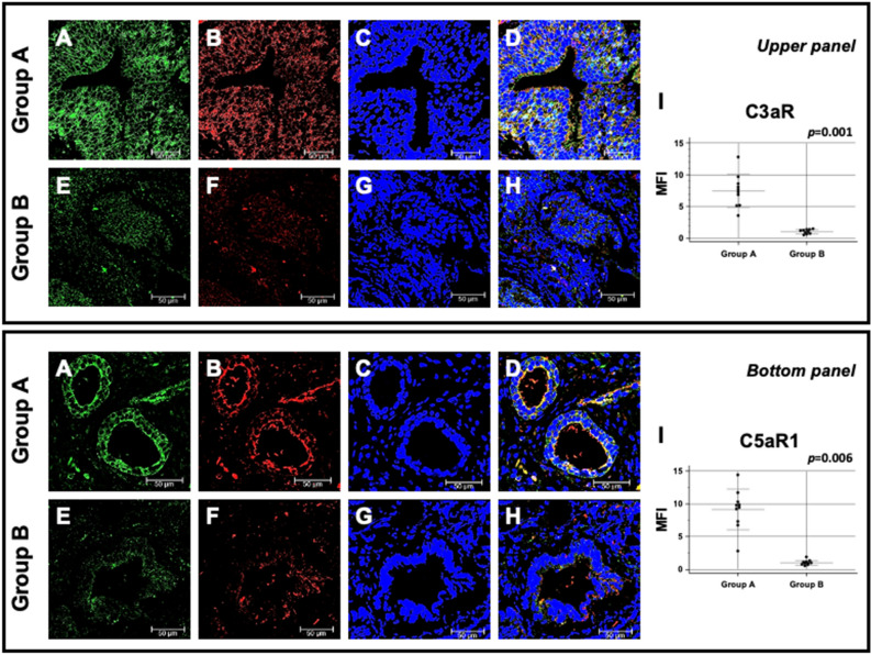
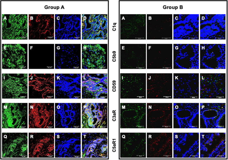
Pentraxin 3 (PTX3) is an essential component of the innate immune system and a recognized modulator of Complement cascade. The role of Complement system in the pathogenesis of prostate cancer has been largely underestimated. The aim of our study was to investigate the role of PTX3 as possible modulator of Complement activation in the development of this neoplasia. We performed a single center cohort study; from January 2017 through December 2018, serum and prostate tissue samples were obtained from 620 patients undergoing prostate biopsy. A group of patients with benign prostatic hyperplasia (BPH) underwent a second biopsy within 12-36 months demonstrating the presence of a prostate cancer (Group A, n = 40) or confirming the diagnosis of BPH (Group B, N = 40). We measured tissue PTX3 protein expression together with complement activation by confocal microscopy in the first and second biopsy in group A and B patients. We confirmed that that PTX3 tissue expression in the first biopsy was increased in group A compared to group B patients. C1q deposits were extensively present in group A patients co-localizing and significantly correlating with PTX3 deposits; on the contrary, C1q/PTX3 deposits were negative in group B. Moreover, we found a significantly increased expression of C3a and C5a receptors within resident cells in group A patient. Interestingly, C1q/PTX3 deposits were not associated with activation of the terminal Complement complex C5b-9; moreover, we found a significant increase of Complement inhibitor CD59 in cancer tissue. Our data indicate that PTX3 might play a significant pathogenic role in the development of this neoplasia through recruitment of the early components of Complement cascade with hampered activation of terminal Complement pathway associated with the upregulation of CD59. This alteration might lead to the PTX3-mediated promotion of cellular proliferation, angiogenesis and insensitivity to apoptosis possible leading to cancer cell invasion and migration.


Conflict of interest statement

The authors declare that the research was conducted in the absence of any commercial or financial relationships that could be construed as a potential conflict of interest.

Figures

Figure 1
Figure 1
PTX3 protein expression and co-localization with PSA in first PBx of patients subsequently developing prostate cancer (group A) or not (group B). PTX3 (green; A and E) and PSA (red; B and F) protein expression were evaluated in the first PBx by double label immunofluorescence and confocal microscopy as detailed in Materials and Methods. TO-PRO-3 was used to stain nuclei (blue; C and G). Merged images (D and H) demonstrate the co-localization of the two protein signals (yellow). Mean fluorescence intensity (MFI) quantification confirmed a statistically significant higher PTX3 expression in group A compared to group B (I, J). Bar length = 50 µ.
Figure 2
Figure 2
Complement factor C1q deposition and co-localization with PTX3 in first PBx of patients subsequently developing prostate cancer (group A) or not (group B). PTX3 protein expression (green; A and E) and C1q deposition (red; B and F) were evaluated in the first PBx by double label immunofluorescence and confocal microscopy as detailed in Materials and Methods. TO-PRO-3 was used to stain nuclei (blue; C and G). Merged images (D and H) demonstrate the co-localization of the two protein signals (yellow). Mean fluorescence intensity (MFI) quantification confirmed a statistically significant higher C1q deposition in group A compared to group B (I). Bar length = 50 µ; (J) significant correlation of PTX3 and C1q deposits in group A.
Figure 3
Figure 3
Terminal Complement complex, C5b-9, deposition and co-localization with PTX3 in first PBx of patients subsequently developing prostate cancer (group A) or not (group B). PTX3 protein expression (green; A and E) and C5b-9 deposition (red; B and F) were evaluated in the first PBx by double label immunofluorescence and confocal microscopy as detailed in Materials and Methods. TO-PRO-3 was used to stain nuclei (blue; C and G). Merged images (D and H) do not demonstrate any co-localization of the two protein signals. Mean fluorescence intensity (MFI) quantification confirmed the absence of C5b-9 deposition in both groups (I). Bar length = 50 µ.
Figure 4
Figure 4
CD59 protein expression and co-localization with PTX3 in first PBx of patients subsequently developing prostate cancer (group A) or not (group B). Protein expression of PTX3 (green; A and E) and CD59 (red; B and F) were evaluated in the first PBx by double label immunofluorescence and confocal microscopy as detailed in Materials and Methods. TO-PRO-3 was used to stain nuclei (blue; C and G). Merged images (D and H) demonstrate the co-localization of the two protein signals (yellow). Mean fluorescence intensity (MFI) quantification confirmed a statistically significant higher CD59 expression in group A compared to group B (I). Bar length = 50 µ.
Figure 5
Figure 5
C3a and C5a receptors protein expression and co-localization with PTX3 in first PBx of patients subsequently developing prostate cancer (group A) or not (group B). UPPER PANEL: Protein expression of PTX3 (green; A and E) and C3a receptor (red; B and F) were evaluated in the first PBx by double label immunofluorescence and confocal microscopy as detailed in Materials and Methods. TO-PRO-3 was used to stain nuclei (blue; C and G). Merged images (D and H) demonstrate the co-localization of the two protein signals (yellow). Mean fluorescence intensity (MFI) quantification confirmed a statistically significant higher C3a receptor expression in group A compared to group B (I). Bar length = 50 µ. BOTTOM PANEL: Protein expression of PTX3 (green; A and E) and C5aR1 (red; B and F) were evaluated in the first PBx by double label immunofluorescence and confocal microscopy. TO-PRO-3 was used to stain nuclei (blue; C and G). Merged images (D and H) demonstrate the co-localization of the two protein signals (yellow). Mean fluorescence intensity (MFI) quantification confirmed a statistically significant higher C5a receptor expression in group A compared to group B (I). Bar length = 50 µ.
Figure 6
Figure 6
C1q and C5b-9 deposition and CD59, C3a and C5a receptor expression and their co-localization with PTX3 in second biopsy of patients who developed prostate cancer (group A) or not (group B). PTX3 (A, E, I, M, Q), C1q (B), C5b-9 (F), CD59 (J), C3a (N) and C5a receptor (R) proteins were investigated by double-label immunofluorescence and confocal microscopy as detailed in Materials and Methods. TO-PRO-3 was employed to counterstain nuclei (blue; C, G, K, O, S). Merged images (yellow; D, H, L, P, T) demonstrated the co-localization (green) of C1q (D), CD59 (L), C3a (P) and C5aR1 (T) with PTX3, whereas no co-localization was evident for C5b-9 (H). Bar length = 50 µ.

References

    1. Jemal A, Siegel R, Xu J, Ward E. Cancer statistics, 2010. CA. Cancer J. Clin. 2010;60:277–300. doi: 10.3322/caac.20073. - DOI - PubMed
    1. De Marzo AM, et al. Inflammation in prostate carcinogenesis. Nat. Rev. Cancer. 2007;7:256–269. doi: 10.1038/nrc2090. - DOI - PMC - PubMed
    1. Garlanda C, Bottazzi B, Bastone A, Mantovani A. Pentraxins at the crossroads between innate immunity, inflammation, matrix deposition, and female fertility. Annu. Rev. Immunol. 2005;23:337–366. doi: 10.1146/annurev.immunol.23.021704.115756. - DOI - PubMed
    1. Mantovani A, et al. The long pentraxin PTX3 in vascular pathology. Vascul. Pharmacol. 2006;45:326–330. doi: 10.1016/j.vph.2006.08.011. - DOI - PubMed
    1. Baruah P, et al. The tissue pentraxin PTX3 limits C1q-mediated complement activation and phagocytosis of apoptotic cells by dendritic cells. J. Leukoc. Biol. 2006;80:87–95. doi: 10.1189/jlb.0805445. - DOI - PubMed